. 2019 Dec 2;216(12):2714-2723.
doi: 10.1084/jem.20180610. Epub 2019 Oct 3.

ILC2s mediate systemic innate protection by priming mucus production at distal mucosal sites

Affiliations

ILC2s mediate systemic innate protection by priming mucus production at distal mucosal sites

Laura Campbell et al. J Exp Med. .

Abstract

Host immunity to parasitic nematodes requires the generation of a robust type 2 cytokine response, characterized by the production of interleukin 13 (IL-13), which drives expulsion. Here, we show that infection with helminths in the intestine also induces an ILC2-driven, IL-13-dependent goblet cell hyperplasia and increased production of mucins (Muc5b and Muc5ac) at distal sites, including the lungs and other mucosal barrier sites. Critically, we show that type 2 priming of lung tissue through increased mucin production inhibits the progression of a subsequent lung migratory helminth infection and limits its transit through the airways. These data show that infection by gastrointestinal-dwelling helminths induces a systemic innate mucin response that primes peripheral barrier sites for protection against subsequent secondary helminth infections. These data suggest that innate-driven priming of mucus barriers may have evolved to protect from subsequent infections with multiple helminth species, which occur naturally in endemic areas.


Figures

Figure 1.
Figure 1.
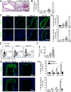
Intestinal T. spiralis infection causes IL-13–mediated goblet cell hyperplasia distally in the lung. (A) Representative PAS staining of lung tissue sections from T. spiralis–infected mice (bar, 100 µm). (B) Quantification of PAS staining over time course of infection. PAS-stained representative sections of five mice/group were analyzed. (C) Representative lung sections from T. spiralis–infected mice stained for either Muc5b (top panel) or Muc5ac (bottom panel; bars, 100 µm). (D) Quantification of Muc5b- and Muc5ac-stained lung sections (n = 5 mice/group). (E) Flow cytometry analysis of lung IL-13eGFP+ cells from infected mice. Representative dot plots are shown (pregate: singlet, live, CD45+ cells). SSC, side scatter. (F) Quantification of total lung IL-13eGFP+ cells (n = 6 mice/group). Naive and T. spiralis–infected mice were injected i.p. with either anti–IL-13 or isotype control (from day −1 and every 2 d until collection; 200 µg/mouse). (G) Representative day 18 p.i. lung sections from anti–IL-13 and isotype-treated mice stained for either Muc5b (top panel) or Muc5ac (bottom panel; bars, 100 µm). (H) Quantification of Muc5b- and Muc5ac-stained lung sections (n = 3 mice/group). Data are representative of two (H) or three (B, D, and F) independent experiments. Error bars indicate the mean ± SEM. Comparisons to either naive group (B, D, and F) or isotype control (H) were calculated using unpaired Student’s t tests. *, P ≤ 0.05; **, P ≤ 0.01; ***, P ≤ 0.001; ****, P ≤ 0.0001.
Figure 2.
Figure 2.
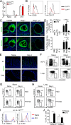
Inflammatory ILC2-derived IL-13 is sufficient to drive lung goblet cell hyperplasia. (A) Analysis of lung-derived IL-13eGFP+ cells from naive and T. spiralis–infected mice. Cell subsets determined by lineage expression (LINneg: CD3, TCRβ, TCRγδ, CD49b, CD19, B220, CD11b, CD11c, TER119, GR1, FcεR1α). (B) Expression of CD25 and CD127 on lung IL-13eGFP+ cells from day 10 T. spiralis–infected mice in the Linneg subset (pregate: singlet, live, CD45+, IL-13eGFP+ cells; n = 6 mice/group). (C and D) Representative lung sections from day 10–infected WT, Rag2−/−, and Rag2−/− γc−/− mice (C) stained for either Muc5b (top panel) or Muc5ac (bottom panel; bars, 200 µm) and corresponding quantification of Muc5b- and Muc5ac-stained lung sections (D; n = 3 mice/group). (E) Representative small intestine (SI) and lung sections from naive and day 4–infected mice stained with DCLK1 (bars, 200 µm). (F) Cells from the small intestinal lamina propria (siLPL) and lung of naive and day 4–infected C57/BL6 mice were analyzed by flow cytometry for iILC2 markers (gated as Lin CD127+ CD90.2lo KLRG1hi). (G and H) C57/BL6 mice were injected i.p. with either FTY720 or H2O (daily from day −1; 1 mg/kg). Cells from the blood (G) and lung (H) of naive and day 4–infected mice were analyzed by flow cytometry for iILC2 markers (gated as Lin CD127+ CD90.2lo KLRG1hi). (I) Expression of CD90.2 and KLRG1 on Linneg IL-13eGFP+ cells from naive (red line) and day 10–infected (blue line) lungs (n = 5 mice/group). (J) Frequency of IL-13eGFP+ cells expressing either low or high levels of CD90.2 from naive and day 10–infected lungs (n = 5 mice/group). Data are representative of two (D, F, G, and H) or four (A, B, and J) independent experiments. Error bars indicate the mean ± SEM. Comparisons to naive group were calculated using unpaired Student’s t tests. **, P ≤ 0.01; ***, P ≤ 0.001; ****, P ≤ 0.0001.
Figure 3.
Figure 3.
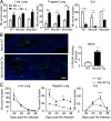
Systemic priming by T. spiralis infection impedes a subsequent Nb infection in the lung. (A) Representative lung sections from day 22 after T. spiralis infection, day 2 after Nb infection, and coinfected mice stained for either Muc5b (top panel) or Muc5ac (bottom panel; bars, 100 µm). (B) Quantification of Muc5b- and Muc5ac-stained lung sections from Nb-infected C57/BL6 mice. Naive, day 22 after T. spiralis (Ts), day 2 after Nb and coinfected (Ts + Nb; n = 3 mice/group). (C–E) Naive (solid line) or day 20 after T. spiralis–infected (dashed line) C57/BL6 mice were i.v. infected with Nb. Total Nb worm numbers were collected from lung tissue (C), collagenase-digested lung tissue (D), and small intestine (E; n = 3 mice/group). Data are representative of three independent experiments. Error bars indicate the mean ± SEM. Comparisons between groups were calculated using either two-way (A) or one-way (B) ANOVA and Sidak’s post-test. *, P ≤ 0.05; **, P ≤ 0.01; ****, P ≤ 0.0001.
Figure 4.
Figure 4.
Lung goblet cell hyperplasia protects against secondary helminth infection in the lung. (A) Naive (white bars) or day 20 after T. spiralis (Ts) infection (black bars) WT, Muc5b−/−, or Muc5ac−/− mice were i.v. infected with Nb. Total Nb worm numbers were collected at day 3 after Nb infection from lung tissue (left), collagenase-digested lung tissue (middle), and small intestine (right; n = 3 mice/group). (B) Representative lung sections from naive WT and Muc5b overexpressing (Muc5b-Tg) mice stained for Muc5b (bars, 400 µm) and corresponding quantification of staining (n = 3–4 mice/group). (C) WT and Muc5b-Tg mice were s.c. infected with Nb, and total Nb worm numbers were collected from lung tissue (left), collagenase-digested lung tissue (middle), and small intestine (right; n = 3–4 mice/group). Data are representative of two independent experiments. Error bars indicate the mean ± SEM. Comparisons between groups were calculated using either unpaired Student’s t tests (A and B) or two-way ANOVA (C) and Sidak’s post-test. *, P ≤ 0.05; **, P ≤ 0.01; ***, P ≤ 0.001; ****, P ≤ 0.0001.

References

    1. Ahn S.J., Ryoo N.-K., and Woo S.J.. 2014. Ocular toxocariasis: clinical features, diagnosis, treatment, and prevention. Asia Pac. Allergy. 4:134–141. 10.5415/apallergy.2014.4.3.134 - DOI - PMC - PubMed
    1. Barlow J.L., Bellosi A., Hardman C.S., Drynan L.F., Wong S.H., Cruickshank J.P., and McKenzie A.N.. 2012. Innate IL-13-producing nuocytes arise during allergic lung inflammation and contribute to airways hyperreactivity. J. Allergy Clin. Immunol. 129:191–8.e1: 4. 10.1016/j.jaci.2011.09.041 - DOI - PubMed
    1. Behnke J.M., Wahid F.N., Grencis R.K., Else K.J., Ben-Smith A.W., and Goyal P.K.. 1993. Immunological relationships during primary infection with Heligmosomoides polygyrus (Nematospiroides dubius): downregulation of specific cytokine secretion (IL-9 and IL-10) correlates with poor mastocytosis and chronic survival of adult worms. Parasite Immunol. 15:415–421. 10.1111/j.1365-3024.1993.tb00626.x - DOI - PubMed
    1. Bouchery T., Kyle R., Camberis M., Shepherd A., Filbey K., Smith A., Harvie M., Painter G., Johnston K., Ferguson P., et al. . 2015. ILC2s and T cells cooperate to ensure maintenance of M2 macrophages for lung immunity against hookworms. Nat. Commun. 6:6970 10.1038/ncomms7970 - DOI - PubMed
    1. Camberis M., Le Gros G., and Urban J.. 2003. Animal Model of Nippostrongylus brasiliensis and Heligmosomoides polygyrus. Curr. Protoc. Immunol. Chapter 19:Unit 19.12. - PubMed

Publication types

MeSH terms