Review
. 2019 Oct 3;132(19):jcs233338.
doi: 10.1242/jcs.233338.

Tissue-specific roles of p73 in development and homeostasis

Affiliations
Review

Tissue-specific roles of p73 in development and homeostasis

Alice Nemajerova et al. J Cell Sci. .

Abstract

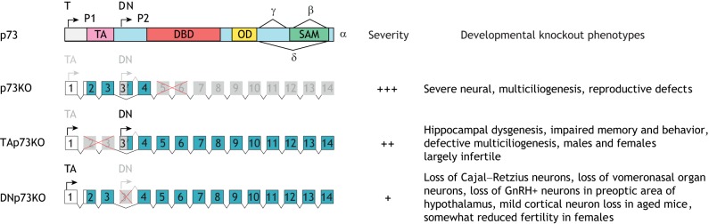
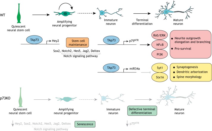
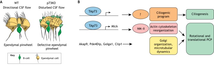
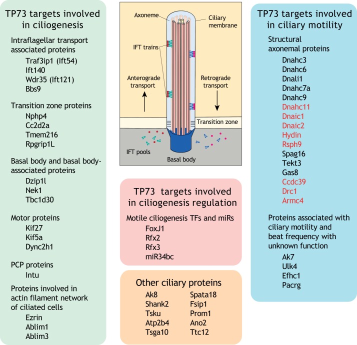
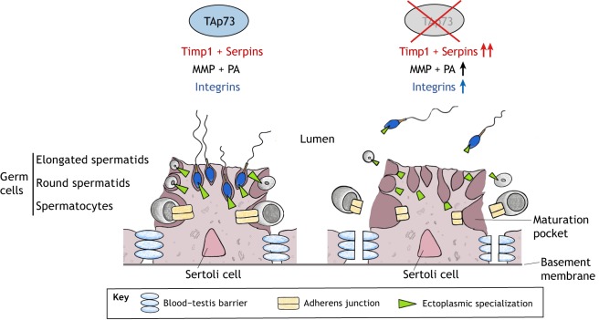
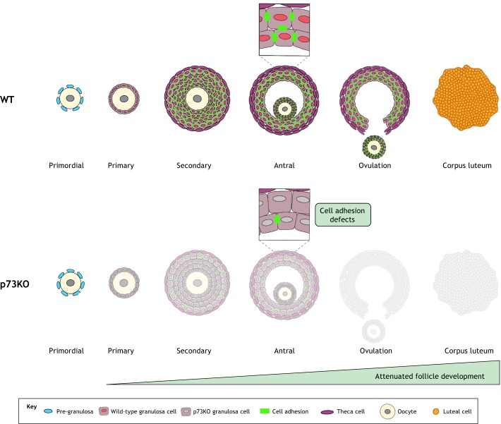
p73 (TP73) belongs to the p53 family of transcription factors. Its gene locus encodes two opposing types of isoforms, the transcriptionally active TAp73 class and the dominant-negative DNp73 class, which both play critical roles in development and homeostasis in an astonishingly diverse array of biological systems within specific tissues. While p73 has functions in cancer, this Review focuses on the non-oncogenic activities of p73. In the central and peripheral nervous system, both isoforms cooperate in complex ways to regulate neural stem cell survival, self-renewal and terminal differentiation. In airways, oviduct and to a lesser extent in brain ependyma, TAp73 is the master transcriptional regulator of multiciliogenesis, enabling fluid and germ cell transport across tissue surfaces. In male and female reproduction, TAp73 regulates gene networks that control cell-cell adhesion programs within germinal epithelium to enable germ cell maturation. Finally, p73 participates in the control of angiogenesis in development and cancer. While many open questions remain, we discuss here key findings that provide insight into the complex functions of this gene at the organismal, cellular and molecular level.

Keywords: TP73; Trp73; p73.


Conflict of interest statement

Competing interestsThe authors declare no competing or financial interests.

Figures

Fig. 1.
Fig. 1.
The p73 gene and its knockout phenotypes. The p73 locus encodes two classes of isoforms. The P1 promoter yields TAp73 variants containing the transactivation (TA) domain. The P2 promoter yields N-terminally truncated variants (deltaN, DNp73). DNA-binding domain (DBD), oligomerization domain (OD) and sterile alpha motif domain (SAM) are shared. C-terminal splice variants generate alternate β, γ and δ isoforms of the TA and DN classes. Global (p73KO) and isoform-specific knockout mice (TAp73KO and DNp73KO) were generated by deleting the indicated exons. They exhibit distinct developmental phenotypes that also differ in severity as discussed in the main text.
Fig. 2.
Fig. 2.
TAp73 plays multiple critical roles in the development of neuronal cells. TAp73 is required for embryonic and adult neural stem cell (NSC) maintenance through its role in the induction of Sox and Notch pathways. In particular, Hey2 is a direct transcriptional target of TAp73 (top). TAp73 also regulates terminal neuronal differentiation through direct induction of p75NTR and miR34a, which in turn signal via RAS–NFκB–PI3K and Syt1–Stx1A pathways, respectively. Loss of TAp73 results in marked reduction of NSCs (bottom). Furthermore, differentiation of mutant NSCs into immature neurons and oligodendrocytes is defective.
Fig. 3.
Fig. 3.
p73 regulates ependymal cell development, ciliogenesis and planar cell polarity. (A) Wild-type (WT) ependymal cells (ECs) form perinatally from GFAP-positive radial glial cells, which transform into a central stem cell (B-cell, remaining GFAP-positive) and surrounding, multi- and bi-ciliated ECs (becoming GFAP-negative). Together they form neuro-regenerative units along the ventricular walls, called pinwheels. p73-deficient ECs fail to organize into pinwheels, thereby disrupting subventricular zone niche architecture and function, and exhibiting defective ciliogenesis with disturbed cerebrospinal fluid (CSF) flow. Wild-type cells have cilia asymmetrically distributed at the anterior apical surface, and all cilia are polarized with the same orientation. p73KO cells have lost their polarization, with basal bodies distributed throughout the apical surface. (B) Other as-yet-undescribed direct TAp73 targets in ciliogenesis might also play a role in this process (indicated by the question mark). p73 is also essential for the polarized junctional assembly of PCP core proteins and the asymmetric localization of their downstream signaling effectors. Mechanistically, TAp73 regulates translational PCP (asymmetrical polarization of cilia clusters at the anterior apical surface) and actin dynamics through the direct transcriptional induction of myosin light-chain kinase (Mlck), which modulates non-muscle myosin-II (NM-II) activity, thereby affecting actin and microtubular transport dynamics, as well as Golgi organization.
Fig. 4.
Fig. 4.
TAp73 is the master transcriptional regulator of motile multiciliogenesis. Distinctive epithelia that line airways, brain ependyma, the female oviduct and the testicular efferent duct undergo motile multiciliogenesis by amplifying their centrioles to convert into basal bodies and then extend hundreds of motile cilia on their apical cell surface. Cilia beat vigorously and synchronously to generate directional fluid flow across tissue surfaces to move fluids. TAp73 occupies a position at the top of the gene network hierarchy of this program. It directly binds to promoter/enhancer regions of genes and activates the key ciliogenic regulators FoxJ1, Rfx2, Rfx3 and miR34bc, as well as nearly 50 structural and functional ciliary genes, eight of which are associated with known hereditary human primary ciliary dyskinesia syndromes (Hydin, Dnaic1, Drc1, Armc4, Dnahc11, Dnaic2, Ccdc39 and Rsph9, in red). The individual factors are grouped by functional class. Many of the ‘other ciliary proteins’ listed are also important for sperm development.
Fig. 5.
Fig. 5.
Role of TAp73 in germ cell maturation. Germ cells (gray cells) mature in pockets of somatic Sertoli cells (large pink cell). This requires the cyclical disassembly and reassembly of blood testis barrier (BTB) and Sertoli–germ cell junctions to allow the upward migration of germ cells on their maturational journey to the tubular lumen. TAp73 is exclusively produced in germ cells and controls a balanced transcriptional program that mediates adhesion to and disadhesion from Sertoli cell pockets, which includes peptidase inhibitors (Timps, Serpins), proteases (MMP, matrix metalloproteinases; PA, plasminogen activator-type serine proteases), receptors and integrins, thereby ensuring germ cell maturation. Upon TAp73 loss, there is broad dysregulation of these adhesion and migration effectors. This leads to junctional defects (adherens junctions, ectoplasmic specialization) and defective BTB with secondary degeneration of Sertoli cells. The end result is a massive loss of immature germ cells (spermatocytes, round and elongated spermatids) and mature spermatozoa, causing impaired fertility.
Fig. 6.
Fig. 6.
Role of TAp73 in follicle development. TAp73 is required for ovarian follicle development and corpus luteum formation. TAp73 is expressed in granulosa cells and regulates a p73-dependent adhesion gene network from cell-to-cell and from cell-to-surrounding basal lamina (focimatrix) that ensures follicle maturation. While primordial follicle numbers are intact in p73KO ovaries, there is attenuated follicle development with a marked decrease in the number of maturing follicles and a >90% decrease in the number of corpora lutea, i.e. residual follicles post-ovum release, which produce progesterone.

References

    1. Agostini M., Tucci P., Chen H., Knight R. A., Bano D., Nicotera P., McKeon F. and Melino G. (2010). p73 regulates maintenance of neural stem cell. Biochem. Biophys. Res. Commun. 403, 13-17. 10.1016/j.bbrc.2010.10.087 - DOI - PMC - PubMed
    1. Agostini M., Niklison-Chirou M. V., Catani M. V., Knight R. A., Melino G. and Rufini A. (2014). TAp73 promotes anti-senescence-anabolism not proliferation. Aging (Albany NY) 6, 921-930. 10.18632/aging.100701 - DOI - PMC - PubMed
    1. Agostini M., Tucci P., Killick R., Candi E., Sayan B. S., Rivetti di Val Cervo P., Nicotera P., McKeon F., Knight R. A., Mak T. W. et al. (2011a). Neuronal differentiation by TAp73 is mediated by microRNA-34a regulation of synaptic protein targets. Proc. Natl. Acad. Sci. USA 108, 21093-21098. 10.1073/pnas.1112061109 - DOI - PMC - PubMed
    1. Agostini M., Tucci P., Steinert J. R., Shalom-Feuerstein R., Rouleau M., Aberdam D., Forsythe I. D., Young K. W., Ventura A., Concepcion C. P. et al. (2011b). microRNA-34a regulates neurite outgrowth, spinal morphology, and function. Proc. Natl. Acad. Sci. USA 108, 21099-21104. 10.1073/pnas.1112063108 - DOI - PMC - PubMed
    1. Alexandrova E. M. and Moll U. M. (2012). Role of p53 family members p73 and p63 in human hematological malignancies. Leuk. Lymphoma 53, 2116-2129. 10.3109/10428194.2012.684348 - DOI - PubMed

Publication types

MeSH terms

Substances