. 2019 Oct 3;9(1):14284.
doi: 10.1038/s41598-019-50763-3.

Investigation of the Guinea fowl and domestic fowl hybrids as potential surrogate hosts for avian cryopreservation programmes

Affiliations

Investigation of the Guinea fowl and domestic fowl hybrids as potential surrogate hosts for avian cryopreservation programmes

Mariann Molnár et al. Sci Rep. .

Abstract

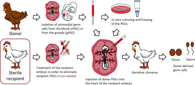
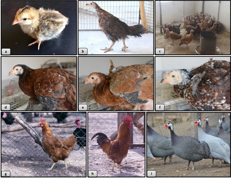
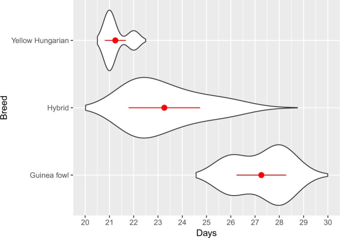
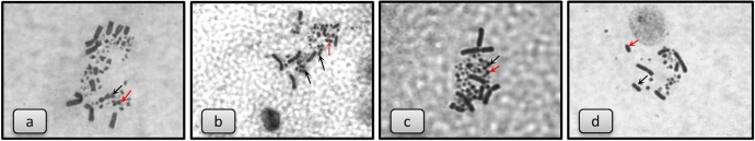
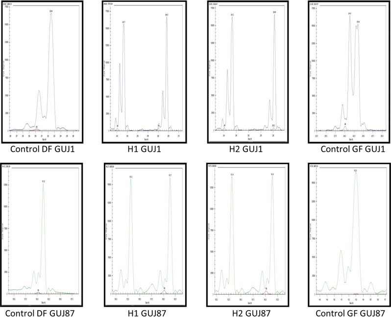
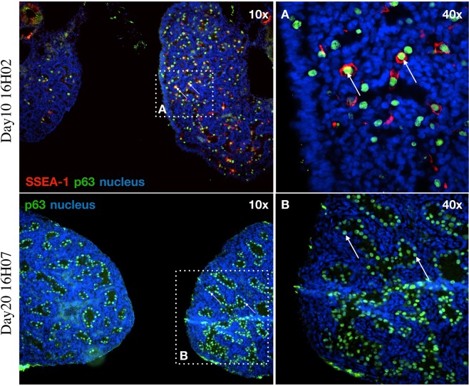
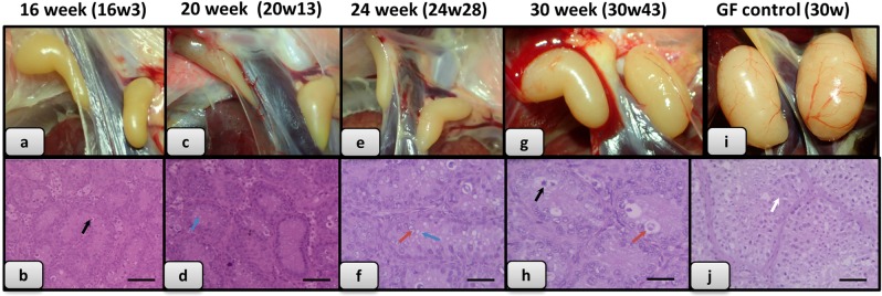
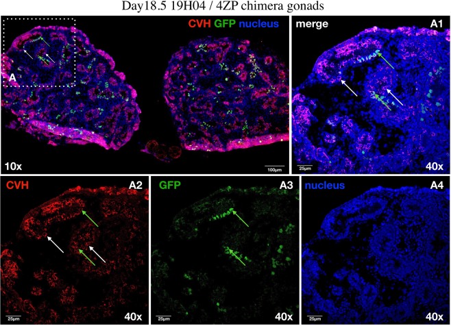
In the last decade, avian gene preservation research has focused on the use of the early precursors of the reproductive cells, the primordial germ cells (PGCs). This is because avian PGCs have a unique migration route through the vascular system which offers easy accessibility. Furthermore, culturing of the cells in vitro, freezing/thawing, reintegration into a recipient embryo and the development of the germ cells can be carried out in well-defined laboratory circumstances. The efficient recovery of the donor genotype and the frequency of germline transmission from the surrogate host animals are still areas which need further development. Thus, the aim of the present study was to investigate an infertile interspecific hybrid (recipient) as an appropriate host for primordial germ cells from native poultry breeds. Guinea fowl × chicken hybrids were produced, the crossing was repeated inversely. The phenotype, the hatching time, the hatching rate, the sex ratio, the presence of own germ cells, the fertility and the phenotype of viable hybrids and the incidence of chromosomal abnormalities of dead hybrid embryos were described. 6.65% viable offspring was obtained with crossing of Guinea fowl females with domestic fowl males. Crossing of domestic fowl hens with Guinea fowl male resulted in lower fertility, 0.14% viable offspring. Based on the investigations, the observed offspring from the successful crossing were sterile male hybrids, thus an extreme form of Haldane's rule was manifested. The sterile hybrid male embryos were tested by injecting fluorescently labeled chicken PGCs. The integration rate of labeled PGCs was measured in 7.5-day, 14.5-day and 18.5-day old embryonic gonads. 50%, 5.3% and 2.4% of the injected hybrid embryos survived and 40%, 5.3% and 2.4% of the examined gonads contained fluorescent labeled donor PGCs. Therefore, these sterile hybrid males may be suitable recipients for male PGCs and possibly for female PGCs although with lower efficiency. This research work shows that the sterility of hybrids can be used in gene conservation to be a universal host for PGCs of different avian species.


Conflict of interest statement

The authors declare no competing interests.

Figures

Figure 1
Figure 1
General outline of producing donor-derived hatchlings with sterile recipients. The migration of PGCs reaches its peak in the bloodstream between HH stages 13–17 (48–65 hours after laying in chicken); thus this is the optimal stage for collecting donor PGCs, and also this is the suitable stage for injecting them back to the recipient embryo. After the isolation, using a selective media, in vitro cultures of PGCs will be possible. For long term storage we cryopreserved the cells and keep them in liquid nitrogen. As a next step, cryopreserved or fresh PG cells are injected into the recipient embryo. After the hatching the presumptive germline chimeras are crossed back with the original breed or with each other to regenerate the donor genotype. With the usage of sterile hybrids, the treatment of the recipient embryos is not needed therefore the process is more efficient.
Figure 2
Figure 2
Phenotype of hybrids, Yellow Hungarian chicken and Guinea fowl. (a) 1 day old hybrid, (b) 8 weeks old hybrid, (c) 16 weeks old hybrids, (d) brownish colour variety, (e) yellow colour variety, (f) white, mixed colour variety, (g) Yellow Hungarian hen, (h) Yellow Hungarian rooster, (i) Guinea fowls.
Figure 3
Figure 3
Hatching time of the hybrid eggs in comparison with the original breeds. The hatching time of hybrid, Yellow Hungarian and Guinea fowl eggs were monitored. The eggs of the Yellow Hungarian chicken and the Guinea fowl hatched after 21–22 days and 26–29 days, respectively. In case of the hybrid eggs, an extended period of time was experienced (from day 21 to 27) which is a transition between the Yellow Hungarian and the Guinea fowl. Mean values and standard deviations are shown with red colour.
Figure 4
Figure 4
Karyotypes of hybrids and abnormalities were found in dead embryos. (a) Karyotype of hybrid female with ZW chromosomes and 5. metacentric chromosome from GF. (b) Karyotype of hybrid male with ZZ chromosomes and 5. metacentric chromosome from GF. (c) Aneuploid hybrid karyotype (2n-1) from a dead embryo. Absence one chromosome from the 1. pair. (d) Haploid karyotype from a dead embryo shows haploid/diploid mosaicism. (Z chromosome indicated with black arrow, 5. chromosome indicated with red arrow).
Figure 5
Figure 5
Allele sizes of hybrid, control domestic fowl and guinea fowl samples (Suppl. Fig. 1–8). Marker GUJ1 resulted allele 262 bp in domestic fowl (DF) and allele 241 bp; 243 bp in guinea fowl (GF). Hybrid 1 (H1) received one allele from domestic fowl (264 bp) and another from guinea fowl (243 bp). Hybrid 2 (H2) also have one allele from chicken (260 bp) and another from guinea fowl (243 bp). In case of marker GUJ87 there is allele 161 bp in domestic fowl (DF) and allele 153 bp in guinea fowl (GF) in homozygous form. Both hybrids (H1, H2) received both alleles (153 bp and 161 bp).
Figure 6
Figure 6
Examination the gonads of the hybrid embryos: p63 and SSEA-1 immunostaining identify the endogenous PGCs in the gonads of hybrid embryo at day-10 (16H02) and p63 at day-20 (16H07). (A) The SSEA-1 expressing PGCs are red colored on the cell surface. The p63 expressing PGCs are green colored in nucleus. White square shows the cells on the picture (A) (right top). (B) The p63 expressing PGCs are green colored in nucleus. White square shows the cells on the picture (B) (right bottom). White arrows demonstrate two host derived PGCs. For nuclear staining (nucleus) we used Hoechst 33342 staining (blue).
Figure 7
Figure 7
Histological analysis of gonads of the raised hybrids. (a) Gonads of 16 week old hybrid (Individual no. 3); (b) Histological section of this gonad (No. 3); (c) Gonads of 20 week old hybrid (No. 13); (d) Histological section of this gonad (No. 13); (e) Gonads of 24 week old hybrid (No. 28); (f) Histological section of this gonad (No. 28); (g) Gonads of 30 week old hybrid (individual no. 43); (h) Histological section of this gonad (No. 43); (i) Gonads of 30 week old Guinea fowl control; (j) Histological section of this gonad. (Degenerate cells with pyknotic nucleus separated from the interstitium indicated with black arrow, foamy cytoplasm indicated with blue arrow, foamy nucleus, loosened chromatin structure indicated with red arrow; spermatozoa indicated with white arrow. Scale bar b, d and j: 50 μm; f and h: 25 μm).
Figure 8
Figure 8
Identification of the endogenous host-derived and injected GFP expressing donor derived PGCs in the left and right gonads of 18.5-day-old hybrid embryo (19H04). (A) CVH expressing PGCs are red colored in the cytoplasm. The donor derived GFP expressing PGCs are green colored. White square shows the cells on the picture (A1–A4). Whyte arrows demonstrate two host derived PGCs. Green arrows indicate two integrated donor derived GFP expressing PGCs. (A1) Confocal merge images of CVH (red), GFP (green) and nuclear stained (blue) PGCs. (A2) Confocal images of CVH (red) stained PGCs. (A3) Confocal images of GFP expressing PGCs. (A4) Confocal images of nuclear stained PGCs. For nuclear staining (nucleus) we used TO-PRO®-3 stain (blue). Scale bars: 100 μm (A), 25 μm (A1–A4).

References

    1. IUCN Red List, https://www.iucnredlist.org [accessed 17.12.18] (2018).
    1. Sztán N, et al. Successful chimera production in the Hungarian goose (Anser anser domestica) by intracardiac injection of blastodermal cells in 3-day-old embryos. Reprod Fertil Dev. 2017;29:2206–16. doi: 10.1071/RD16289. - DOI - PubMed
    1. Sawicka D, Brzeziñska J, Bednarczyk M. Cryoconservation of embryonic cells and gametes as a poultry biodiversity preservation method. Folia biol-Kraków. 2011;59:1–5. doi: 10.3409/fb59_1-2.01-05. - DOI - PubMed
    1. Blesbois E, et al. Development of avian reproductive biotechnologies for the management of genetic diversity: CRYOBIRDS. Worlds. Poult Sci J. 2012;68(Suppl. 1):281–84.
    1. Tajima A. Conservation of avian genetic resources. Rewiev. Jap Poult Sci. 2013;50:1–8. doi: 10.2141/jpsa.0120083. - DOI

Publication types