. 2019 Oct 3;9(1):14281.
doi: 10.1038/s41598-019-50656-5.

Stimulator of interferon genes (STING) activation exacerbates experimental colitis in mice

Affiliations

Stimulator of interferon genes (STING) activation exacerbates experimental colitis in mice

Gary R Martin et al. Sci Rep. .

Abstract

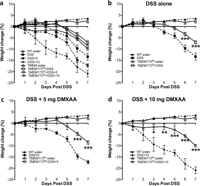
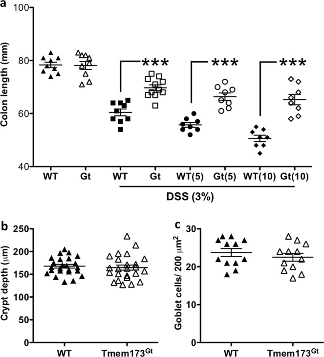
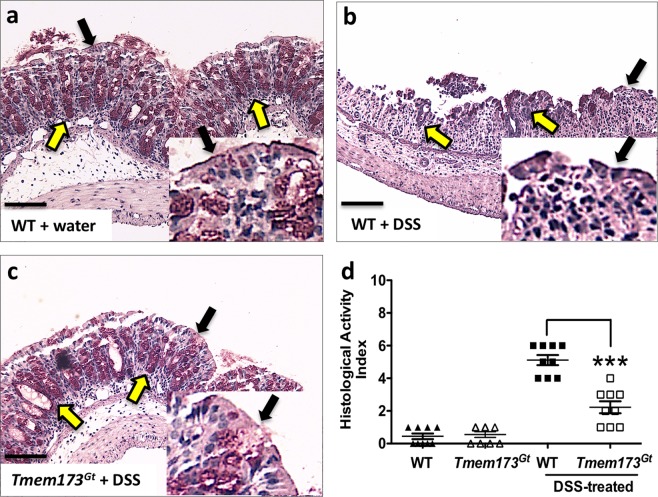
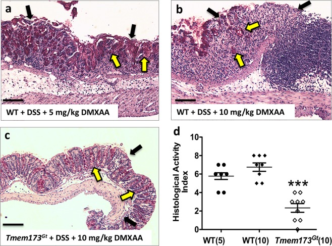
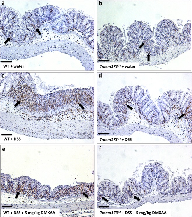
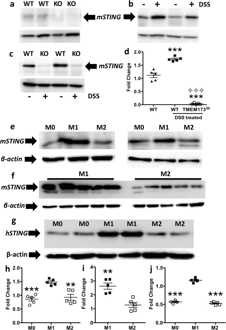
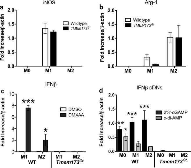
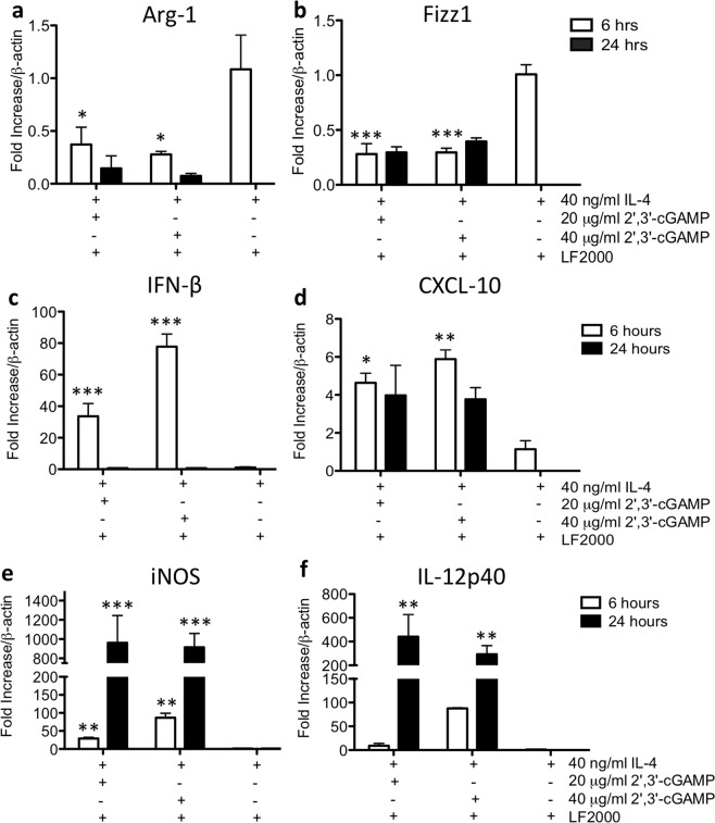
Detection of cytoplasmic DNA by the host's innate immune system is essential for microbial and endogenous pathogen recognition. In mammalian cells, an important sensor is the stimulator of interferon genes (STING) protein, which upon activation by bacterially-derived cyclic dinucleotides (cDNs) or cytosolic dsDNA (dsDNA), triggers type I interferons and pro-inflammatory cytokine production. Given the abundance of bacterially-derived cDNs in the gut, we determined whether STING deletion, or stimulation, acts to modulate the severity of intestinal inflammation in the dextran sodium sulphate (DSS) model of colitis. DSS was administered to Tmem173gt (STING-mutant) mice and to wild-type mice co-treated with DSS and a STING agonist. Colitis severity was markedly reduced in the DSS-treated Tmem173gt mice and greatly exacerbated in wild-type mice co-treated with the STING agonist. STING expression levels were also assessed in colonic tissues, murine bone marrow derived macrophages (BMDMs), and human THP-1 cells. M1 and M2 polarized THP-1 and murine BMDMs were also stimulated with STING agonists and ligands to assess their responses. STING expression was increased in both murine and human M1 polarized macrophages and a STING agonist repolarized M2 macrophages towards an M1-like subtype. Our results suggest that STING is involved in the host's response to acutely-induced colitis.


Conflict of interest statement

The authors declare no competing interests.

Figures

Figure 1
Figure 1
Weight losses associated with DSS-induced colitis in Tmem17Gt and wild-type mice. (a) Weight losses were exacerbated in the DSS mice that were co-treated with the murine STING agonist, DMXAA, and attenuated in the Tmem173Gt group of mice. To examine the effects of a given treatment regime on weight change, we isolated groups and compared the effects of (b) DSS-induced colitis either alone or following co-treatments with (c) 5 mg/kg or (d)10 mg/kg of DMXAA (i.p.). Statistical significance between data sets was assessed by one-way ANOVA followed by Tukey’s multiple comparisons post-hoc test between all groups. Values are means ± SEM, n ≥ 8 mice per group with differences denoted by *P < 0.05,**P < 0.01, ***P < 0.001 relative to DSS-treated WT.
Figure 2
Figure 2
Colonic shortening. (a) DSS treatment induced colonic shortening in all treatment groups, but relative to DSS-treated WT mice, was significantly greater in the WT DSS mice that had also received DMXAA. In contrast, this effect was significantly reduced in both the Tmem173gt and the Tmem173gt mice co-treated with the STING agonist. Black-filled dots are for WT, or WT + 5 or + 10 mg/kg DMXAA; white-clear dots are for the Tmem173gt receiving similar treatments. Statistical significance between data sets was assessed by one-way ANOVA followed by Tukey’s multiple comparisons post-hoc test between all groups. Values are means ± SEM, n ≥ 8 mice per group, ***P < 0.001 longer relative to DSS-treated WT. Analysis of (b) crypt depth or (c) goblet cell numbers in the colons of the untreated WT and TMEM173Gt mice. To assess crypt depth, 4 random locations from 6 untreated WT or TMEM173Gt mice (n = 24 total) measurements were plotted. Upon statistical comparison, there were no significant differences in crypt height or goblet cell numbers when sections from untreated WT and Tmem173gt mice were compared. Goblet cell counts represent the number of goblet cells per 200 um2 area, n = 12, Student’s T-test was used to determine possible differences between data sets.
Figure 3
Figure 3
STING-deficient mice demonstrate attenuation of DSS-induced acute colitis. Representative histological sections harvested from (a) WT, (b) DSS-treated WT and (c) DSS-treated Tmem173gt mice. After 7 days of DSS treatment, crypt loss, as well as infiltration and hypertrophy, was reduced in the DSS-treated Tmem173gt mice when compared to WT mice. In addition, epithelial brush border (black arrowheads) and goblet cell (yellow arrowheads) losses were reduced in the colonic tissues of DSS-treated Tmem173gt mice when compared to similarly treated WT controls. (d) Although the HAI was elevated in all DSS-treated mice when compared to non-treated WT mice, colonic damage in the DSS-treated Tmem173gt mice was markedly reduced. PAS stain. Sections imaged using a 20x objective. Statistical significance was assessed by one-way ANOVA followed by Tukey’s multiple comparisons post-hoc test between all groups. Values represent means ± SEM, n ≥ 8 mice per group, ***P < 0.001 relative to DSS-treated WT.
Figure 4
Figure 4
Administration of a STING agonist exacerbates DSS-induced colitis. Sections were harvested from DSS-treated WT mice that were co-treated with (a) 5 mg/kg/day of DMXAA or from (b) WT or (c) Tmem173gt mice co-treated with 10 mg/kg/day of DMXAA every other day. (d) Inflammation-as assessed using the histological activity index (HAI), an index that provides a damage score-in the WT mice that were administered DMXAA was more severe than that of DMXAA-treated Tmem173gt mice. Note the loss of the epithelial brush border (black arrowheads) and goblet cells (yellow arrowheads). Colonic damage in the Tmem173gt mice co-treated with DMXAA was markedly reduced when compared to both WT groups. Sections were harvested on day 7. PAS stain. All sections imaged using a 20x objective; black bars at the bottom left of the panels represent 200 μM. Statistical significance was assessed by one-way ANOVA followed by Tukey’s multiple comparisons post-hoc test between all groups. Values for (D) are the means ± SEM, n ≥ 7 mice per group, ***P < 0.001 relative to both the WT(5) and WT(10) groups of mice.
Figure 5
Figure 5
DSS-induced colitis increased the numbers of activated macrophages in WT mice. To assess macrophage activation, paraffin sections from the various treatment groups were stained with Iba-1, a protein upregulated during activation of macrophages and possibly neutrophils (black arrows). Representative sections harvested from vehicle-treated (a) WT or (b) Tmem173gt mice, or from DSS-treated (c) WT or (d) Tmem173gt mice or from DSS treated (e) WT or (f) Tmem173gt mice that were co-treated with DMXAA (5 mg/kg). Although there were no discernable differences in macrophage activation between the untreated WT and Tmem173gt groups of mice, Iba-1 staining of colonic tissues from the DSS-treated WT and DSS-treated WT mice co-treated with DMXAA looked to be increased when compared with similarly-treated Tmem173gt mice. All sections were harvested on day 7 from mice treated with 3% DSS. Anti-Iba-1 immuno-histochemical stain. Sections imaged using a 20x objective.
Figure 6
Figure 6
Colitis and M1 polarization of murine and human macrophages increased STING protein expression. (a) A representative blot of STING expression from the colonic lysates of two untreated WT or Tmem173gt (KO) mice. (b–d) DSS-induced colitis increased STING protein expression in WT mice (+ = DSS treated) relative to that observed in both tap water treated control mice (− = tap water only) and STING deficient mice. One-way ANOVA followed by Tukey’s multiple comparisons post-hoc test between all groups was used. Values for (d) are means ± SEM, n = 5 mice per group, ***P < 0.001 relative to both the WT and Tmem173gt groups and ⟡⟡⟡P < 0.001 relative to DSS-treated WT mice. (c,h) Representative blots and a histogram from two mice to show that STING protein expression in murine BMDM lysates was increased in M1 polarized macrophages when compared to M2 polarized or M0 macrophages. (d) To confirm the previous finding in (c), BMDMs from 5 mice were polarized into M1 or M2 macrophages. (i) Again, there was a significant increase in mSTING expression in the M1 polarized macrophages relative to the expression levels found in the M2 cells. (g,j) This also occurred in human THP-1 monocytic cells as M1 polarization markedly increased STING protein expression. Immunoblots of polarized murine BMDMs: M0, untreated; M1, 50 ng/ml of LPS and IFNγ; M2, 40 ng/ml IL-4 for 48 hrs. 50 μg of protein/lane was loaded, and β-actin was used as the loading control for all immunoblot analyses. For (h,j) one-way ANOVA followed by Tukey’s multiple comparisons post-hoc test between all groups was used, n = 5, **P < 0.01 or ***P < 0.001 relative to M0 or M2; for (i), a Student’s T-test was applied with **P < 0.01 vs. M2.
Figure 7
Figure 7
STING agonist induction of IFN-β. Bone marrow-derived macrophages (BMDMs) derived from the femurs of WT or Tmem173gt mice were polarized in vitro into M1 or M2 subtypes. (a,b) Using iNOS/NOS2 mRNA induction as a marker for M1 cells, and Arg-1 mRNA induction as a marker for the M2 subtype, we observed no differences in the expression of either marker when these cells from the WT and Tmem173gt were compared. (c) Increased IFN-β expression or secretion in response to STING agonists (or cytoplasmic DNA exposure) is a commonly used marker to show that the STING pathway has been activated. In this regard, when M1 or M2 polarized cells from the WT or Tmem17Gt were incubated for 6-hr with DMXAA, it was observed that IFN-β mRNA expression was only induced in the WT macrophages. (d) Similarly, when the 3 subtypes of macrophages were exposed to the non-canonical ligand 2′3′-cGAMP or the bacterial second messenger c-di-AMP, IFN-β expression was increased solely in the WT macrophage polarized subsets. Significance was assessed by one-way ANOVA followed by Tukey’s multiple comparisons post-hoc test between all groups. Values represent the means ± SEM of the fold changes in the respective mRNAs; *P < 0.05, **P < 0.01, or ***P < 0.001 vs. the corresponding Tmem17Gt group (eg. WT M1, DMXAA treated vs. Tmem17Gt M1, DMXAA treated). For macrophage polarization: M0, untreated; M1, 50 ng/ml of LPS and IFNγ; M2, 40 ng/ml IL-4 for 48 hrs. STING agonists: DMXAA 20 μg/ml; c-di-AMP 20 μg/ml plus lipofectamine; 2′3′-cGAMP 20 μg/ml plus lipofectamine for 6 hrs. Data was normalized to β-actin mRNA and experimental transcripts expressed as the relative fold change in each mRNA selected.
Figure 8
Figure 8
cDN-induced repolarization of M2 polarized macrophages towards the M1 pro-inflammatory subtype. To establish M2 subtype of macrophages, cells were pre-treated with 40 ng/ml IL-4 for 48 hrs prior to the addition of cDNs. (a,b) A 6-hr treatment with either 2′3′-cGAMP or c-di-AMP significantly decreased Arg-1 and Fizz1 mRNA expression as compared to the IL-4-alone treated cells. (cf) Moreover, IFN-β, CXCL-10, iNOS and IL-12p40 mRNAs were also increased at either the 6 hr or the 24 hr timepoint post-cDN exposure. All cDN treatments were carried out using lipofectamine permeabilization of the cultured cells. Significance was assessed by one-way ANOVA followed by Tukey’s multiple comparisons post-hoc test between all groups. Values represent the means ± SEM of the fold changes in the respective mRNAs; *P < 0.05, **P < 0.01, ***P < 0.001 vs. the control groups treated with IL-4 alone. Data was normalized to β-actin mRNA and experimental transcripts expressed as the relative fold change in each mRNA tested.

References

    1. Woodward JJ, Iavarone AT, Portnoy DA. c-di-AMP secreted by intracellular Listeria monocytogenes activates a host type I interferon response. Science. 2010;328:1703–1705. doi: 10.1126/science.1189801. - DOI - PMC - PubMed
    1. Danilchanka O, Mekalanos JJ. Cyclic dinucleotides and the innate immune response. Cell. 2013;154:962–970. doi: 10.1016/j.cell.2013.08.014. - DOI - PMC - PubMed
    1. Sun L, Wu J, Du F, Chen X, Chen ZJ. Cyclic GMP-AMP synthase is a cytosolic DNA sensor that activates the type I interferon pathway. Science. 2013;339:786–791. doi: 10.1126/science.1232458. - DOI - PMC - PubMed
    1. Wu J, et al. Cyclic GMP-AMP is an endogenous second messenger in innate immune signaling by cytosolic DNA. Science. 2013;339:826–830. doi: 10.1126/science.1229963. - DOI - PMC - PubMed
    1. Chandra D, et al. STING ligand c-di-GMP improves cancer vaccination against metastatic breast cancer. Cancer Immunol Res. 2014;2:901–910. doi: 10.1158/2326-6066.CIR-13-0123. - DOI - PMC - PubMed

Publication types

MeSH terms