. 2019 Sep 8:2019:2165918.
doi: 10.1155/2019/2165918. eCollection 2019.

Inhibition of Synovial Macrophage Pyroptosis Alleviates Synovitis and Fibrosis in Knee Osteoarthritis

Affiliations

Inhibition of Synovial Macrophage Pyroptosis Alleviates Synovitis and Fibrosis in Knee Osteoarthritis

Li Zhang et al. Mediators Inflamm. .

Abstract

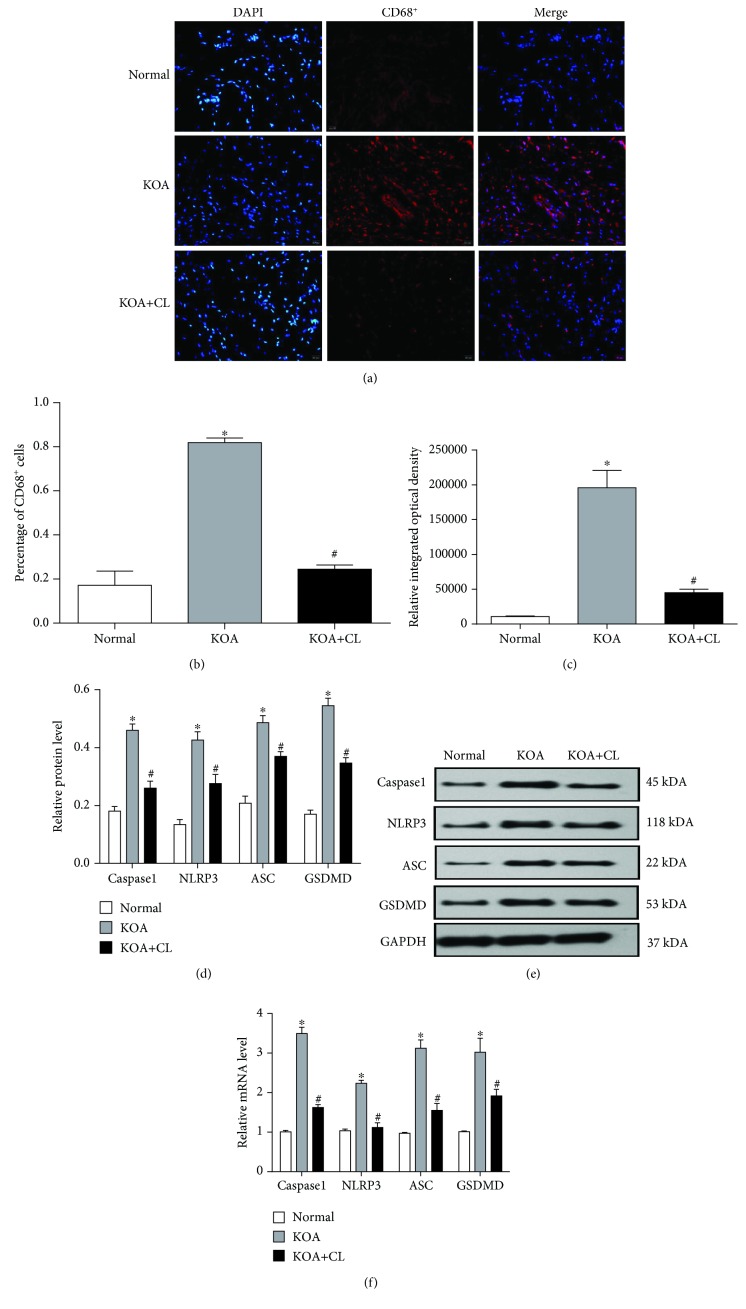
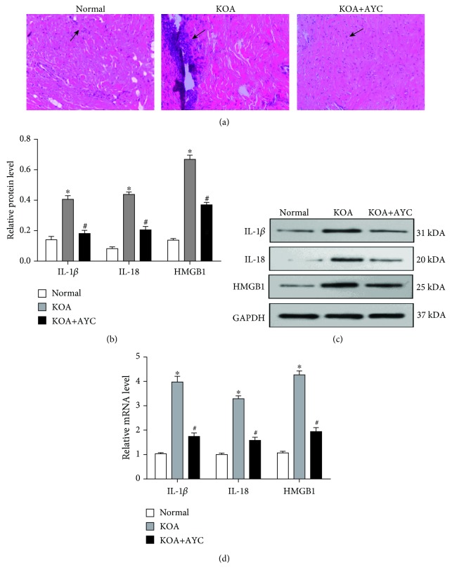
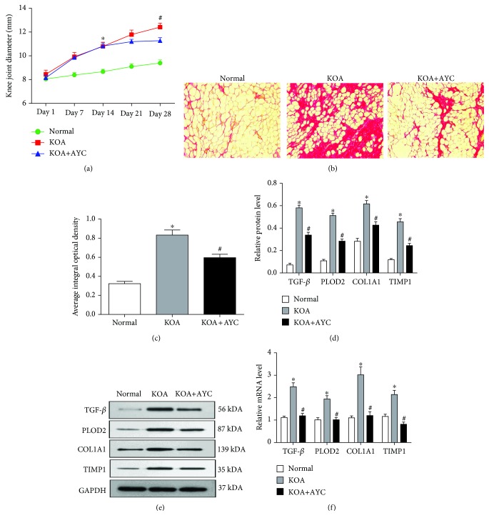
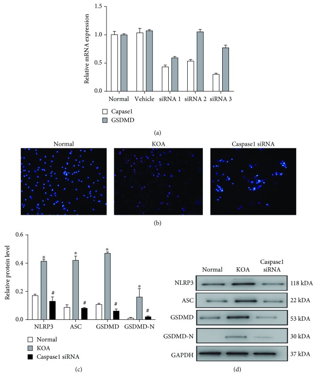
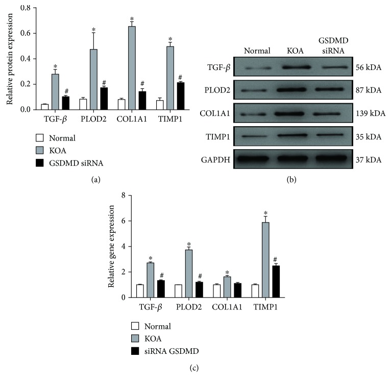
Increasing evidence has shown that macrophage pyroptosis in different tissues participates in chronic aseptic inflammation and is related to tissue fibrosis. Our last studies also revealed the vital role of synovial fibroblast pyroptosis in the onset and development of knee osteoarthritis (KOA). In this study, we aimed to investigate whether synovial macrophage pyroptosis did occur and whether this form of cell death should be related to synovitis and fibrosis of KOA. In the synovial tissue of KOA model rats, we observed a decrease of caspase1, NLRP3, ASC, and GSDMD caused by macrophage depletion in both the mRNA and protein expressions. Besides, rats treated with the specific caspase1 inhibitor Ac-YVAD-CMK showed less inflammatory reaction and fibrosis, not only in the expression of proinflammatory factors IL-1β, IL-18, and HMGB1 and fibrosis markers TGF-β, PLOD2, COL1A1, and TIMP1 but also in the observation of HE staining, Sirius Red staining, and the transverse diameters of the right knees. Subsequently, we established an LPS+ATP-induced model in macrophages mimicking the inflammatory environment of KOA and inducing macrophage pyroptosis. Macrophages transfected with caspase1 siRNA showed reduced cell death; meanwhile, the relative expression of pyroptosis-related proteins were also downregulated. In addition, the level of fibrotic markers in synovial fibroblasts were significantly decreased after coculture with siRNA GSDMD-transfected macrophages. To conclude, synovial macrophage pyroptosis may occur in the pathological processes of KOA and inhibition of synovial macrophage pyroptosis alleviates synovitis and fibrosis in KOA model rats.


Conflict of interest statement

All authors declare that there are no actual or potential conflicts of interest, including any financial, personal, or other relationships with other people or organizations that could inappropriately influence, or be perceived to influence, our work.

Figures

Figure 1
Figure 1
Synovial macrophage pyroptosis in KOA model rats. (a) Representative synovial tissues stained with DAPI and TRITC. Scale bar = 20 μm (×100). (b) Percentage of CD68+ cells in each group. P < 0.05, in comparison with the Normal group. #P < 0.05, in comparison with the KOA group. (c) Relative integrated optical density (IOD) value in each group. Data were analyzed by ImageJ. (d) Protein expressions of caspase1, NLRP3, ASC, and GSDMD between groups. P < 0.05, in comparison with the Normal group. #P < 0.05, in comparison with the KOA group (n = 6). Western blot data are presented from three independent experiments. (e) Typical protein bands for each group. (f) mRNA level of caspase1, NLRP3, ASC, and GSDMD between groups. Trend is consistent with protein expression.
Figure 2
Figure 2
Inhibition of synovial macrophage pyroptosis may alleviate synovitis in KOA rats. (a) Representative synovial tissues of each group stained with HE staining (200×). Arrow: inflammatory cell infiltration. (b) Protein expressions of IL-1β, IL-18, and HMGB1 between groups. P < 0.05, in comparison with the Normal group. #P < 0.05, in comparison with the KOA group (n = 6). (c) Typical protein bands for each group. (d) mRNA levels of IL-1β, IL-18, and HMGB1 between groups. The trend is consistent with protein expression.
Figure 3
Figure 3
Inhibition of synovial macrophage pyroptosis may alleviate fibrosis in KOA rats. (a) The severity of synovial fibrosis evaluated by the transverse diameters of the right knees. P < 0.05 vs. the Normal group; #P < 0.05 vs. the KOA+AYC group. (b) Collagen deposition was revealed through Sirius Red staining (200×). (c) Semiquantification of Sirius Red staining was evaluated using the average integral optical density (AIOD). The results were expressed as the mean ± SD (n = 6). P < 0.05, in comparison with the Normal group. #P < 0.05, in comparison with the KOA group. Data were analyzed by ImageJ. (d) Comparison of TGF-β, PLOD2, COL1A1, and TIMP1 protein expressions between groups. P < 0.05, in comparison with the Normal group. #P < 0.05, in comparison with the KOA group (n = 6). (e) Typical protein bands for each group. (f) The mRNA levels of TGF-β, PLOD2, COL1A1, and TIMP1 between groups.
Figure 4
Figure 4
Pyroptosis is induced in LPS+ATP-treated macrophages. (a) The silencing effects of the caspase1 siRNA and GSDMD siRNA were both confirmed by PCR. (b) Pyroptotic cells observed by Hoechst and PI staining (200×). (c) Comparison of NLRP3, ASC, GSDMD, and GSDMD-N protein expressions between groups. P < 0.05, in comparison with the Normal group. #P < 0.05, in comparison with the KOA group. (d) Typical protein bands for each group.
Figure 5
Figure 5
Macrophage pyroptosis upregulates the expression of fibrosis markers cocultured with synovial fibroblasts. (a) Comparison of TGF-β, PLOD2, COL1A1, and TIMP1 protein expressions between groups. P < 0.05, in comparison with the Normal group. #P < 0.05, in comparison with the KOA group. (b) Typical protein bands for each group. (c) Relative mRNA expression of TGF-β, COL1A1, PLOD2, and TIMP1 in synovial fibroblasts in each group. P < 0.05, in comparison with the Normal group. #P < 0.05, in comparison with the KOA group.

References

    1. Hügle T., Geurts J. What drives osteoarthritis?—Synovial versus subchondral bone pathology. Rheumatology. 2017;56(9):1461–1471. doi: 10.1093/rheumatology/kew389. - DOI - PubMed
    1. Glyn-Jones S., Palmer A. J. R., Agricola R., et al. Osteoarthritis. The Lancet. 2015;386(9991):376–387. doi: 10.1016/S0140-6736(14)60802-3. - DOI - PubMed
    1. Smith M. D. The normal synovium. The Open Rheumatology Journal. 2011;5(1):100–106. doi: 10.2174/1874312901105010100. - DOI - PMC - PubMed
    1. Lopes E. B. P., Filiberti A., Husain S. A., Humphrey M. B. Immune contributions to osteoarthritis. Current Osteoporosis Reports. 2017;15(6):593–600. doi: 10.1007/s11914-017-0411-y. - DOI - PubMed
    1. Kraus V. B., McDaniel G., Huebner J. L., et al. Direct in vivo evidence of activated macrophages in human osteoarthritis. Osteoarthritis and Cartilage. 2016;24(9):1613–1621. doi: 10.1016/j.joca.2016.04.010. - DOI - PMC - PubMed

Substances