Review
. 2019 Oct 1:21:19.
doi: 10.1186/s12575-019-0107-z. eCollection 2019.

DNA Methylation Validation Methods: a Coherent Review with Practical Comparison

Affiliations
Review

DNA Methylation Validation Methods: a Coherent Review with Practical Comparison

Šárka Šestáková et al. Biol Proced Online. .

Abstract

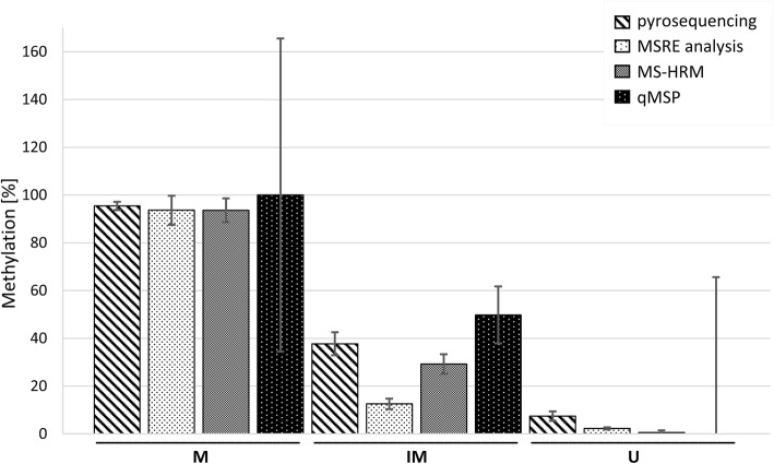
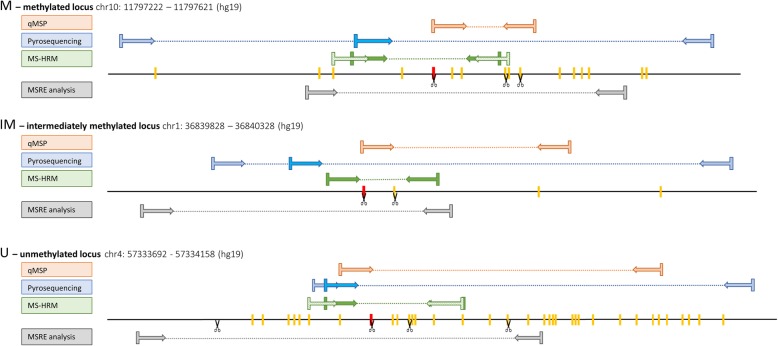
Here, we present a practical overview of four commonly used validation methods for DNA methylation assessment: methylation specific restriction endonucleases (MSRE) analysis, pyrosequencing, methylation specific high-resolution DNA melting (MS-HRM) and quantitative methylation specific polymerase chain reaction (qMSP). Using these methods, we measured DNA methylation levels of three loci in human genome among which one was highly methylated, one intermediately methylated and one unmethylated. We compared the methods in terms of primer design demands, methods' feasibility, accuracy, time and money consumption, and usability for clinical diagnostics. Pyrosequencing and MS-HRM proved to be the most convenient methods. Using pyrosequencing, it is possible to analyze every CpG in a chosen region. The price of the instrument may represent the main limitation of this methodology. MS-HRM is a simple PCR-based method. The measurement was quick, cheap and very accurate. MSRE analysis is based on a methylation specific digestion of DNA. It does not require a bisulfite conversion of DNA as the other methods. MSRE analysis was very easy to perform, however, it was not suitable for intermediately methylated regions and it was also quite expensive. qMSP is a qPCR-based method that uses primers designed specifically for methylated and unmethylated alleles of a chosen region. This was the least accurate method and also the primer design and optimization of PCR conditions were highly demanding.

Keywords: DNA methylation; MS-HRM; MSRE; Pyrosequencing; Validation methods; qMSP.


Conflict of interest statement

Competing InterestsThe authors declare that they have no competing interests.

Figures

Fig. 1
Fig. 1
The influence of digestion time on measured methylation levels in MSRE analysis. Error bars represent the standard deviation (n = 4). M - methylated locus, IM – intermediately methylated locus, U – unmethylated locus
Fig. 2
Fig. 2
Summary of DNA methylation levels measured by the investigated methods. The average methylation of 10 samples is shown, the error bars represent the SD. Displayed MSRE data were measured after 2-h digestion. Displayed MS-HRM data were acquired using HRM M, HRM IM and HRM U Wojdacz primers. qMSP data shown were calculated using ΔΔCt approach and multiplied by 100 to gain the percentage. For the M locus in qMSP, all values were higher than 100% so we set the mean to 100% to make the figure more comprehensible, the SD was calculated from the original values multiplied by 100. For the IM and U loci in qMSP, we calculated the methylation percentage as 1-(Unm ΔΔCt), the SD was also calculated from the original ΔΔCt values multiplied by 100. M – methylated locus, IM – intermediately methylated locus, U – unmethylated locus
Fig. 3
Fig. 3
Positions of primer pairs, CpGs and restriction sites within studied regions. CpGs are shown as red and yellow bars on a line representing the DNA sequence. The red CpG is the one originally chosen from Infinium MethylationEPIC BeadChip. The scissors indicate sites that are cut by MSREs. The lighter blue primers were used for initial pyrosequencing PCR and the darker blue primers represent the sequencing primers. The patterned light green HRM primers were designed with a CpG on its 5’end (M/U Wojdacz primers)

References

    1. Schubeler D. Function and information content of DNA methylation. Nature. 2015;517(7534):321. doi: 10.1038/nature14192. - DOI - PubMed
    1. Jin Z, Liu Y. DNA methylation in human diseases. Genes & Diseases. 2018;5(1):1–8. doi: 10.1016/j.gendis.2018.01.002. - DOI - PMC - PubMed
    1. Levenson VV. DNA methylation as a universal biomarker. Expert Rev Mol Diagn. 2010;10(4):481–488. doi: 10.1586/erm.10.17. - DOI - PMC - PubMed
    1. Heyn H, Esteller M. DNA methylation profiling in the clinic: applications and challenges. Nat Rev Genet. 2012;13(10):679–692. doi: 10.1038/nrg3270. - DOI - PubMed
    1. Pidsley R, Zotenko E, Peters TJ, Lawrence MG, Risbridger GP, Molloy P, et al. Critical evaluation of the Illumina MethylationEPIC BeadChip microarray for whole-genome DNA methylation profiling. Genome Biol. 2016;17(1):208. doi: 10.1186/s13059-016-1066-1. - DOI - PMC - PubMed