. 2019 Sep 9:2019:2168709.
doi: 10.1155/2019/2168709. eCollection 2019.

Transplantation of Human Urine-Derived Stem Cells Ameliorates Erectile Function and Cavernosal Endothelial Function by Promoting Autophagy of Corpus Cavernosal Endothelial Cells in Diabetic Erectile Dysfunction Rats

Affiliations

Transplantation of Human Urine-Derived Stem Cells Ameliorates Erectile Function and Cavernosal Endothelial Function by Promoting Autophagy of Corpus Cavernosal Endothelial Cells in Diabetic Erectile Dysfunction Rats

Chi Zhang et al. Stem Cells Int. .

Abstract

Aims: Cavernosal endothelial dysfunction is one of the factors in developing diabetic erectile dysfunction (DED), but the mechanism of cavernosal endothelial dysfunction is unclear. The present study is aimed at determining the contribution of autophagy in cavernosal endothelial dysfunction of DED rats and explaining the therapeutic effect of urine-derived stem cells (USCs).

Methods: After rat corpus cavernosal vascular endothelial cells (CCECs) were isolated and cultured in vitro, CCECs were treated with advanced glycation end products (AGEs) to mimic the diabetic situation. Autophagy flux, proliferation, and apoptosis of CCECs were determined by mRFP-GFP-LC3 adenovirus infection combined with fluorescence observation and western blot analysis. USCs were isolated from the urine of six healthy male donors, and coculture systems of USCs and CCECs were developed to assess the protective effect of USCs for CCECs in vitro. The contribution of autophagy to the cellular damage in CCECs was evaluated by the autophagic inhibitor, 3-methyladenine (3-MA). Then, DED rats were induced by streptozotocin (50 mg/kg) and screened by apomorphine test (100 μg/kg). In DED rats, USCs or PBS as vehicle was administrated by intracavernous injection (n = 15 per group), and another 15 normal rats served as normal controls. Four weeks after injection, erectile function was evaluated by measuring the intracavernosal pressure (ICP) and mean arterial pressure (MAP). Cavernosal endothelial function and autophagic activity were examined by western blot, immunofluorescence, and transmission electron microscopy.

Results: In vitro, AGE-treated CCECs displayed fewer LC3 puncta formation and expressed less LC3-II, Beclin1, and PCNA but expressed more p62 and cleaved-caspase3 than controls (p < 0.05). Coculture of USCs with CCECs demonstrated that USCs were able to protect CCECs from AGE-induced autophagic dysfunction and cellular damage, which could be abolished by 3-MA (p < 0.05). DED rats showed lower ratio of ICP/MAP, reduced expression of endothelial markers, and fewer autophagic vacuoles in the cavernosal endothelium when compared with normal rats (p < 0.05). Intracavernous injection of USCs improved erectile function and cavernosal endothelial function of DED rats (p < 0.05). Most importantly, our data showed that the repaired erectile function and cavernosal endothelial function were the result of restored autophagic activity of the cavernosal endothelium in DED rats (p < 0.05).

Conclusions: Impaired autophagy is involved in the cavernosal endothelial dysfunction and erectile dysfunction of DED rats. Intracavernous injection of USCs upregulates autophagic activity in the cavernosal endothelium, contributing to ameliorating cavernosal endothelial dysfunction and finally improving the erectile dysfunction induced by diabetes.


Conflict of interest statement

The authors declare that there is no duality of interest associated with this manuscript.

Figures

Figure 1
Figure 1
Characterization of urine-derived stem cells (USCs). (a) The typical rice-shaped appearance of USCs (p1). (b) Osteogenic- and (c) adipogenic-induced USCs with Alizarin red S staining or Oil red O staining, respectively. (d) Flow cytometry analysis showed that USCs (p3) were strongly positive for mesenchymal stem cell markers (CD24, CD44, CD73, and CD90), weakly positive for CD105, and negative for hematopoietic stem cell markers (CD31, CD34, and CD45).
Figure 2
Figure 2
Characterization of corpus cavernosal vascular endothelial cells (CCECs). (a) The typical cobblestone-like appearance of CCECs (p1). (b) Immunofluorescent staining showed that more than 90% of CCECs (p2) were positive for endothelial markers (CD31, red fluorescence).
Figure 3
Figure 3
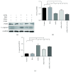
USCs protected CCECs from AGE-induced autophagic dysfunction in vitro and inhibition of autophagy attenuated the protective effect of USCs. (a) Western blot and (b–d) quantification for western blot of autophagy positive-related proteins (LC3 and Beclin1) and autophagy negative-related proteins (p62) in CCECs for each group. (e) Representative images of LC3 staining in CCECs for each group after infection with mRFP-GFP-LC3 adenovirus. Autophagosomes were shown as yellow puncta (RFP+GFP+), and autolysosomes were shown as red puncta (RFP+GFP-). (f, g) Quantification for autophagosome and autolysosome formation representing puncta staining sites per cell of 30 cells from each group. Treatment groups: control, BSA, AGEs, AGEs+USCs, and AGEs+USCs+3-MA. The concentration of BSA or AGEs was 200 μg/ml. The concentration of 3-MA was 2 mM, and the treat time was 24 h. n = 3. p < 0.05. C-caspase3: cleaved-caspase3; BSA: bovine serum albumin; AGEs: advanced glycation end products; 3-MA: 3-methyladenine.
Figure 4
Figure 4
USCs protected CCECs from AGE-induced cellular damage in vitro and inhibition of autophagy attenuated the protective effect of USCs. (a) Western blot and (b, c) quantification for western blot of proliferation-related protein (PCNA) and apoptosis-related protein (C-caspase3) in CCECs for each group. Treatment groups: control, BSA, AGEs, AGEs+USCs, and AGEs+USCs+3-MA. The concentration of BSA or AGEs was 200 μg/ml. The concentration of 3-MA was 2 mM, and the treat time was 24 h. n = 3. p < 0.05. C-caspase3: cleaved-caspase3; BSA: bovine serum albumin; AGEs: advanced glycation end products; 3-MA: 3-methyladenine.
Figure 5
Figure 5
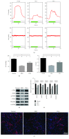
USCs restored autophagic activity of the cavernosal endothelium in DED rats. (a) Western blot and (b) quantification for western blot of autophagy positive-related proteins (LC3 and Beclin1) and autophagy negative-related proteins (p62) in cavernous tissue of normal rats and DED rats 4 weeks after intracavernous injection of PBS or USCs (n = 6 per group). (c) Representative TEM images of autophagic vacuoles (arrows) in cavernosal endothelial cells for each group. (d) Quantification for average autophagic vacuoles in 10 cells randomly selected from every specimen in each group (n = 3 per group). p < 0.05. TEM: transmission electron microscopy.
Figure 6
Figure 6
USCs improved erectile function and cavernosal endothelial function in diabetic erectile dysfunction (DED) rats. (a) Representative ICP tracing response to the stimulation of cavernous nerve (2 ms, 5 V, 25 Hz, and duration of 60 s) in normal rats and DED rats 4 weeks after intracavernous injection of PBS or USCs. (b) The USC injection increased ICP of DED rats, and (c) the ratio of ICP to MAP was calculated for each group (n = 8 per group). (d) Western blot and (e) quantification for western blot revealed that CD31, eNOS, p-eNOS, VEGFRA, and VEGFR2 expressions were all increased in the USC-treated group 4 weeks after cell transplantation (n = 6 per group). (f) Immunofluorescent staining confirmed the significantly higher expression of CD31 in cavernous tissue. p < 0.05. ICP: intracavernous pressure; MAP: mean arterial pressure; p-eNOS: phospho-eNOS.

References

    1. Malavige L. S., Levy J. C. Erectile Dysfunction in Diabetes Mellitus. The Journal of Sexual Medicine. 2009;6(5):1232–1247. doi: 10.1111/j.1743-6109.2008.01168.x. - DOI - PubMed
    1. Thorve V. S., Kshirsagar A. D., Vyawahare N. S., Joshi V. S., Ingale K. G., Mohite R. J. Diabetes-induced erectile dysfunction: epidemiology, pathophysiology and management. Journal of Diabetes and its Complications. 2011;25(2):129–136. doi: 10.1016/j.jdiacomp.2010.03.003. - DOI - PubMed
    1. Penson D. F., Latini D. M., Lubeck D. P., Wallace K. L., Henning J. M., Lue T. F. Do impotent men with diabetes have more severe erectile dysfunction and worse quality of life than the general population of impotent patients? Results from the Exploratory Comprehensive Evaluation of Erectile Dysfunction (ExCEED) database. Diabetes Care. 2003;26(4):1093–1099. doi: 10.2337/diacare.26.4.1093. - DOI - PubMed
    1. De Tejada I. S., Angulo J., Cellek S., et al. Pathophysiology of Erectile Dysfunction. The Journal of Sexual Medicine. 2005;2(1):26–39. doi: 10.1111/j.1743-6109.2005.20103.x. - DOI - PubMed
    1. Hidalgo-Tamola J., Chitaley K. Type 2 Diabetes Mellitus and Erectile Dysfunction. The Journal of Sexual Medicine. 2009;6(4):916–926. doi: 10.1111/j.1743-6109.2008.01116.x. - DOI - PubMed

LinkOut - more resources