A challenging case of concurrent multiple sclerosis and anaplastic astrocytoma
- PMID: 31583163
- PMCID: PMC6763678
- DOI: 10.25259/SNI_176_2019
A challenging case of concurrent multiple sclerosis and anaplastic astrocytoma
Abstract
Background: Cases of gliomas coexisting with multiple sclerosis (MS) have been described over the past few decades. However, due to the complex clinical and radiological traits inherent to both entities, this concurrent phenomenon remains difficult to diagnose. Much has been debated about whether this coexistence is incidental or mirrors a poorly understood neoplastic phenomenon engaging glial cells in the regions of demyelination.
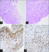
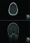
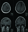
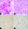
Case description: We present the case of a 41-year-old patient diagnosed with a left-sided frontal contrast enhancing lesion initially assessed as a tumefactive MS. Despite systemic treatment, the patient gradually developed signs of mass effect, which led to decompressive surgery. The initial microscopic evaluation demonstrated the presence of MS and oligodendroglioma; the postoperative evolution proved complex due to a series of MS-relapses and tumor recurrence. An ulterior revaluation of the samples for the purpose of this report showed an MS-concurrent anaplastic astrocytoma. We describe all relevant clinical aspects of this case and review the medical literature for possible causal mechanisms.
Conclusion: Although cases of concurrent glioma and MS remain rare, we present a case illustrating this phenomenon and explore a number of theories behind a potential causal relationship.
Keywords: Anaplastic astrocytoma; Gamma knife radiosurgery; Magnetic resonance imaging; Multiple sclerosis; Oligodendroglioma; Tumefactive multiple sclerosis.
Copyright: © 2019 Surgical Neurology International.
Conflict of interest statement
None of the authors has any conflicts of interest to disclose.
Figures


References
-
- Aarli JA, Mørk SJ, Myrseth E, Larsen JL. Glioblastoma associated with multiple sclerosis: Coincidence or induction ? Eur Neurol. 1989;29:312–6. - PubMed
-
- Acqui M, Caroli E, Di Stefano D, Ferrante L. Cerebral ependymoma in a patient with multiple sclerosis case report and critical review of the literature. Surg Neurol. 2008;70:414–20. - PubMed
-
- Brendle C, Hempel JM, Schittenhelm J, Skardelly M, Reischl G, Bender B, et al. Glioma grading by dynamic susceptibility contrast perfusion and 11C-methionine positron emission tomography using different regions of interest. Neuroradiology. 2018;60:381–9. - PubMed
-
- Butteriss DJ, Ismail A, Ellison DW, Birchall D. Use of serial proton magnetic resonance spectroscopy to differentiate low grade glioma from tumefactive plaque in a patient with multiple sclerosis. Br J Radiol. 2003;76:662–5. - PubMed
Publication types
LinkOut - more resources
Full Text Sources
Miscellaneous
