Review
. 2019:1175:15-44.
doi: 10.1007/978-981-13-9913-8_2.

Evolution of Neuroglia

Affiliations
Review

Evolution of Neuroglia

Alexei Verkhratsky et al. Adv Exp Med Biol. 2019.

Abstract

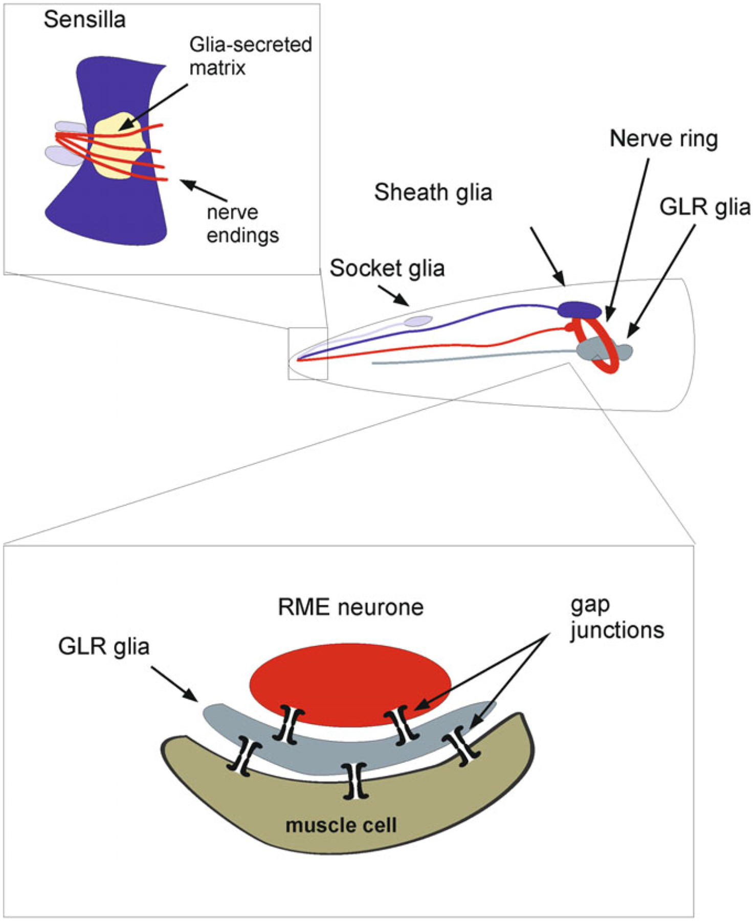
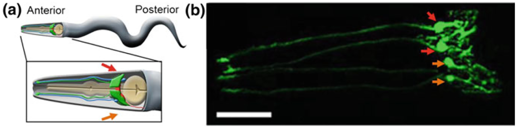
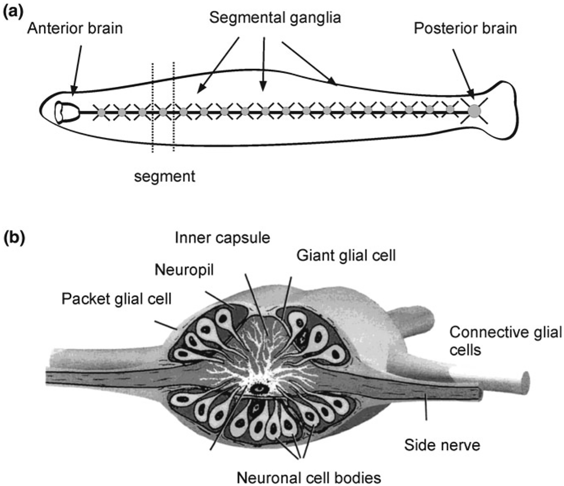
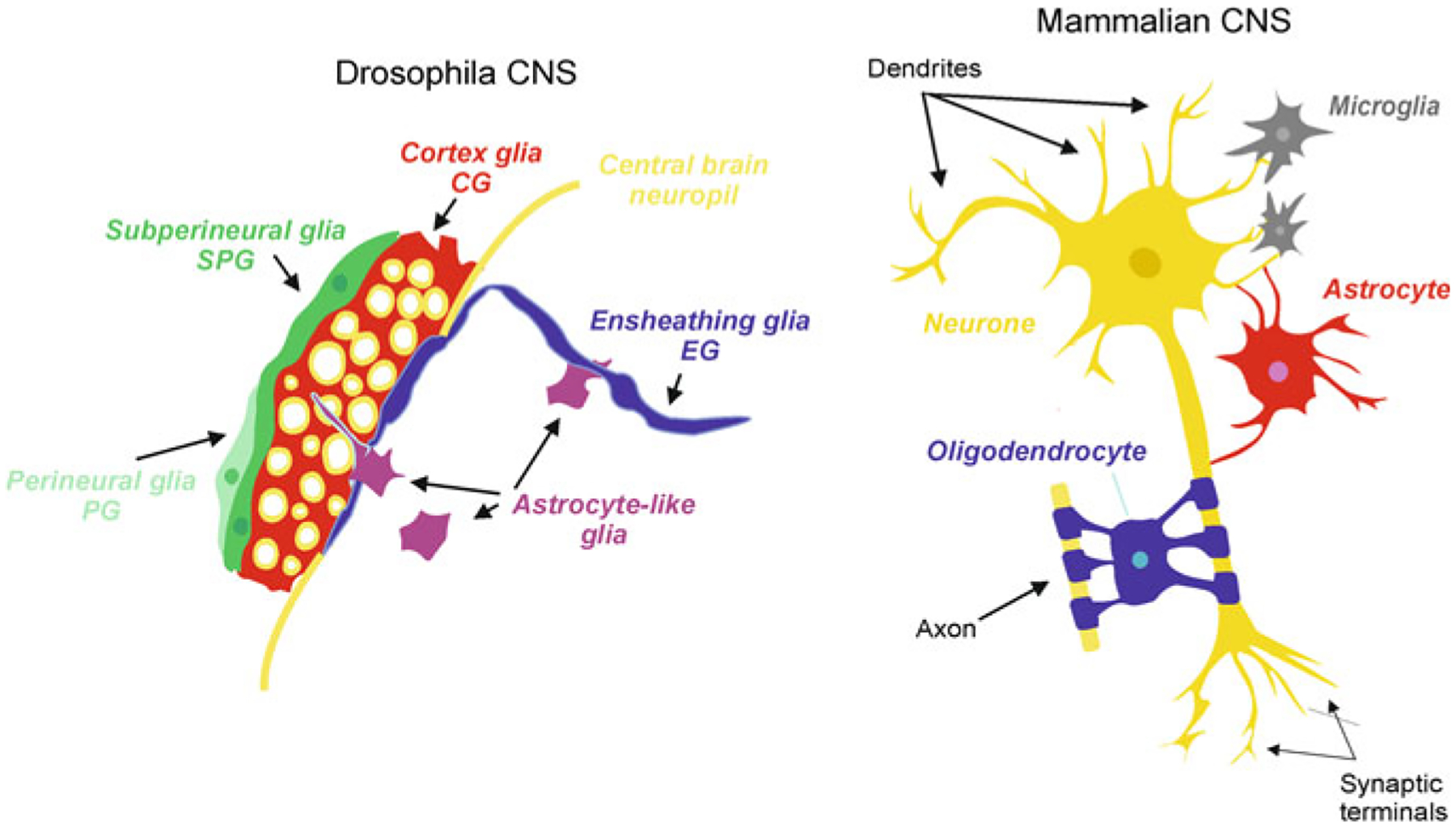
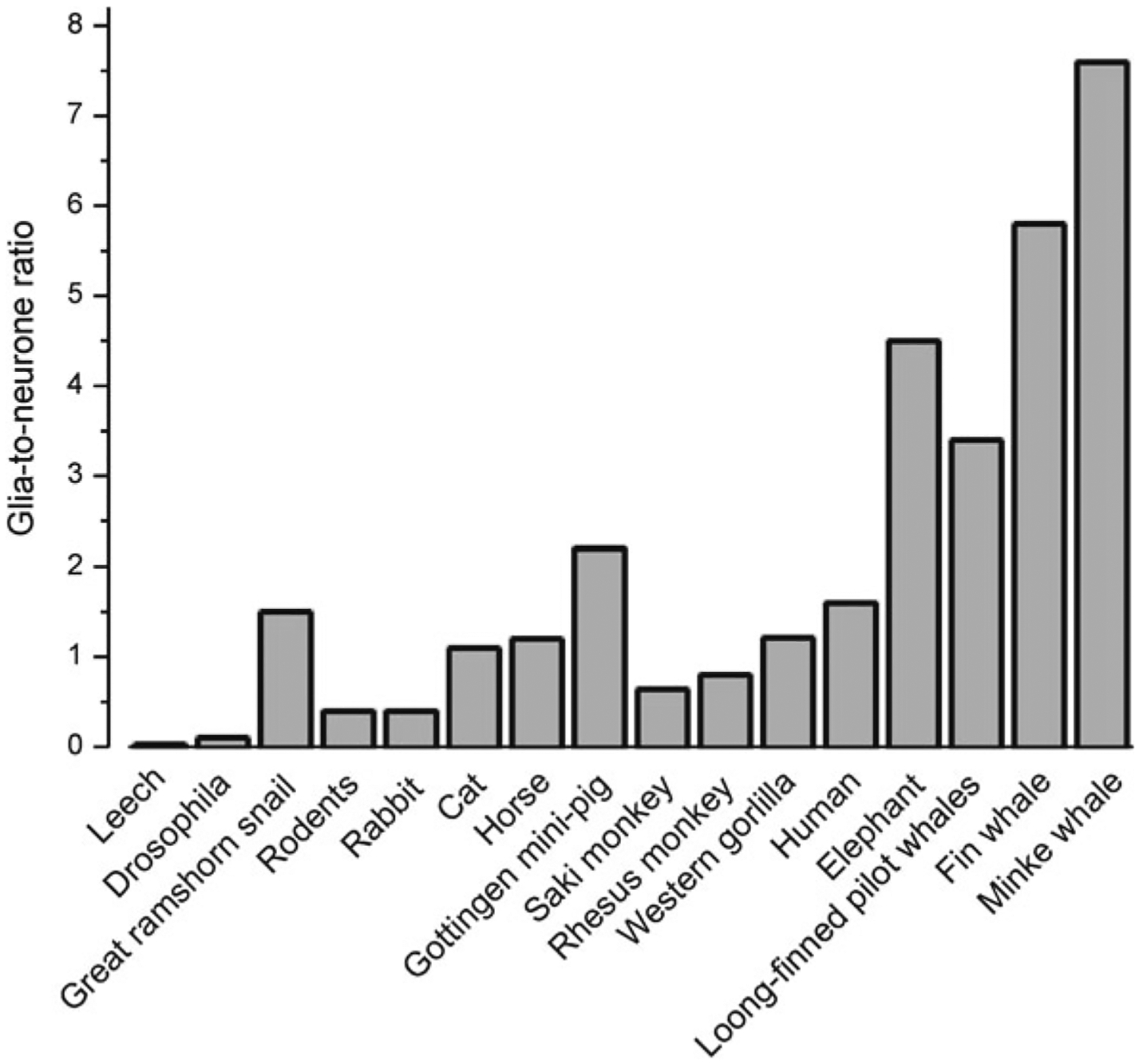
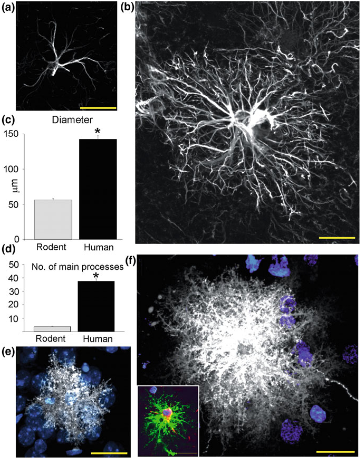
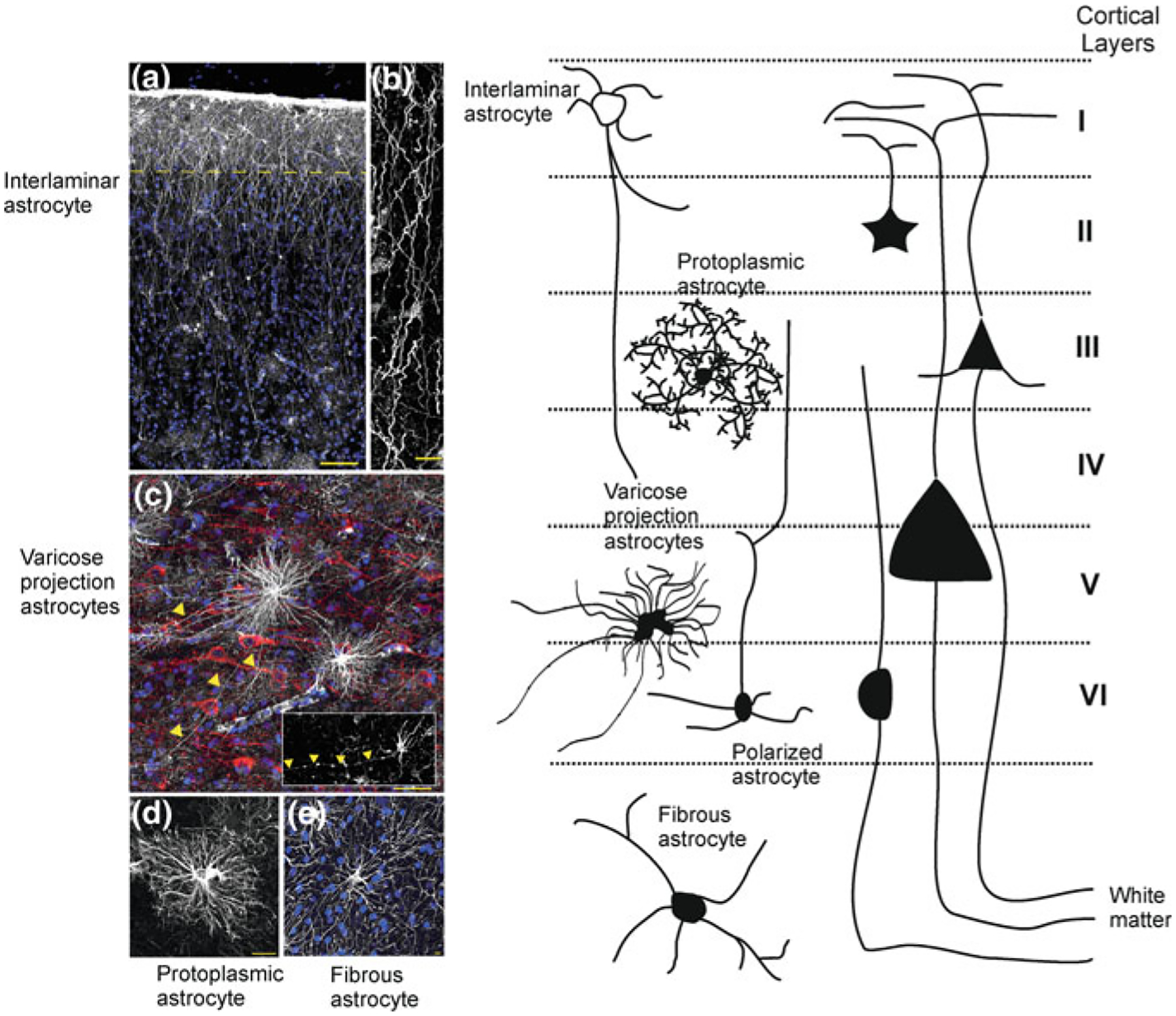
As the nervous system evolved from the diffused to centralised form, the neurones were joined by the appearance of the supportive cells, the neuroglia. Arguably, these non-neuronal cells evolve into a more diversified cell family than the neurones are. The first ancestral neuroglia appeared in flatworms being mesenchymal in origin. In the nematode C. elegans proto-astrocytes/supportive glia of ectodermal origin emerged, albeit the ensheathment of axons by glial cells occurred later in prawns. The multilayered myelin occurred by convergent evolution of oligodendrocytes and Schwann cells in vertebrates above the jawless fishes. Nutritive partitioning of the brain from the rest of the body appeared in insects when the hemolymph-brain barrier, a predecessor of the blood-brain barrier was formed. The defensive cellular mechanism required specialisation of bona fide immune cells, microglia, a process that occurred in the nervous system of leeches, bivalves, snails, insects and above. In ascending phylogeny, new type of glial cells, such as scaffolding radial glia, appeared and as the bran sizes enlarged, the glia to neurone ratio increased. Humans possess some unique glial cells not seen in other animals.

Keywords: Astrocytes; Blood/haemolymph-brain barrier; Brain size; Complexity of glia; Glia to neuron ratio; Microglia; Myelination; Oligodendrocytes; Radial glia.


Figures

Fig. 2.1
Fig. 2.1
Tree of life and evolution of the nervous system and of neuroglia. Adapted from Verkhratsky and Butt [143]
Fig. 2.2
Fig. 2.2
Glial cells in Caenorhabditis elegans. The “brain” of C. elegance is represented by the nerve ring. Most of the glial cells are part of sensory organs known as sensilla. Each sensilla has two glial cells: the sheath cell and socket cell. In the anterior part there are 4 CEP (cephalic) glial cells that ensheath nerve ring. The nerve ring also has 6 GLR glial cells which establish gap junctional contacts between ring motor neurones (RME) and muscle cells
Fig. 2.3
Fig. 2.3
The CEPsh glia. a A cartoon of an adult worm showing the four CEPsh glial cells (green) positioned in the anterior of the worm (inset). The CEPsh cell bodies with their velate processes are positioned around the central nerve ring (red) which they enwrap along with the proximal section of the ventral nerve cord. Additionally, each CEPsh glial cell possesses a long anterior process, emanating to the anterior sensory tip, which closely interacts with the dendritic extension of a nearby CEP neuron (blue). Arrows indicate the dorsal (red arrow) and ventral (orange arrow) side of the worm. b A confocal image showing green fluorescent protein expression driven by the hlh-17 promoter to visualize the four CEPsh glial cells (worm strain VPR839). The anterior (head) of a juvenile (larval stage 4) worm is shown; the worm is turned ~45° from “upright” such that all four CEP sheath cells are visible. The sheath portion of the cells that form a tube around the dendritic endings of the CEP neurones are seen at the left of the image. The dorsal (red arrow) and ventral (orange arrow) CEPsh cell bodies are seen. The thin sheet-like extensions that surround and invade the nerve ring are seen in the rightmost part of the image. Scale bar, 20 μm. Image adapted from [134]
Fig. 2.4
Fig. 2.4
L-type voltage-gated Ca2+channels (VGCCs) play a role in depolarization-induced intracellular Ca2+ elevations in CEPsh glial cells. a The hlh-17 promoter can be used to drive expression of a red fluorescent protein marker (red, mCherry) in the CEPsh glia along with a fluorescent-protein based Ca2+ sensor (green, GCaMP2.0). DIC, differential interference contrast. An anterior portion of an L4 stage worm (VPR108 strain) is shown. b CEPsh glial cells in mixed culture prepared from embryos can be identified based on their mCherry/GCaMP2.0 expression. c Time-lapse of GCaMP2.0 fluorescence emission from CEPsh glial cells. Paired-pulse application of a depolarization stimulus, high extracellular potassium (HiK+, 100 mM; horizontal bar), to CEPsh glial cells results in an elevation of intracellular Ca2+ levels (black squares). Nemadipine-A (NemA), a pharmacological L-type VGCC blocker, can be used to test the channels present in glial cells in culture; Con, sham stimulated control. (right, bar graph). Ratio of the peak Ca2+ level in response to the second HiK+ application (P2) over the first application (P1). *Indicates a significant difference. Adapted from [133]
Fig. 2.5
Fig. 2.5
Neuroglia in medicinal leech Hirudo medicinalis. a General structure of the nervous system. b Structure of a segmental ganglion, which contains three types of glial cells: the giant glial cell; packet glial cells and connective glial cells. Adapted from Verkhratsky and Butt [143]
Fig. 2.6
Fig. 2.6
Neuroglia in Drosophila and mammals
Fig. 2.7
Fig. 2.7
Phylogenetical advance of neuroglia. Glia-to-neurone ratio in the nervous system of invertebrates and in the cortex of vertebrates. Glia-to-neurone ratio is generally increased in phylogeny; more or less this ratio linearly follows an increase in the size of the brain
Fig. 2.8
Fig. 2.8
Comparison of rodent and human protoplasmic astrocytes. a Typical mouse protoplasmic astrocyte. GFAP, White. Scale bar, 20 μm. b Typical human protoplasmic astrocyte at the same scale. Scale bar, 20 μm. c, d Human protoplasmic astrocytes are 2.55-fold larger and have 10-fold more main GFAP processes than mouse astrocytes (human, n = 50 cells from 7 patients; mouse, n = 65 cells from 6 mice; mean ± SEM; *p < 0.005, t test). e Mouse protoplasmic astrocyte diolistically labelled with DiI (white) and sytox (blue) revealing the full structure of the astrocyte including its numerous fine processes. Scale bar, 20 μm. f Human astrocyte demonstrates the highly complicated network of fine process that defines the human protoplasmic astrocyte. Scale bar, 20 μm. Inset, Human protoplasmic astrocyte diolistically labelled as well as immunolabelled for GFAP (green) demonstrating colocalisation. Scale bar, 20 μm. Reproduced, with permission from [99, 144]
Fig. 2.9
Fig. 2.9
Morphological heterogeneity and subtypes of astrocytes in the cortex of higher primates. a Pial surface and layers 1–2 of human cortex. GFAP staining in white; DAPI, in blue. Scale bar, 100 μm. Yellow line indicates border between layer I and II. b Interlaminar astrocyte processes. Scale bar, 10 μm. c Varicose projection astrocytes reside in layers V and VI and extend long processes characterized by evenly spaced varicosities. Inset: Varicose projection astrocyte from chimpanzee cortex. Yellow arrowheads indicate varicose projections. Scale bar, 50 μm. d Typical human protoplasmic astrocyte. Scale bar, 20 μm. e Human fibrous astrocytes in white matter. Scale bar, 10 μm. (modified with permission from [98]. Left panel schematically shows different astrocytes and their relatuons to cortical layers. Adapted from [143]

References

    1. Altun ZF, Chen B, Wang ZW, Hall DH (2009) High resolution map of Caenorhabditis elegans gap junction proteins. Dev Dyn 238:1936–1950 - PMC - PubMed
    1. Ari C, Kalman M (2008) Evolutionary changes of astroglia in Elasmobranchii comparing to amniotes: a study based on three immunohistochemical markers (GFAP, S-100, and glutamine synthetase). Brain Behav Evol 71:305–324 - PubMed
    1. Awasaki T, Lai SL, Ito K, Lee T (2008) Organization and postembryonic development of glial cells in the adult central brain of Drosophila. J Neurosci 28:13742–13753 - PMC - PubMed
    1. Bacaj T, Tevlin M, Lu Y, Shaham S (2008) Glia are essential for sensory organ function in C. elegans. Science 322:744–747 - PMC - PubMed
    1. Bahney J, von Bartheld CS (2018) The cellular composition and glia-neuron ratio in the spinal cord of a human and a nonhuman primate: comparison with other species and brain regions. Anat Rec (Hoboken) 301:697–710 - PMC - PubMed