Review
. 2019:1175:45-91.
doi: 10.1007/978-981-13-9913-8_3.

Physiology of Astroglia

Affiliations
Review

Physiology of Astroglia

Alexei Verkhratsky et al. Adv Exp Med Biol. 2019.

Abstract

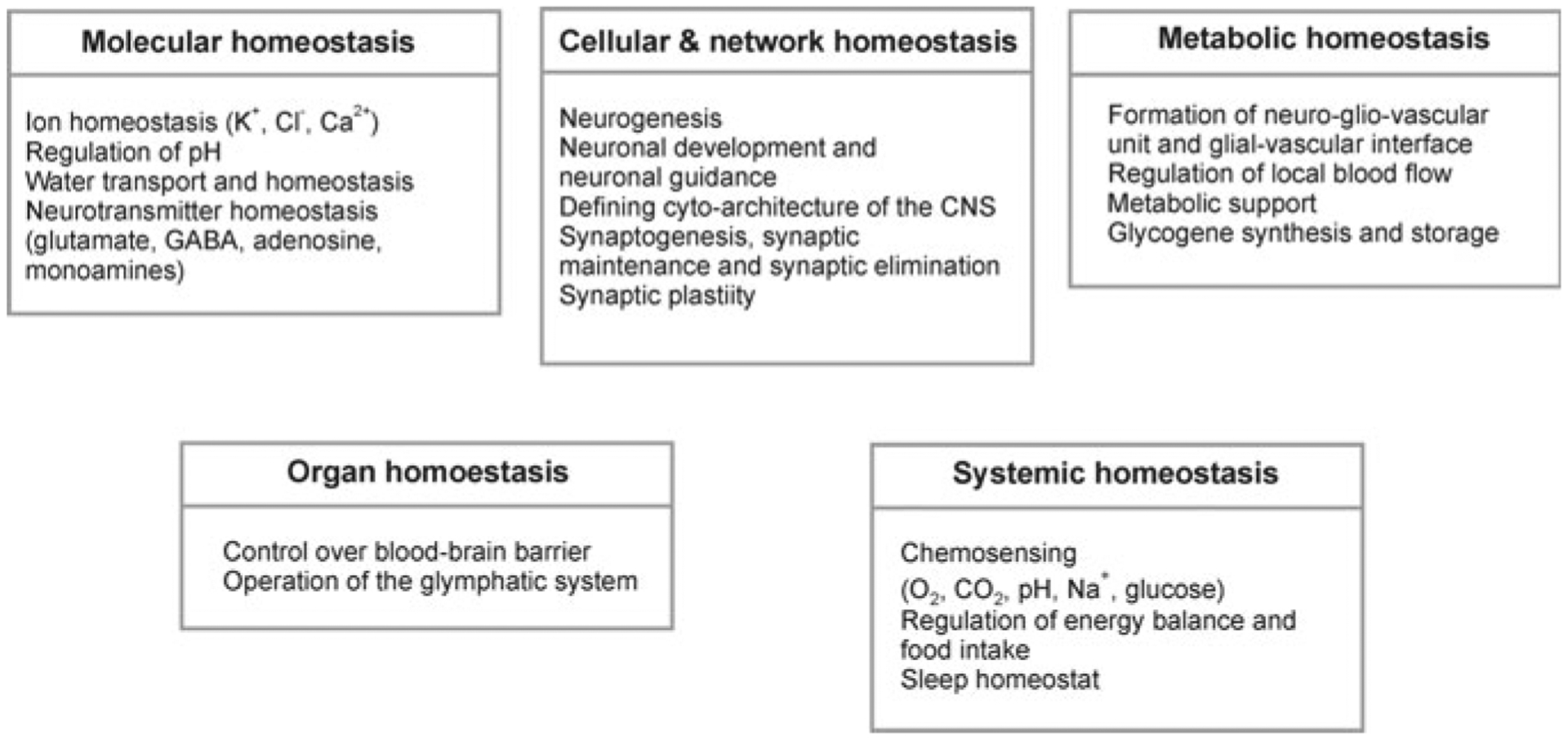
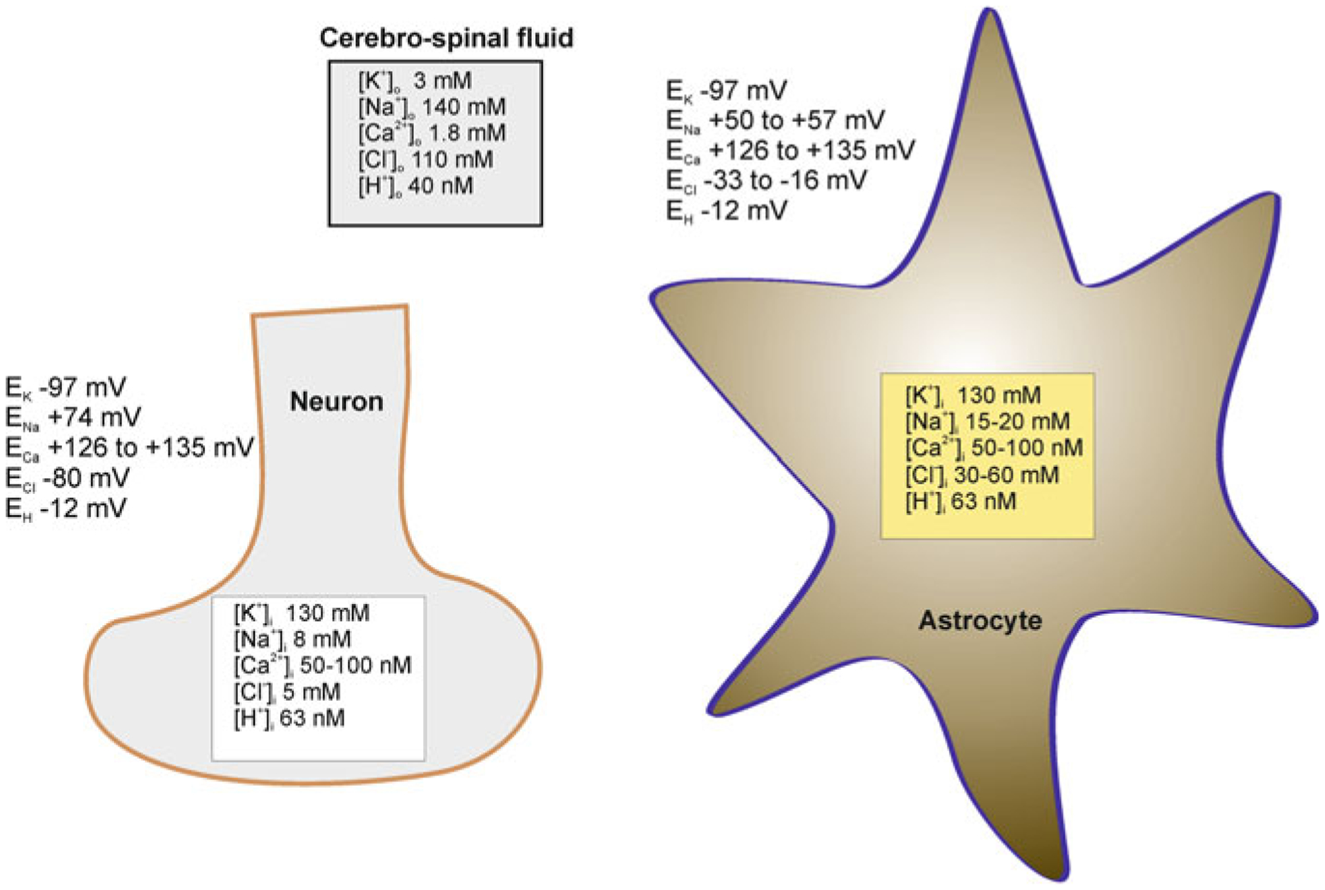
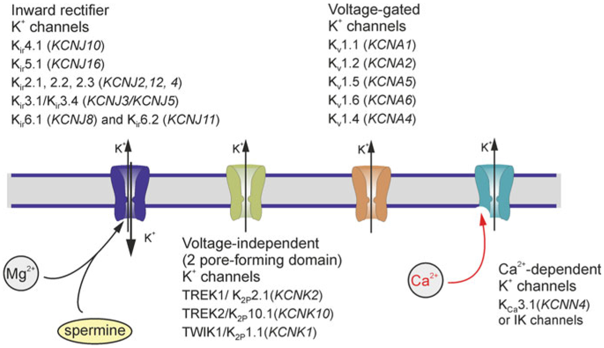
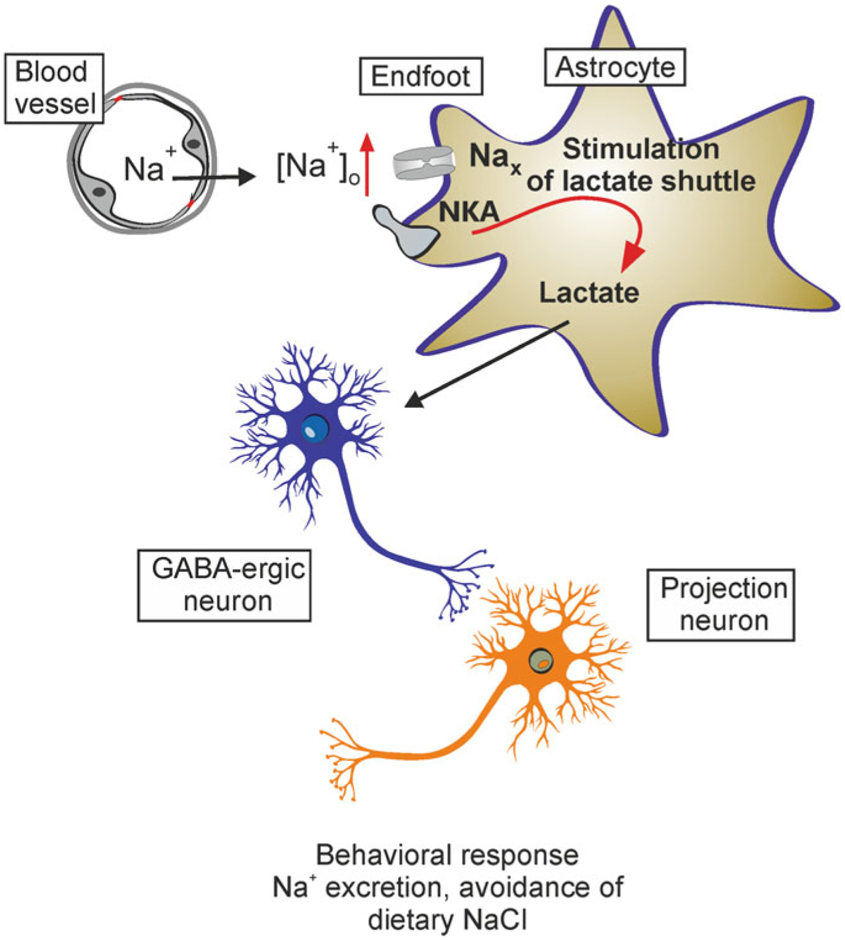
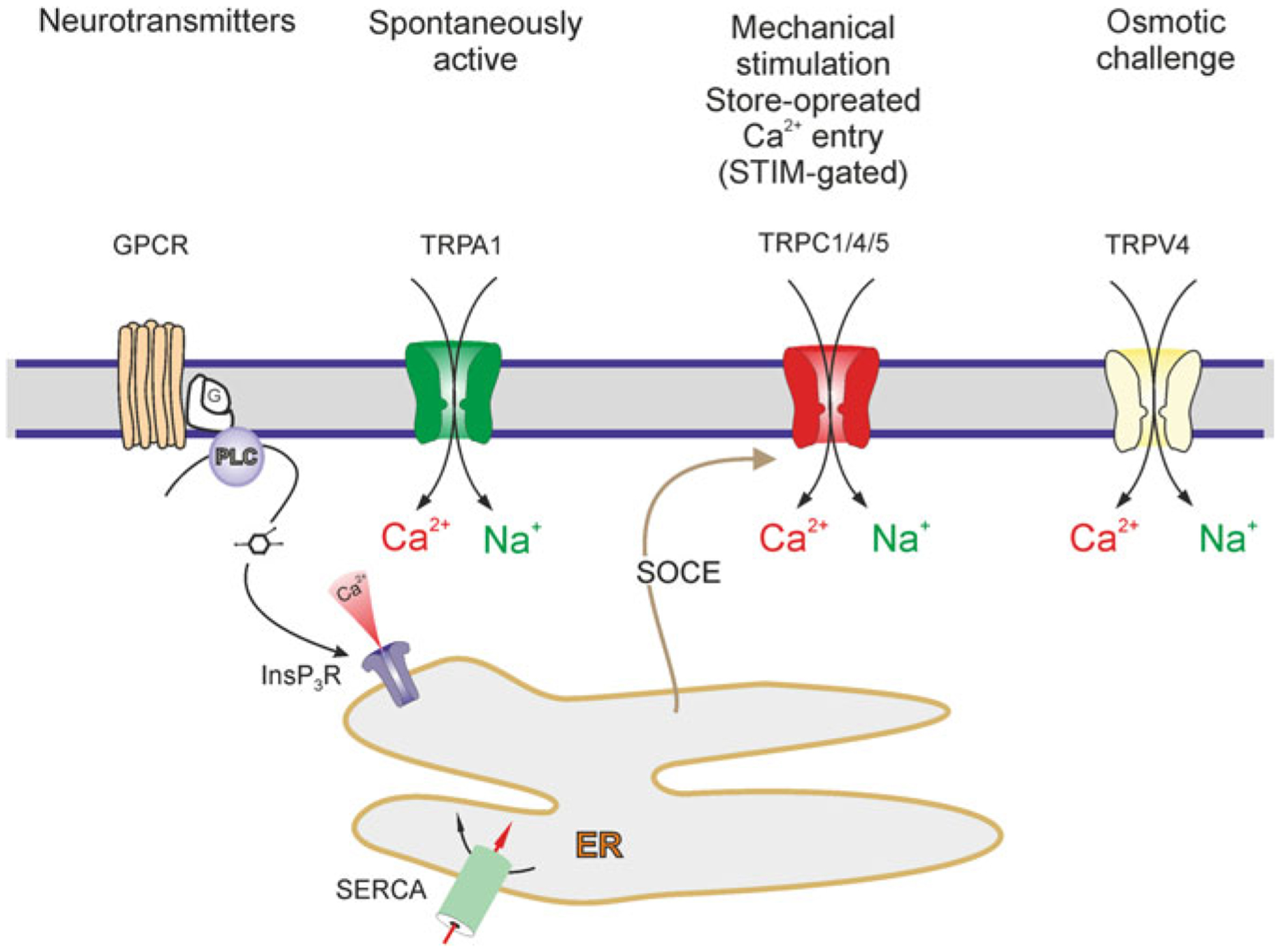
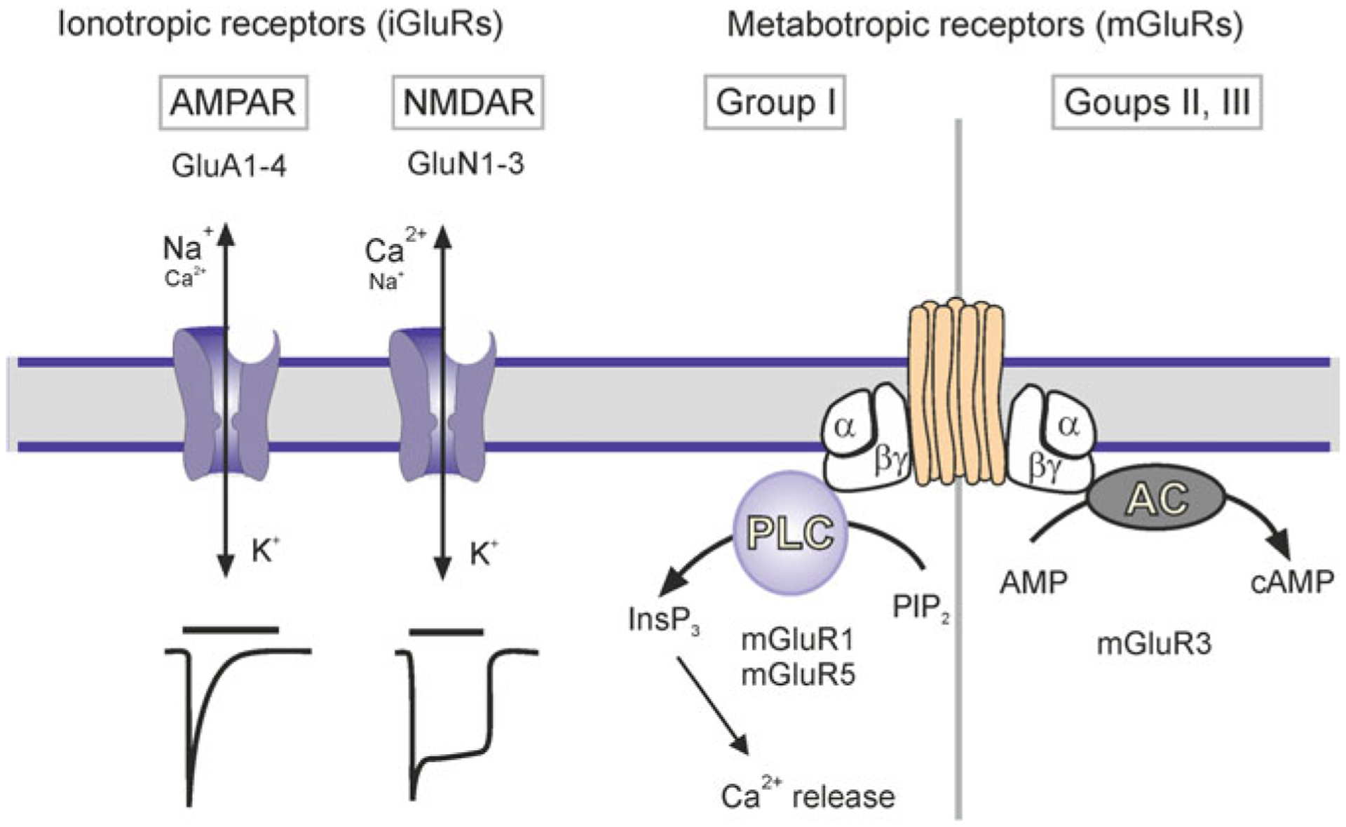
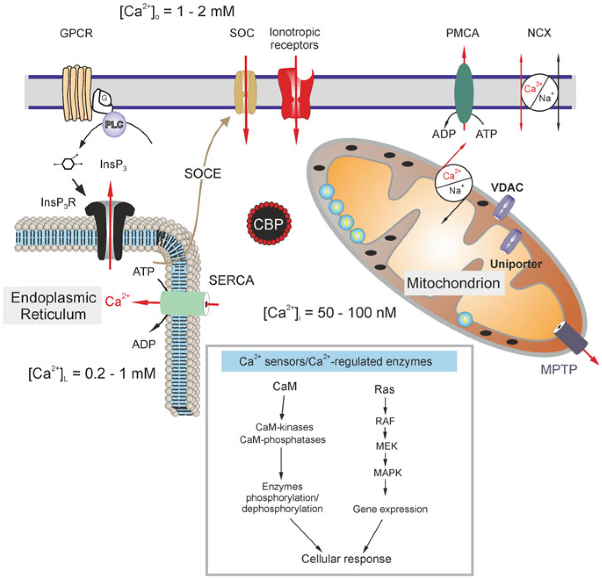
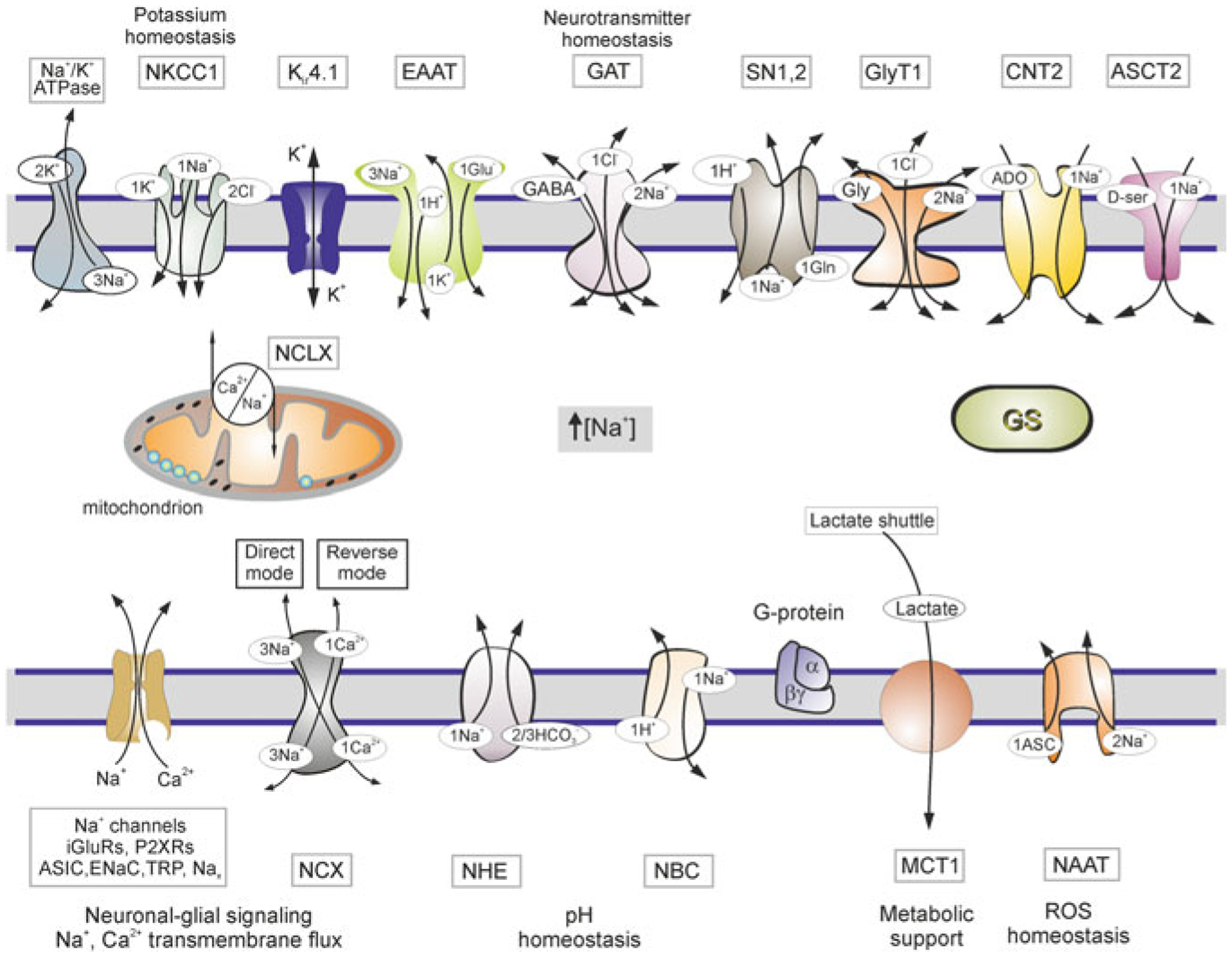
Astrocytes are principal cells responsible for maintaining the brain homeostasis. Additionally, these glial cells are also involved in homocellular (astrocyte-astrocyte) and heterocellular (astrocyte-other cell types) signalling and metabolism. These astroglial functions require an expression of the assortment of molecules, be that transporters or pumps, to maintain ion concentration gradients across the plasmalemma and the membrane of the endoplasmic reticulum. Astrocytes sense and balance their neurochemical environment via variety of transmitter receptors and transporters. As they are electrically non-excitable, astrocytes display intracellular calcium and sodium fluctuations, which are not only used for operative signalling but can also affect metabolism. In this chapter we discuss the molecules that achieve ionic gradients and underlie astrocyte signalling.

Keywords: Astrocytes; Brain homoeostasis; Ca2+ signalling; Ion channels; Na+ signalling; Neurotransmitter receptors; SLC transporters.


Figures

Fig. 3.1
Fig. 3.1
Homoeostatic functions of astroglia
Fig. 3.2
Fig. 3.2
Ion distribution (and corresponding values of equilibrium potentials for different ions) between the cerebrospinal fluid and cytosol of astrocytes and neurones. Modified from [413]
Fig. 3.3
Fig. 3.3
Potassium channels in astroglia. Gene names for a given channels are shown in parentheses. Modified from [413]
Fig. 3.4
Fig. 3.4
Astroglial Nax channels in systemic Na+ regulation. Increases in blood Na+ concentration activate Nax sodium channels localised in astrocytes residing in the subfornical organ. This leads to an increase in cytosolic Na+ concentration, which in turn increases astroglial production of lactate. Lactate released by astrocytes is accumulated by neighbouring neurones (release and uptake carried by MCT1 in astrocytes and MCT4 in neurones, respectively), thus increasing ATP production in neurones. Increased ATP in turn closes neuronal ATP-sensitive K+ channels, which results in depolarisation and subsequent activation of neuronal networks responsible for systemic Na+ homeostasis. NKA, sodium-potassium ATPase. Modified from [413]
Fig. 3.5
Fig. 3.5
Astroglial TRP channels. Activation of G-protein coupled receptors (GPCR), i.e. metabotropic stimulation, can lead to production of InsP3 and release of Ca2+ from the ER store. The Ca2+ content of the ER store is refilled by Sarco(Endo)Plasmic Reticulum Ca2+ ATPase, i.e. SERCA. Depletion of the ER Ca2+ store activates (via STIM) TRPC channels in astrocytes which are therefore acting as a store-operated channel, contributing to capacitative Ca2+ entry. Activation of all TRP channels mediates Ca2+ and Na+ influx. Modified from [413]
Fig. 3.6
Fig. 3.6
Classes of purinoreceptors. ATP after being released from neurones and glia is rapidly degrading by ectonucleotidases into ADP, AMP and adenosine, which act on P1 metabotropic adenosine receptors, P2X ionotropic and P2Y metabotropic nucleotide receptors. Adenine stimulates A0 adenine metabotorpic receptors, which hitherto have not been detected in astrocytes. Modified from [406]
Fig. 3.7
Fig. 3.7
Classes of glutamate receptors expressed in astrocytes. Current traces show a faster time course for AMPAR than NMDAR. AC, adenylyl cyclase; AMP, adenosine monophosphate; cAMP, cyclic AMP; InsP3, inositol 1,4,5-trisphosphate; PIP2, phosphatidylinositol 4,5-bisphosphate; PLC, phospholipase C. Modified from [406]
Fig. 3.8
Fig. 3.8
Calcium distribution and calcium signalling cascades in intracellular compartments. Stimuli-induced increases in [Ca2+]i could be caused by the entry of Ca2+ from the extracellular space through ionotropic receptors or store-operated channels (SOC). Plasmalemmal Ca2+ pumps/ATPases (PMCA) can extrude cytosolic Ca2+, while the plasmalemmal sodium-calcium exchanger (NCX) can operate in both directions depending on intercellular Na+ concentration and membrane potential. An additional source of Ca2+ is available from the ER internal store that possesses inositol 1,4,5 trisphosphate (InsP3) receptors, which can be activated by the activity of metabotropic G-protein coupled receptors (GPCRs) and phospholipase C (PLC). The ER store is (re)filled by the activity of the store-specific Ca2+-ATPase (SERCA). Cytosolic Ca2+ levels can be affected by a variety of cytosolic Ca2+-binding proteins (CBPs) and by the action of mitochondria. A negative membrane potential exists across the inner mitochondrial membrane. Mitochondrial Ca2+ uptake occurs through voltage-dependent anion channels (VDACs) present in the outer membrane and by the uniporter in the inner membrane as the electrochemical gradient drives Ca2+ into the matrix, while free Ca2+ exits the mitochondrial matrix through the mitochondrial Na+/Ca2+ exchanger and transient opening of the mitochondrial permeability transition pore (MPTP). Concentrations of free Ca2+ in different compartments are indicated on the scheme. Inset shows various Ca2+ effector molecules, sensors and enzymes. CaM, calmodulin; RAF, Rapidly Accelerated Fibrosarcoma, MAPK, mitogen-activated protein kinase (MAPK), MEK, MAPK kinase. Modified from [413]
Fig. 3.9
Fig. 3.9
Molecules of Na+ homeostasis and targets of Na+ signalling in astroglia. Schematic diagram showing receptors and transporters involved in and sensitive to changes in [Na+]i and their relations to main homeostatic functions of astroglia. Abbreviations ASCT2, alanineserine-cysteine transporter 2; ASIC-acid sensing ion channels; CNT2, concentrative nucleoside transporters; EAAT-excitatory amino acid transporters; ENaC-epithelial sodium channels; GAT-GABA transporters; GS-glutamine synthetase, GlyT1-glycine transporter. iGluRs-ionotropic glutamate receptors; Nax-Na+ channels activated by extracellular Na+; NAAT-Na+-dependent ascorbic acid transporter; NBC-Na+/HCO3 (sodium-bicarbonate) cotransporter; NCX-Na+/Ca2+ exchanger; NCLX-mitochondrial Na+/Ca2+ exchanger; NHE-Na+/H+ exchanger; NKCC1-Na+/K+/Cl cotransporter, MCT1-monocarboxylase transporter 1; P2XRs-ionotropic purinoceptors; SN1,2-sodium-coupled neutral amino acid transporters which underlie exit of glutamine; TRP-transient receptor potential channels. Reactive oxygen species (ROS). Modified from [413]

References

    1. Abbracchio MP, Ceruti S (2006) Roles of P2 receptors in glial cells: focus on astrocytes. Purinergic Signal 2:595–604 - PMC - PubMed
    1. Adermark L, Lovinger DM (2008) Electrophysiological properties and gap junction coupling of striatal astrocytes. Neurochem Int 52:1365–1372 - PMC - PubMed
    1. Agarwal A, Wu PH, Hughes EG, Fukaya M, Tischfield MA, Langseth AJ, Wirtz D, Bergles DE (2017) Transient opening of the mitochondrial permeability transition pore induces microdomain calcium transients in astrocyte processes. Neuron 93(587–605):e587 - PMC - PubMed
    1. Agulhon C, Fiacco TA, McCarthy KD (2010) Hippocampal short- and long-term plasticity are not modulated by astrocyte Ca2+ signaling. Science 327:1250–1254 - PubMed
    1. Allbritton NL, Meyer T, Stryer L (1992) Range of messenger action of calcium ion and inositol 1,4,5-trisphosphate. Science 258:1812–1815 - PubMed