Review
. 2019 Oct;25(5):1236-1264.
doi: 10.1212/CON.0000000000000768.

Optic Neuritis

Review

Optic Neuritis

Jeffrey L Bennett. Continuum (Minneap Minn). 2019 Oct.

Abstract

Purpose of review: This article discusses the clinical presentation, evaluation, and management of the patient with optic neuritis. Initial emphasis is placed on clinical history, examination, diagnostic testing, and medical decision making, while subsequent focus is placed on examining specific inflammatory optic neuropathies. Clinical clues, examination findings, neuroimaging, and laboratory testing that differentiate autoimmune, granulomatous, demyelinating, infectious, and paraneoplastic causes of optic neuritis are assessed, and current treatments are evaluated.

Recent findings: Advances in technology and immunology have enhanced our understanding of the pathologies driving inflammatory optic nerve injury. Clinicians are now able to interrogate optic nerve structure and function during inflammatory injury, rapidly identify disease-relevant autoimmune targets, and deliver timely therapeutics to improve visual outcomes.

Summary: Optic neuritis is a common clinical manifestation of central nervous system inflammation. Depending on the etiology, visual prognosis and the risk for recurrent injury may vary. Rapid and accurate diagnosis of optic neuritis may be critical for limiting vision loss, future neurologic disability, and organ damage. This article will aid neurologists in formulating a systematic approach to patients with optic neuritis.


Figures

FIGURE 3–1
FIGURE 3–1
Fat-suppressed postcontrast T1-weighted orbital imaging of optic neuritis. A, Axial image shows bilateral longitudinally extensive lesions involving the orbital and intracanalicular optic nerves in a patient with myelin oligodendrocyte glycoprotein antibody (MOG-IgG) optic neuritis. Note the enhancement of the optic discs suggestive of disc edema. B, Coronal image of optic nerves in panel A showing both sheath (right eye) and nerve (left eye) enhancement. Gadolinium contrast fills both superior orbital veins (arrows). C, Coronal image showing an enlarged, enhancing optic chiasm in a patient with neuromyelitis optica (NMO) spectrum disorder-associated optic neuritis. Additional cloudlike enhancement is seen in the left thalamus/basal ganglia due to NMO inflammation. D, Bilateral enhancing lesions of the orbital and intracanalicular optic nerves in a patient with chronic relapsing immune-mediated optic neuropathy.
FIGURE 3–2
FIGURE 3–2
Mild optic disc edema associated with multiple sclerosis.
FIGURE 3–3
FIGURE 3–3
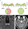
Findings of the patient in CASE 3–1. A, Spectral domain optical coherence tomography of the peripapillary retinal nerve fiber layer showing severe thinning on the right (mean right: 51 microns; mean left: 104 microns). B, Axial fat-suppressed postcontrast T1-weighted MRI showing a subtle longitudinally extensive enhancing lesion of right orbital nerve and sheath. C, Axial T2-weighted MRI showing subtle signal abnormality (arrowhead) in the posterior right optic nerve.
FIGURE 3–4
FIGURE 3–4
Findings of the patient in CASE 3–2. A, Axial fat-saturated T2-weighted turbo inversion recovery magnitude images showing enlarged optic nerves with long T2-hyperintense orbital lesions. B, Coronal short tau inversion recovery (STIR) images showing T2-hyperintense enlarged optic nerves. C, Spectral domain optical coherence tomography of the peripapillary retinal nerve fiber layer showing normal mean thickness in both eyes despite two prior episodes of optic neuritis. Mild segmental retinal nerve fiber layer thickening is seen in the right eye due to edema as well as focal temporal thinning due to prior episodes of optic neuritis, together leading to the normal mean thickness (pseudo-normal mean thickness). The patient had no clinical evidence of disc edema on examination.
FIGURE 3–5
FIGURE 3–5
Optic disc edema and retinal inflammation in optic neuritis. Optic disc edema with nerve fiber layer (splinter) hemorrhages due to myelin oligodendrocyte glycoprotein (MOG)-IgG optic neuritis.
FIGURE 3–6
FIGURE 3–6
Perivascular infiltrates (candle wax droppings) due to sarcoidosis.
FIGURE 3–7
FIGURE 3–7
Findings of the patient in CASE 3–3. A, Fundus photography of the left eye revealing optic disc pallor. B, Axial fat-suppressed postcontrast T1-weighted MRI showing longitudinally extensive enhancement of left optic nerve. C, Low-magnification image of hilar lymph node biopsy stained with hematoxylin and eosin (H&E) showing replacement of the normal lymph node architecture by multiple small, well-defined, non-necrotizing granulomas of relatively uniform sizes and shapes that coalesce with variable degrees of fibrosis. D, High-magnification image of granuloma showing cytologic features of the epithelioid histiocytes. Panels C and D courtesy of Jeffrey Schowinsky, MD.
FIGURE 3–8
FIGURE 3–8
Optic disc edema with partial macular star (macular fan) due to neuroretinitis. Note the dilated venules due to venous compression from disc edema.
FIGURE 3–9
FIGURE 3–9
Funduscopy of the patient in CASE 3–4. Fundus photos demonstrate severe disc edema with nerve fiber layer hemorrhages in the right eye. The left optic nerve is normal. The diagnosis was optic neuritis due to syphilis.

References

    1. Beck RW, Cleary PA, Anderson MM, et al. A randomized, controlled trial of corticosteroids in the treatment of acute optic neuritis. The Optic Neuritis Study Group. N Engl J Med 1992;326(9): 581–588. doi:10.1056/NEJM199202273260901. - DOI - PubMed
    1. Jarius S, Paul F, Aktas O, et al. MOG encephalomyelitis: international recommendations on diagnosis and antibody testing. J Neuroinflammation 2018;15(1):134. doi:10.1186/s12974-018-1144-2. - DOI - PMC - PubMed
    1. Jurynczyk M, Messina S, Woodhall MR, et al. Clinical presentation and prognosis in MOG-antibody disease: a UK study. Brain 2017;140(12): 3128–3138. doi:10.1093/brain/awx276. - DOI - PubMed
    1. Ramanathan S, Prelog K, Barnes EH, et al. Radiological differentiation of optic neuritis with myelin oligodendrocyte glycoprotein antibodies, aquaporin-4 antibodies, and multiple sclerosis. Mult Scler 2016;22(4):470–482. doi:10.1177/1352458515593406. - DOI - PubMed
    1. The clinical profile of optic neuritis. Experience of the Optic Neuritis Treatment Trial. Optic Neuritis Study Group. Arch Ophthalmol 1991; 109(12):1673–1678. - PubMed