. 2019 Nov 1;43(6):674-683.
doi: 10.1093/femsre/fuz023.

Epithelial cell infection by Epstein-Barr virus

Affiliations

Epithelial cell infection by Epstein-Barr virus

Jia Chen et al. FEMS Microbiol Rev. .

Abstract

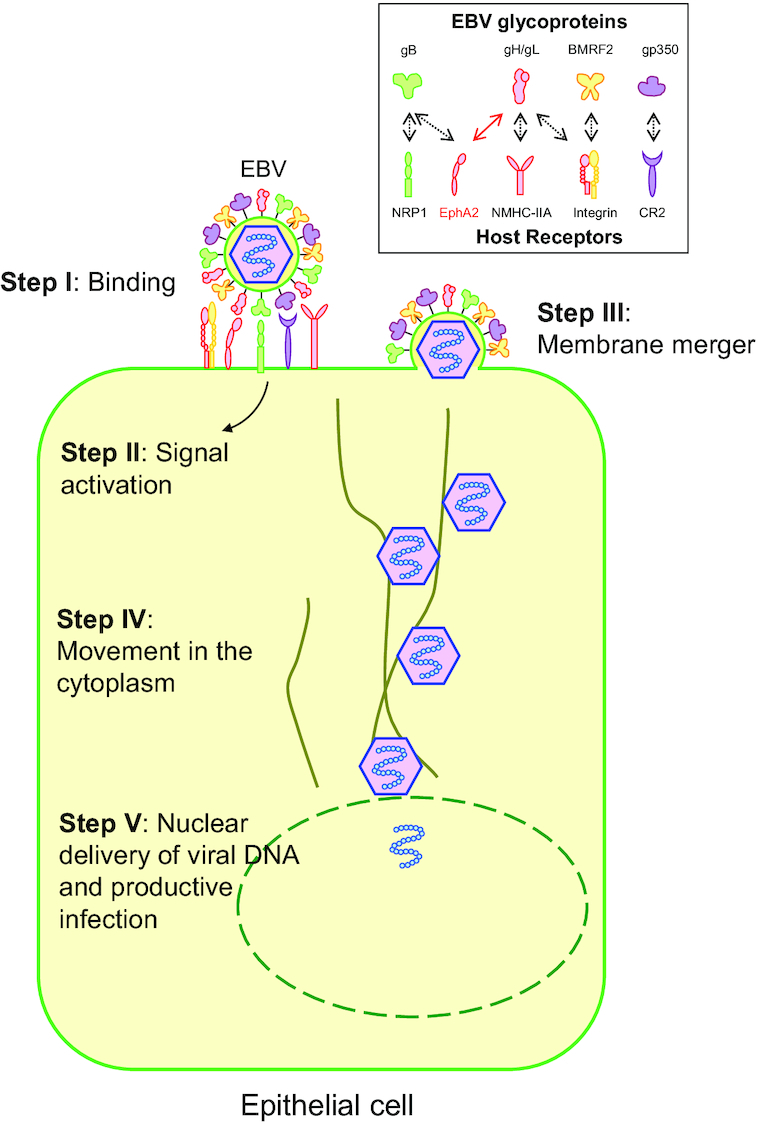
Epstein-Barr Virus (EBV) is etiologically associated with multiple human malignancies including Burkitt lymphoma and Hodgkin disease as well as nasopharyngeal and gastric carcinoma. Entry of EBV into target cells is essential for virus to cause disease and is mediated by multiple viral envelope glycoproteins and cell surface associated receptors. The target cells of EBV include B cells and epithelial cells. The nature and mechanism of EBV entry into these cell types are different, requiring different glycoprotein complexes to bind to specific receptors on the target cells. Compared to the B cell entry mechanism, the overall mechanism of EBV entry into epithelial cells is less well known. Numerous receptors have been implicated in this process and may also be involved in additional processes of EBV entry, transport, and replication. This review summarizes EBV glycoproteins, host receptors, signal molecules and transport machinery that are being used in the epithelial cell entry process and also provides a broad view for related herpesvirus entry mechanisms.

Keywords: Epstein–Barr virus; epithelial cells; gamma-herpesviruses.


Figures

Figure 1.
Figure 1.
Model illustrating the steps of EBV infection of epithelial cells. In the first step (Step I), EBV binds to target cells using variable host cell surface receptors and multiple viral envelope glycoproteins. In some cases, binding of EBV virions can induce signaling pathway activation (Step II). After binding to the cell surface receptors, (either by direct membrane fusion or fusion with the endosomal membrane) (Step III), the viral capsid is then transported in the cytosol to the nuclear periphery (Step IV). Once at a nuclear pore, the viral genome is released into the nucleus through a nuclear pore (Step V). Integrins (αvβ5, αvβ6 and αvβ8), NRP1 and NMHC-IIA interact with corresponding glycoproteins indicated as black-dashed arrows. EphA2 binds to both gH/gL (red solid arrow) and gB (black-dashed arrow). EphA2 is the most important entry receptor for EBV epithelial cell infection and is therefore indicated in red.
Figure 2.
Figure 2.
Different anti-gH/gL antibodies target different regions of gH/gL and inhibit EBV epithelial cell infection. The structure of gH/gL is shown as cartoon (Matsuura et al. ; Connolly et al. 2011). gH consists of four domains: D-I (blue), D-II (magenta), D-III (green) and D-IV (yellow). gL is colored red and interacts with gH in D-I. The E1D1 antibody binding region on gH/gL is shown as red surface representation in D-I gL (gL 27–33, gL 72–79 and gL 127–131; D-I is also the potential EphA2 binding region). The most important AMMO1 antibody-binding residues on EBV gH/gL are represented by green spheres in D-I (gH K73 and gH Y76). The CL40 binding amino acids on gH/gL are represented by magenta spheres (gH R184, gH H239, gH V243, gH D284 and gH E286). The CL59 antibody binding regions on gH/gL are shown as green and yellow surface representations in D-III and D-IV (gH406–415, gH456–468, gH494–503, gH623–626 and gH645–656).

References

    1. Aaron PA, Jamklang M, Uhrig JPet al. .. The blood–brain barrier internalises Cryptococcus neoformans via the EphA2-tyrosine kinase receptor. Cell Microbiol. 2018;20:e12811. - PMC - PubMed
    1. Arii J, Goto H, Suenaga Tet al. .. Non-muscle myosin IIA is a functional entry receptor for herpes simplex virus-1. Nature. 2010;467:859–62. - PubMed
    1. Babcock GJ, Decker LL, Volk Met al. .. EBV persistence in memory B cells in vivo. Immunity. 1998;9:395–404. - PubMed
    1. Backovic M, Jardetzky TS, Longnecker R. Hydrophobic residues that form putative fusion loops of Epstein–Barr virus glycoprotein B are critical for fusion activity. J Virol. 2007;81:9596–600. - PMC - PubMed
    1. Backovic M, Longnecker R, Jardetzky TS. Structure of a trimeric variant of the Epstein–Barr virus glycoprotein B. P Natl Acad Sci USA. 2009;106:2880–5. - PMC - PubMed

Publication types