. 2019 Dec;33(12):13632-13643.
doi: 10.1096/fj.201901163R. Epub 2019 Oct 4.

Formyl peptide receptor 2 regulates monocyte recruitment to promote intestinal mucosal wound repair

Affiliations

Formyl peptide receptor 2 regulates monocyte recruitment to promote intestinal mucosal wound repair

Dorothee Birkl et al. FASEB J. 2019 Dec.

Abstract

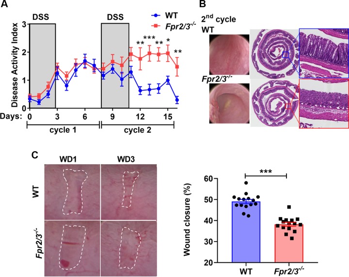
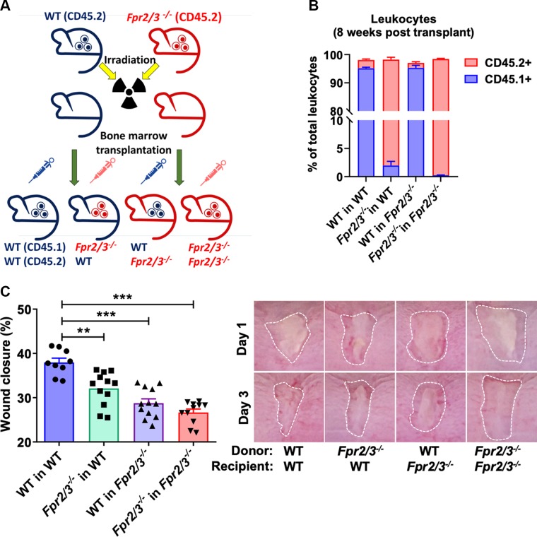
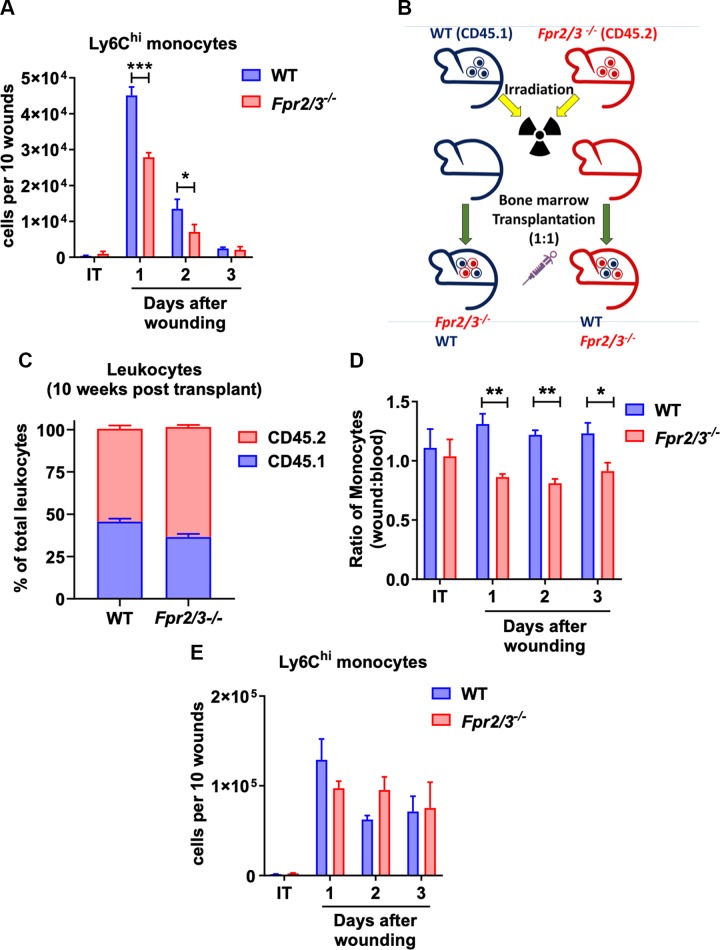
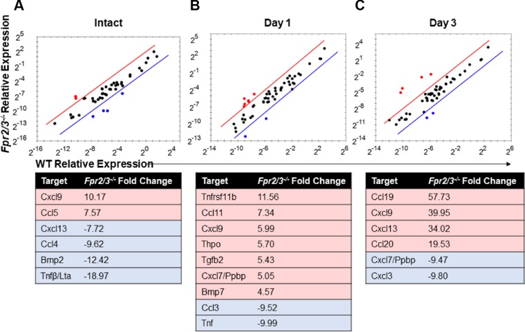
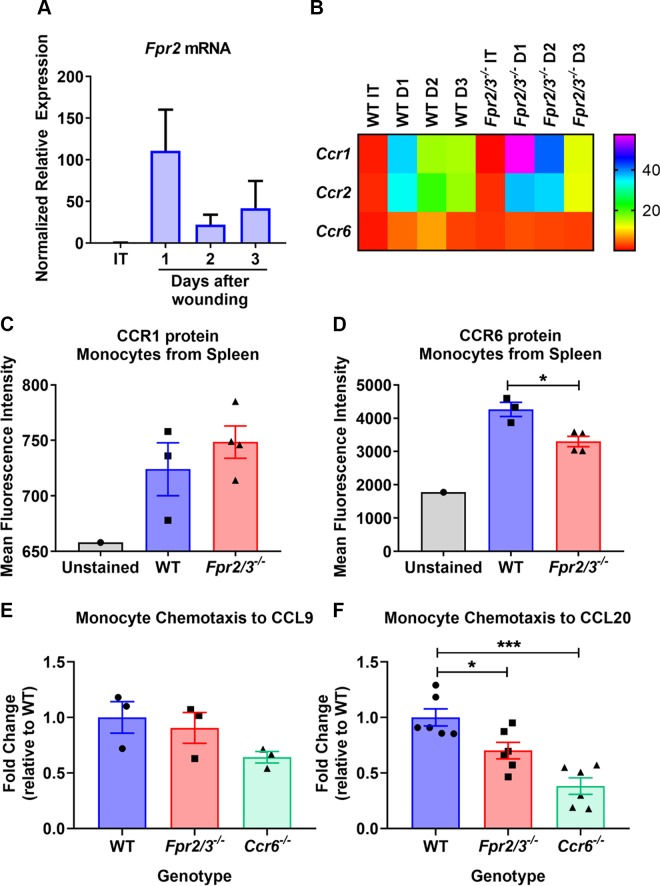
Mucosal wound repair is coordinated by dynamic crosstalk between endogenous and exogenous mediators and specific receptors on epithelial cells and infiltrating immune cells. One class of such receptor-ligand pairs involves formyl peptide receptors (FPRs) that have been shown to influence inflammatory response and repair. Here we explored the role of murine Fpr2/3, an ortholog of human FPR2/receptor for lipoxin A4 (ALX), in orchestrating intestinal mucosal repair. Compared with wild-type (WT) mice, Fpr2/3-/- mice exhibited delayed recovery from acute experimental colitis and perturbed repair after biopsy-induced colonic mucosal injury. Decreased numbers of infiltrating monocytes were observed in healing wounds from Fpr2/3-/- mice compared with WT animals. Bone marrow transplant experiments revealed that Fpr2/3-/- monocytes showed a competitive disadvantage when infiltrating colonic wounds. Moreover, Fpr2/3-/- monocytes were defective in chemotactic responses to the chemokine CC chemokine ligand (CCL)20, which is up-regulated during early phases of inflammation. Analysis of Fpr2/3-/- monocytes revealed altered expression of the CCL20 receptor CC chemokine receptor (CCR)6, suggesting that Fpr2/3 regulates CCL20-CCR6-mediated monocyte chemotaxis to sites of mucosal injury in the gut. These findings demonstrate an important contribution of Fpr2/3 in facilitating monocyte recruitment to sites of mucosal injury to influence wound repair.-Birkl, D., O'Leary, M. N., Quiros, M., Azcutia, V., Schaller, M., Reed, M., Nishio, H., Keeney, J., Neish, A. S., Lukacs, N. W., Parkos, C. A., Nusrat, A. Formyl peptide receptor 2 regulates monocyte recruitment to promote intestinal mucosal wound repair.

Keywords: FPR2; GPCRs; epithelium; inflammation; inflammatory bowel disease.


Conflict of interest statement

The authors thank Prof. Mauro Perretti (Queen Mary University of London London, United Kingdom) for providing the Fpr2/3−/− mice, and Madeline R. Barron and Darius Feier (University of Michigan) for experimental help during their experimental rotation in the researchers’ laboratory. This work was supported by U.S. National Institutes of Health (NIH) National Institute of Diabetes and Digestive and Kidney Diseases Grants DK055679, DK089763, and DK059888 (to A.N.) and DK072564, DK079392, and DK061379 (to C.A.P.); German Research Foundation (DFG) Research Fellowship SI 2282/1-1 (to D.B.); and Crohn’s and Colitis Foundation Career Development Award 544599 (to M.Q.). The authors declare no conflicts of interest.

Figures

Figure 1
Figure 1
Fpr2−/− mice show decreased acute and chronic wound healing. A) Disease activity index comparing C57BL/6J (WT) with Fpr2/3−/− mice after 2 cycles of DSS treatment consisting of 3 d of 2.5% DSS followed by 5 d of recovery with water consumption. B) Representative images of endoscopic videos and hematoxylin and eosin–stained histologic sections of WT or Fpr2/3−/− colonic mucosa after the second cycle of DSS treatment. Boxed areas are magnified in insets equidistant from the rectum. The total magnification of the photomicrographs is original magnification, ×2, and the inset is original magnification, ×40. C) Endoscopic images of colonic mucosal wounds 1 and 3 d after biopsy-induced injury in Fpr2/3−/− (n = 15) or WT control (n = 15) mice. Wound area was quantified using ImageJ. WD, wound day. Graph shows quantification of wound closure. Statistical comparisons were performed using an unpaired 2-tailed Student’s t test with Welch’s correction. ***P < 0.001 (means ± sem).
Figure 2
Figure 2
Expression of Fpr2/3 on epithelial and immune cells is necessary for colonic mucosal wound repair. A) Illustration of BM transplant experiment. B) Engraftment verification after BM transplantation by flow cytometry analysis of leukocytes in irradiated WT or Fpr2/3−/− host mice that were reconstituted with BM from either WT (CD45.1+) or Fpr2/3−/− (CD45.2+) donor mice (WT→WT; Fpr2/3−/− →WT; WT→ Fpr2/3−/−; Fpr2/3−/−Fpr2/3−/−; n = 6; means ± sem). C) Quantification and endoscopic images of colonic mucosal wound repair after biopsy-induced injury, comparing irradiated WT or Fpr2/3−/− host mice that were reconstituted with BM from either WT or Fpr2/3−/− donor mice (WT→WT, n = 9; Fpr2/3−/− →WT, n = 12; WT→ Fpr2/3−/−, n = 12; Fpr2/3−/−Fpr2/3−/−, n = 11).
Figure 3
Figure 3
FPR2 expression is required for competitive advantage of monocytes to migrate to sites of mucosal injury. A) Analysis of monocytes from intact (IT) or wounded lamina propria tissue on different postinjury days isolated from WT or Fpr2/3−/− (n = 6). B) Illustration of competitive BM transplant experiment. C) Engraftment verification after competitive BM transplantation by flow cytometry analysis of leukocytes in irradiated WT or Fpr2/3−/− host mice that were reconstituted with a 1:1 mixture of BM from WT (CD45.1) and Fpr2/3−/− (CD45.2) donor mice (n = 19–20, means ± sem). WT, C57BL/6J. D, E) Analysis of monocytes isolated from IT or wounded lamina propria tissue on different postinjury days isolated from irradiated WT or Fpr2/3−/− host mice were reconstituted with a 1:1 mixture of BM from WT (CD45.1) and Fpr2/3−/− (CD45.2) donor mice. D) Graphs represent the ratio of WT (CD45.1) or Fpr2/3−/− (CD45.2) Ly6Chi cells in the wound vs. in the blood (n = 10) of combined WT and Fpr2/3−/− mice submitted to a competitive BM transplant. E) Numbers of Ly6Chi monocytes on different postinjury days and in IT lamina propria tissue (n = 5). IT, intact tissue; WT, C57BL/6J. Statistical comparisons performed using 2-way ANOVA with Bonferroni’s multiple comparison. *P < 0.5, **P < 0.01, ***P < 0.001 (means ± sem).
Figure 4
Figure 4
Cytokine and chemokine expression analysis in intact and injured tissue. Scatter plots comparing gene expression changes determined by PCR array performed on RNA isolated from 3-mm punch biopsies of WT or Fpr2/3−/− intact tissue (A) or resealing colonic wounds on d 1 (B) and d 3 (C) postinjury isolated from mice. Tables detail the fold change in expression observed in Fpr2/3−/− samples relative to WT (C57BL/6J).
Figure 5
Figure 5
Monocytes lacking FPR2 expression have a deficit in their chemotactic response. A) Fpr2 mRNA levels were determined by qPCR of RNA isolated from monocytes collected from the lamina propria of WT intact tissue or resealing colonic wounds isolated from mice (n = 3–4). B) Heat map representation of expression levels of Ccr1, Ccr2, and Ccr6 mRNA in monocytes was determined by qPCR analysis of RNA (n = 3–4). C, D) Flow cytometric analysis of CCR1 (C) and CCR6 (D) expression on monocytes isolated from spleens of WT or Fpr2/3−/− mice. E, F) In vitro migration of spleen monocytes isolated from WT or Fpr2/3−/− mice toward CCL9 (n = 3) (E) or CCL20 (n = 6) (F). The rate of migration is represented as the fold change relative to WT based on the ratio of total monocytes added to the upper chamber of the Transwell to the number of cells migrated into the bottom. MFI, mean fluorescence intensity. Statistical comparisons performed using 1-way ANOVA with Bonferroni’s multiple comparison or paired Student’s t test. *P < 0.5, ***P < 0.001 (means ± sem).

References

    1. Kagnoff M. F. (2014) The intestinal epithelium is an integral component of a communications network. J. Clin. Invest. 124, 2841–2843 - PMC - PubMed
    1. Leoni G., Neumann P. A., Kamaly N., Quiros M., Nishio H., Jones H. R., Sumagin R., Hilgarth R. S., Alam A., Fredman G., Argyris I., Rijcken E., Kusters D., Reutelingsperger C., Perretti M., Parkos C. A., Farokhzad O. C., Neish A. S., Nusrat A. (2015) Annexin A1-containing extracellular vesicles and polymeric nanoparticles promote epithelial wound repair. J. Clin. Invest. 125, 1215–1227 - PMC - PubMed
    1. Nathan C. (2002) Points of control in inflammation. Nature 420, 846–852 - PubMed
    1. Serhan C. N., Savill J. (2005) Resolution of inflammation: the beginning programs the end. Nat. Immunol. 6, 1191–1197 - PubMed
    1. Fullerton J. N., Gilroy D. W. (2016) Resolution of inflammation: a new therapeutic frontier. Nat. Rev. Drug Discov. 15, 551–567 - PubMed

Publication types

MeSH terms