. 2019 Oct 3;179(2):417-431.e19.
doi: 10.1016/j.cell.2019.09.009.

An Allosteric Anti-tryptase Antibody for the Treatment of Mast Cell-Mediated Severe Asthma

Affiliations

An Allosteric Anti-tryptase Antibody for the Treatment of Mast Cell-Mediated Severe Asthma

Henry R Maun et al. Cell. .

Erratum in

  • An Allosteric Anti-tryptase Antibody for the Treatment of Mast Cell-Mediated Severe Asthma.
    Maun HR, Jackman JK, Choy DF, Loyet KM, Staton TL, Jia G, Dressen A, Hackney JA, Bremer M, Walters BT, Vij R, Chen X, Trivedi NN, Morando A, Lipari MT, Franke Y, Wu X, Zhang J, Liu J, Wu P, Chang D, Orozco LD, Christensen E, Wong M, Corpuz R, Hang JQ, Lutman J, Sukumaran S, Wu Y, Ubhayakar S, Liang X, Schwartz LB, Babina M, Woodruff PG, Fahy JV, Ahuja R, Caughey GH, Kusi A, Dennis MS, Eigenbrot C, Kirchhofer D, Austin CD, Wu LC, Koerber JT, Lee WP, Yaspan BL, Alatsis KR, Arron JR, Lazarus RA, Yi T. Maun HR, et al. Cell. 2020 Jan 23;180(2):406. doi: 10.1016/j.cell.2020.01.003. Cell. 2020. PMID: 31978350 No abstract available.

Abstract

Severe asthma patients with low type 2 inflammation derive less clinical benefit from therapies targeting type 2 cytokines and represent an unmet need. We show that mast cell tryptase is elevated in severe asthma patients independent of type 2 biomarker status. Active β-tryptase allele count correlates with blood tryptase levels, and asthma patients carrying more active alleles benefit less from anti-IgE treatment. We generated a noncompetitive inhibitory antibody against human β-tryptase, which dissociates active tetramers into inactive monomers. A 2.15 Å crystal structure of a β-tryptase/antibody complex coupled with biochemical studies reveal the molecular basis for allosteric destabilization of small and large interfaces required for tetramerization. This anti-tryptase antibody potently blocks tryptase enzymatic activity in a humanized mouse model, reducing IgE-mediated systemic anaphylaxis, and inhibits airway tryptase in Ascaris-sensitized cynomolgus monkeys with favorable pharmacokinetics. These data provide a foundation for developing anti-tryptase as a clinical therapy for severe asthma.

Keywords: allosteric protease inhibitor; anti-IgE; anti-tryptase; antibody engineering; asthma; mast cell; non-type 2 asthma; serine protease; tryptase; tryptase genetics.


Conflict of interest statement

Declaration of interests

All authors except N.N.T., L.B.S., M.B., P.G.W., J.V.F., R.A., and G.H.C. are current or past employees of Genentech, Inc., a member of the Roche group, and may hold Roche stock or stock options. A patent application, entitled “anti-tryptase antibodies, compositions thereof, and uses thereof”, relating to the subject matter of this manuscript, has been filed by Genentech Inc. L.B.S. and G.H.C. are consultants for Genentech for anti-tryptase antibody development.

Figures

Figure 1.
Figure 1.. Elevated total tryptase in severe asthma patients independent of type 2 inflammation.
(A, B) Tryptase in BAL fluid (A) and plasma (B). Albumin-normalized BAL tryptase levels (mean ± SE) to account for dilution. Patient numbers are in methods. (C) Normalized tryptase in BAL fluid of mild asthma patients stratified by blood eosinophil counts (300 cells/μL as cutoff). (D, E) Tryptase in BAL fluid or plasma of healthy subjects and severe asthma patients stratified as in (C). Each dot is an individual patient. Mann-Whitney tests were used to compare differences between groups *p<0.05, **p<0.01, ***p<0.001.
Figure 2
Figure 2. Active tryptase allele count is correlated with systemic tryptase levels.
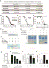
(A) Schematic diagram of tryptase gene loci. (B) Percentage of healthy controls and asthma patients with 1, 2, 3, or 4 active tryptase allele count. (C) The level of tryptase activity per primary MC from foreskins of human donors (N=101) was assessed ex vivo with respect to active tryptase allele count. (D) Serum tryptase levels in healthy subjects with different active tryptase allele count. Serum (E) and plasma (F) tryptase levels from severe uncontrolled asthma subjects with different active tryptase allele count. P-value, correlation estimate (β), and r2 from linear regression are annotated on plots. (G) Histamine concentrations in culture supernatants in LAD2 cells degranulated by inactive β-tryptase (S195A), wild-type (WT) β-tryptase, or β-tryptase + anti-tryptase. (H) Histamine, prostaglandin D2 (PGD2) and percentage of total β-hexosaminidase release in culture supernatant of LAD2 cells treated with saline or IgE/anti-IgE immune complex with or without indicated tryptase SMI or two different clones of inhibitory anti-tryptase IgG antibodies (31A.v11 or E104); mean ± SD (n=3).
Figure 3.
Figure 3.. Treatment benefit from anti-IgE therapy in asthma patients based on active tryptase copy number
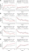
(A) FEV1 percent. (B) Daily number of puffs of albuterol change. (C) Total asthma symptom severity scores (TASS). (D) Overall asthma quality of life with standardized activities (AQLQ[S]). Changes from baseline were assessed in patients from the EXTRA study on the basis of active tryptase copy number (1 or 2 left, 3 or 4 right). Least squares (mean ± SE) are plotted and the mean post-treatment placebo controlled treatment effect estimated by linear contrast are annotated in the plot margins.
Figure 4.
Figure 4.. Identification of a high affinity inhibitory anti-tryptase IgG4 antibody.
(A) Binding kinetics of anti-tryptase to human tryptase. (B, C) Tryptase activity using S-2288 peptide substrate for βI-, βII-, βIII-tryptases with anti-tryptase (B) and for βI-tryptase with anti-tryptase Fab or IgG4 antibody (C). (D) Tryptase activity using ES002 peptide substrate with degranulated foreskin MC releasate with anti-tryptase Fab or IgG4. (E, F) VIP peptide degradation by βI-tryptase (E) or degranulated MC supernatant (F) with saline control, anti-tryptase, or a tryptase small molecule inhibitor (SMI). (G) Fibrinogen α chain degradation by βI-tryptase with or without anti-tryptase at pH 7.5 or pH 6.0. (H) Primary human SMC proliferation stimulated by βI-tryptase with or without anti-tryptase. (I) Primary human SMC contraction induced by βI-tryptase with or without anti-tryptase. (J) Collagen secretion by primary human lung fibroblasts induced by recombinant βI-tryptase treated with anti-tryptase or medium. Data for A-D and H-J are mean ± SE (n=3); student’s t-test was used to compare between two groups. *p<0.05, **p<0.01, ***p<0.001.
Figure 5.
Figure 5.. A crystal structure reveals an allosteric mode of anti-tryptase binding and a tetramer dissociation mechanism.
(A) SEC of anti-tryptase Fab (31A.v11) in complex with βI-tryptase with or without heparin. Tetrameric βI-tryptase alone (Peak 1, black) and Fab (Peak 4, blue and red) eluted at 13.0 mL and 15.5 mL, respectively. A complex of tetrameric βI-tryptase incubated with excess Fab in the presence (Peak 2, red) or absence (Peak 3, blue) of 0.1 mg/mL heparin eluted at 13.8 mL and 14.05 mL, respectively. (B) Non-reducing SDS-PAGE of peak fractions. (C) Depictions in surface (left) and cartoon (right) of slower H-D exchange areas in green (large surface cluster) and slate when Fab is bound to βI-tryptase; catalytic triad residues are in red. (D) Crystal structure of βI-tryptase monomer (white) bound to anti-tryptase Fab (31A.v11) and soybean trypsin inhibitor (STI, in slate) determined at 2.15 Å resolution; HC and LC are in green and blue, respectively. Catalytic triad residues are in red. (E) The βI-tryptase monomer in complex with anti-tryptase was superposed with protomers A and C (beige) of the β-tryptase tetramer (1A0L); protomers B and D in the tetramer are white and orange. Residue clashes between the LCs (blue and purple) of the Fab are labeled and shown as spheres; identical residues in each LC clash with each other. (F, G) Conformational changes in the 60s (F) and 30s (G) loop of βI-tryptase upon Fab binding that affect the large (Protomers B and C in white and beige) or small (Protomers B and A in white and magenta) interface, respectively. (Left panels) βI-tryptase (cyan) from the Fab complex was superposed with Protomer B (white) from the tetramer (1A0L). Right panels show closeup interface views in the circle. Key interface residues and catalytic triad residue H57 are shown in sticks (F). (H, I) SEC of anti-tryptase Fab in complex with WT and two different disulfide-locked variants (Y75C and I99C) of tetrameric βI-tryptase. Tetrameric βI-tryptase alone (Peak 3, black) and Fab (Peak 5, red, blue and green) had elution volumes (Ve) of 13.0 mL and 15.5 mL, respectively. Complexes of tetrameric βI-tryptase incubated with excess Fab for WT (Peak 4, red), Y75C (Peak 1, green) and I99C (Peak 2, blue) eluted at 14.0 mL (Monomer/Fab), 11.3 mL (Dimer/2 Fabs) and 11.9 mL (Dimer/2Fabs), respectively. Non-reducing SDS-PAGE of peak fractions for Y75C βI-tryptase and Fab (I, left panel) and I99C βI-tryptase and Fab (I, right panel) are shown. Dimer refers to βI-tryptases Y75C (I, left panel) and I99C (I, right panel). M, L, P1, P2 and P5 refer to Markers, Load, Peak 1, Peak 2 and Peak 5, respectively.
Figure 6.
Figure 6.. Anti-tryptase antibody inhibits tryptase activity in vivo and attenuates MC-induced anaphylaxis response.
(A, B) FACS analysis of human MCs (defined as KIT+FcεRI+) cells in spleen and lung of control NOD.scid.SGM3 mice or NOD.scid.SGM3 mice reconstituted with human CD34+ stem cells (A). Intracellular tryptase staining in MCs from spleen and lung (B). (C, D) NOD.scid.SGM3 animals reconstituted human CD34+ stem cells were injected with anti-NP IgE and NP-BSA conjugates. (C) Total tryptase concentration after anti-NP IgE and NP-BSA conjugates crosslinking; mean ± SE (n=4). (D) Anti-tryptase or anti-gD (isotype control) injected 24 h before the anti-IgE administration. Body temperature measurement after NP-BSA conjugate injection. mean ± SE (n=4). (E) Active tryptase concentration in BAL in humanized mice at baseline or 10 min after anti-NP IgE/NP-BSA crosslinking; mean ± SE (n=4). Lower limit of quantification (LLOQ) is 0.3 ng/mL. (F) Tryptase enzymatic activity measured with S-2288 substrate in the presence of α1-antitrypsin and STI in BAL fluid 10 min after NP-BSA conjugate injection; mean ± SE (n=3) *p<0.05, ***p<0.001.
Figure 7.
Figure 7.. Anti-tryptase IgG4 antibody exhibits favorable pharmacokinetics and reduces tryptase activity in BAL in nonhuman primates.
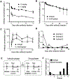
(A) Antibody concentration in NOD.scid mice dosed IV with anti-tryptase IgG4 antibody. (B, C) Antibody concentration in blood (B) or BAL fluid (C) of cynomolgus monkey injected SC with a single dose (30 mg/kg) of anti-tryptase or anti-gD human IgG4. (D) Tryptase activity quantified by an activity-based probe assay in BAL fluid of cynomolgus monkeys on study days -6, 1 (8 h post-dose), 3, 8, 15, 22, and 29. The dotted line is the lower limit of quantification. (E) Study schema for treatment and sampling of sensitized cynomolgus monkeys after inhaled challenge with Ascaris suum. Each animal underwent a vehicle and drug treatment phase, thereby serving as its own control. (F) Tryptase activity quantified using an activity-based probe assay in BAL fluid of Ascaris suum sensitized cynomolgus monkeys after inhaled Ascaris suum challenge. “ND” denotes samples below the lower limit of detection. p<0.05, paired Student’s t-test.

References

    1. Adams PD, Afonine PV, Bunkoczi G, Chen VB, Davis IW, Echols N, Headd JJ, Hung LW, Kapral GJ, Grosse-Kunstleve RW, et al. (2010). PHENIX: a comprehensive Python-based system for macromolecular structure solution. Acta Crystallogr D Biol Crystallogr 66, 213–221. - PMC - PubMed
    1. Alter SC, Kramps JA, Janoff A, and Schwartz LB (1990). Interactions of human mast cell tryptase with biological protease inhibitors. Archives of biochemistry and biophysics 276, 26–31. - PubMed
    1. Alter SC, Metcalfe DD, Bradford TR, and Schwartz LB (1987). Regulation of human mast cell tryptase. Effects of enzyme concentration, ionic strength and the structure and negative charge density of polysaccharides. The Biochemical journal 248, 821–827. - PMC - PubMed
    1. Arena TA, Chou B, Harms PD, and Wong AW (2019). An anti-apoptotic HEK293 cell line provides a robust and high titer platform for transient protein expression in bioreactors. MAbs 11, 977–986. - PMC - PubMed
    1. Babina M, Guhl S, Artuc M, Trivedi NN, and Zuberbier T (2016). Phenotypic variability in human skin mast cells. Exp Dermatol 25, 434–439. - PubMed

Substances