. 2019 Oct 3;105(4):734-746.
doi: 10.1016/j.ajhg.2019.09.002.

Diagnostic Utility of Next-Generation Sequencing for Disorders of Somatic Mosaicism: A Five-Year Cumulative Cohort

Affiliations

Diagnostic Utility of Next-Generation Sequencing for Disorders of Somatic Mosaicism: A Five-Year Cumulative Cohort

Samantha N McNulty et al. Am J Hum Genet. .

Abstract

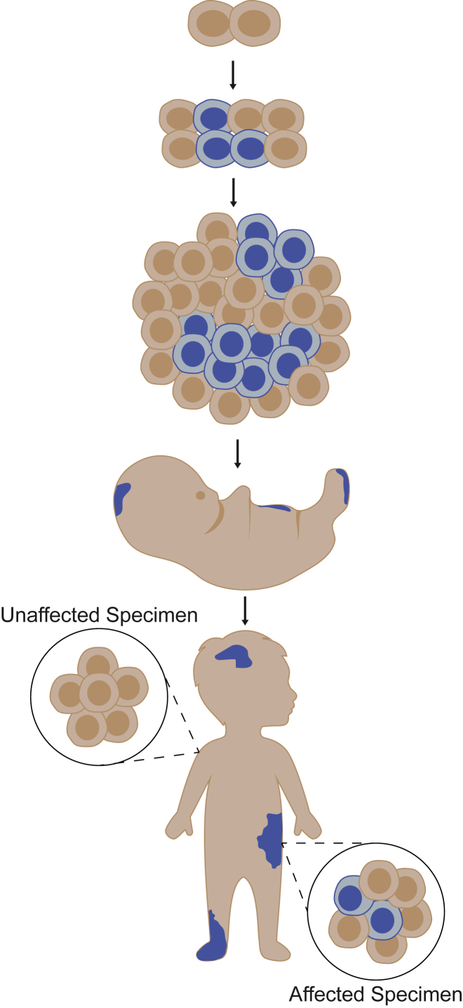
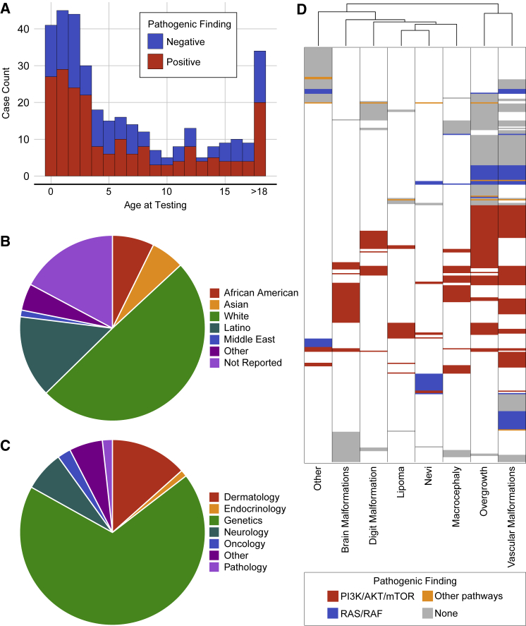
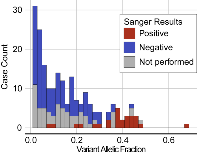
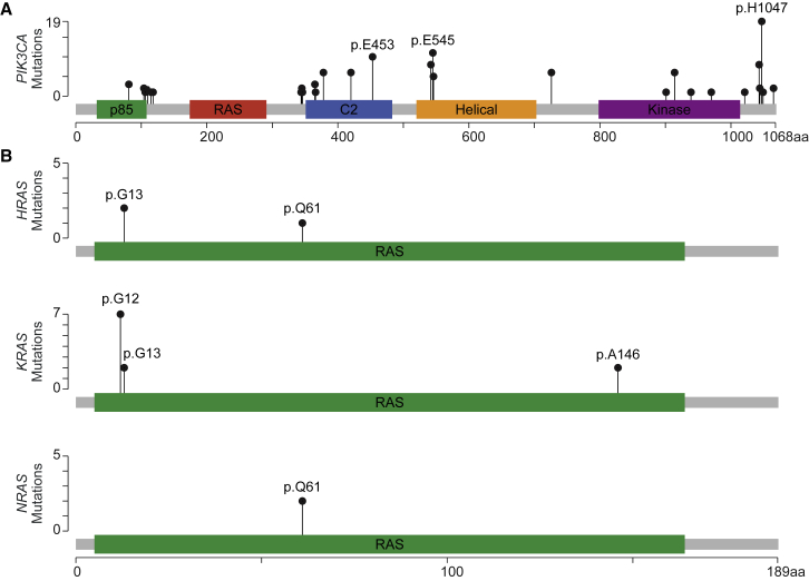
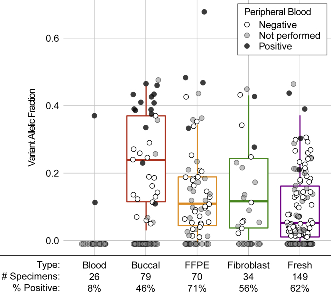
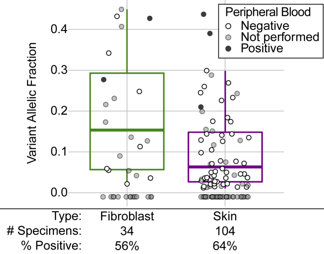
Disorders of somatic mosaicism (DoSM) are a diverse group of syndromic and non-syndromic conditions caused by mosaic variants in genes that regulate cell survival and proliferation. Despite overlap in gene space and technical requirements, few clinical labs specialize in DoSM compared to oncology. We adapted a high-sensitivity next-generation sequencing cancer assay for DoSM in 2014. Some 343 individuals have been tested over the past 5 years, 58% of which had pathogenic and likely pathogenic (P/LP) findings, for a total of 206 P/LP variants in 22 genes. Parameters associated with the high diagnostic yield were: (1) deep sequencing (∼2,000× coverage), (2) a broad gene set, and (3) testing affected tissues. Fresh and formalin-fixed paraffin embedded tissues performed equivalently for identification of P/LP variants (62% and 71% of individuals, respectively). Comparing cultured fibroblasts to skin biopsies suggested that culturing might boost the allelic fraction of variants that confer a growth advantage, specifically gain-of-function variants in PIK3CA. Buccal swabs showed high diagnostic sensitivity in case subjects where disease phenotypes manifested in the head or brain. Peripheral blood was useful as an unaffected comparator tissue to determine somatic versus constitutional origin but had poor diagnostic sensitivity. Descriptions of all tested individuals, specimens, and P/LP variants included in this cohort are available to further the study of the DoSM population.

Keywords: brain malformation; macrocephaly; megalencephaly; mosaicism; nevus syndrome; overgrowth syndrome; somatic overgrowth; vascular malformation.


Conflict of interest statement

B.A.D. serves as a consultant and member of the medical advisory board for Venthera. M.C.S. and J.W.H. consult for Pierian Diagnostics.

Figures

Figure 1
Figure 1
Disorders of Somatic Mosaicism Somatic variants can arise during or after the 2-cell stage of embryogenesis. They are restricted to the specific lineage of cells derived from the original mutant (blue) and are absent from other cells and tissues (tan). Proliferative disorders arise when somatic variants impact the function of key genes involved in the regulation of cell growth, division, and senescence. Their phenotypic manifestations are highly variable, depending on the timing of the mutation during or after embryonic development, the location of the variant within the developing body, allelic diversity, and the function and expression of the altered gene(s). Phenotypes can appear before or after birth and may include soft tissue overgrowth, skin lesions, bone malformations, and brain abnormalities, among others.
Figure 2
Figure 2
Cohort Demographics (A) A total of 358 unique specimens from 343 people were submitted for testing between October 2013 and December 2018. Individuals ranged in age from birth to 62 years with an average age of 7.77 ± 9.41 years. P/LP findings were not concentrated in any particular age group. (B) Racial ancestry was listed on the test requisition for most people: 25 African American, 20 Asian, 170 white, 49 Latino, 4 Middle Easterner, 16 of other backgrounds; 59 people did not report their racial background. (C) Individuals were referred for testing by physicians that represent a total of 18 sub-specialties, including: genetics, dermatology, neurology, oncology, and pathology. (D) Phenotypes reported for each person were binned into eight discrete categories. People were clustered based on the eight phenotypic categories and the biochemical pathway associated with any P/LP finding. Variants in the PI3K/AKT/mTOR pathway (red) are associated with a wide variety of symptoms, while variants in the RAS/RAF pathway (blue) were often associated with overgrowth, vascular malformations, brain malformations, and nevi.
Figure 3
Figure 3
Allelic Frequency of P/LP Variants P/LP variants were found with VAF ranging from 1% to 68% with an average VAF of 16.2% ± 13.5%. One hundred sixty of the 206 P/LP variants were found at VAF < 25% and 87 were found with VAF < 10%. Blood specimens were submitted as a comparative sample determine the distribution of the P/LP variant in the body for 139 of the 199 individuals with P/LP findings; 26 of the secondary blood specimens tested positive for the P/LP variant by Sanger sequencing (red) while 113 tested negative (blue). P/LP findings in the peripheral blood were enriched in case subjects where the variant was found at a high VAF in the original specimen used in NGS testing.
Figure 4
Figure 4
P/LP Variants in PIK3CA and RAS Genes (A) P/LP variants were identified along the length of PIK3CA (MIM: 171834) including four of the five outlined protein domains. (B) Mutational hotspots p.Glu453, p.Glu545, and p.His1047 are common in both DoSM and cancer. In contrast, most of the variants identified in HRAS (MIM: 190020), KRAS (MIM: 190070), and NRAS (MIM: 164790) were found at the p.G12 position, which is commonly mutated in non-small cell lung cancer.
Figure 5
Figure 5
P/LP Variants Identified from Five Specimen Types Five specimen types are accepted for testing: peripheral blood (n = 26), buccal swab (n = 79), formalin fixed paraffin embedded (FFPE, n = 70), cultured fibroblasts (n = 34), and fresh, unfixed tissues (n = 149). Case subjects where P/LP variant was identified are plotted under the 0% VAF line, and the 25th, 50th, and 75th percentiles of VAFs of P/LP variants are displayed by boxplots. Shading of dots reflects results of Sanger sequencing of peripheral blood: black dots represent P/LP findings in blood (multi-tissue distribution), white dots represent case subjects from which the P/LP variant was found only in the affected sample (confirmed somatic), and gray dots represent case subjects for which no peripheral blood was submitted. The percent of specimens with P/LP findings are listed for each specimen type.
Figure 6
Figure 6
P/LP Variants Identified from Cultured Fibroblasts Compared to Skin Biopsies Fibroblasts are typically cultured from skin biopsies, so the P/LP variants found in cultured fibroblasts (n = 34) were compared to those found in fresh tissue skin biopsies (n = 104). Cases where no P/LP variant was identified are plotted under the 0% VAF line. The 25th, 50th, and 75th percentiles of VAFs of P/LP variants are displayed by boxplots. Shading of dots reflects results of Sanger sequencing of peripheral blood: black dots represent P/LP findings in blood (multi-tissue distribution), white dots represent case subjects from which the P/LP variant was found only in the affected sample (confirmed somatic), and gray dots represent case subjects for which no peripheral blood was submitted. The percent of specimens with P/LP findings are listed for each specimen type.

References

    1. Keppler-Noreuil K.M., Parker V.E., Darling T.N., Martinez-Agosto J.A. Somatic overgrowth disorders of the PI3K/AKT/mTOR pathway & therapeutic strategies. Am. J. Med. Genet. C. Semin. Med. Genet. 2016;172:402–421. - PMC - PubMed
    1. Kinsler V.A., Thomas A.C., Ishida M., Bulstrode N.W., Loughlin S., Hing S., Chalker J., McKenzie K., Abu-Amero S., Slater O. Multiple congenital melanocytic nevi and neurocutaneous melanosis are caused by postzygotic mutations in codon 61 of NRAS. J. Invest. Dermatol. 2013;133:2229–2236. - PMC - PubMed
    1. Keppler-Noreuil K.M., Sapp J.C., Lindhurst M.J., Parker V.E., Blumhorst C., Darling T., Tosi L.L., Huson S.M., Whitehouse R.W., Jakkula E. Clinical delineation and natural history of the PIK3CA-related overgrowth spectrum. Am. J. Med. Genet. A. 2014;164A:1713–1733. - PMC - PubMed
    1. Rauen K.A. The RASopathies. Annu. Rev. Genomics Hum. Genet. 2013;14:355–369. - PMC - PubMed
    1. di Blasio L., Puliafito A., Gagliardi P.A., Comunanza V., Somale D., Chiaverina G., Bussolino F., Primo L. PI3K/mTOR inhibition promotes the regression of experimental vascular malformations driven by PIK3CA-activating mutations. Cell Death Dis. 2018;9:45. - PMC - PubMed

Publication types