. 2019 Oct 3;105(4):854-868.
doi: 10.1016/j.ajhg.2019.09.005.

De Novo Pathogenic Variants in N-cadherin Cause a Syndromic Neurodevelopmental Disorder with Corpus Collosum, Axon, Cardiac, Ocular, and Genital Defects

Collaborators, Affiliations

De Novo Pathogenic Variants in N-cadherin Cause a Syndromic Neurodevelopmental Disorder with Corpus Collosum, Axon, Cardiac, Ocular, and Genital Defects

Andrea Accogli et al. Am J Hum Genet. .

Abstract

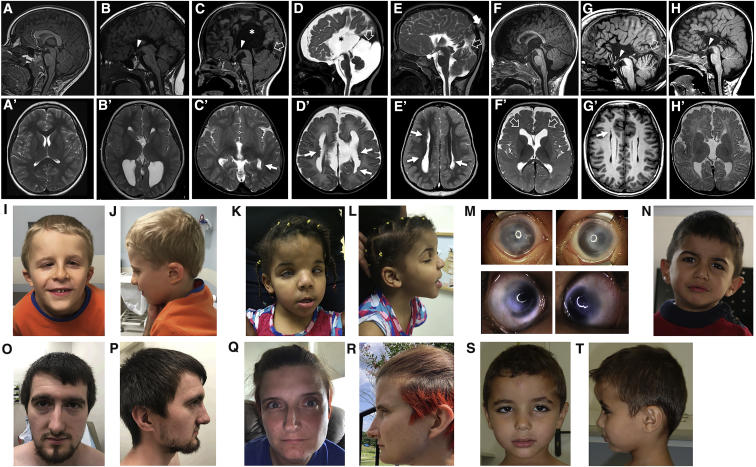
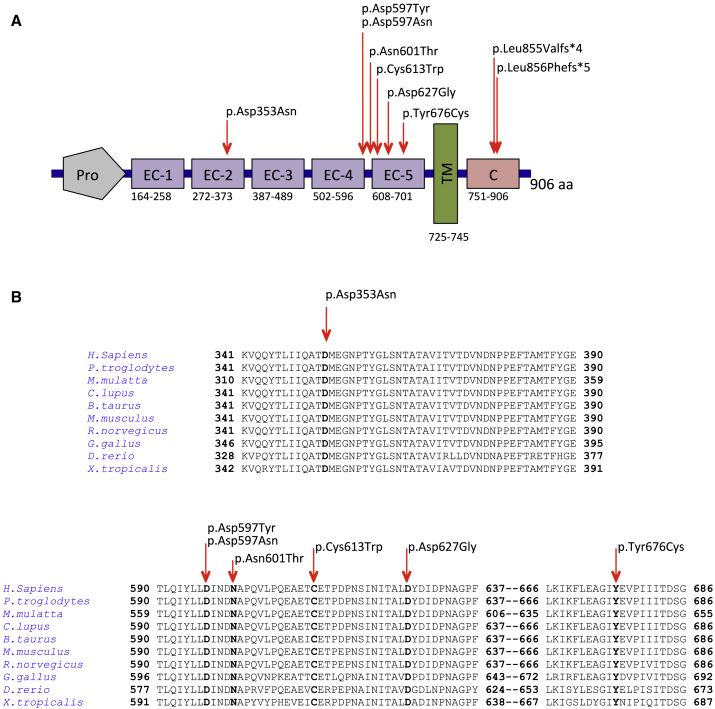
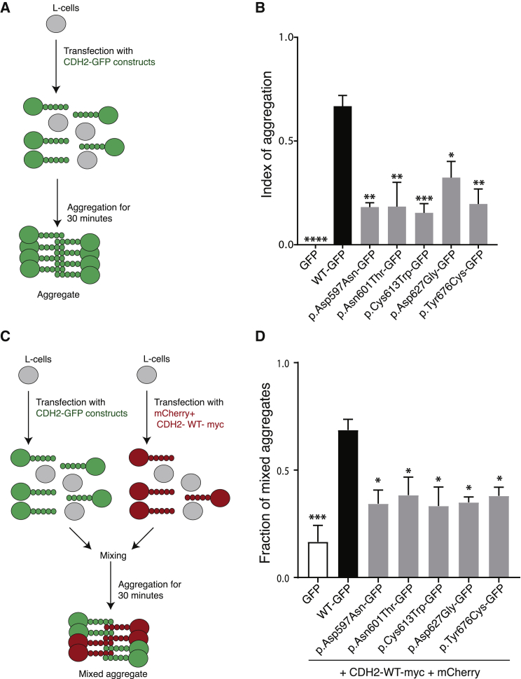
Cadherins constitute a family of transmembrane proteins that mediate calcium-dependent cell-cell adhesion. The extracellular domain of cadherins consists of extracellular cadherin (EC) domains, separated by calcium binding sites. The EC interacts with other cadherin molecules in cis and in trans to mechanically hold apposing cell surfaces together. CDH2 encodes N-cadherin, whose essential roles in neural development include neuronal migration and axon pathfinding. However, CDH2 has not yet been linked to a Mendelian neurodevelopmental disorder. Here, we report de novo heterozygous pathogenic variants (seven missense, two frameshift) in CDH2 in nine individuals with a syndromic neurodevelopmental disorder characterized by global developmental delay and/or intellectual disability, variable axon pathfinding defects (corpus callosum agenesis or hypoplasia, mirror movements, Duane anomaly), and ocular, cardiac, and genital anomalies. All seven missense variants (c.1057G>A [p.Asp353Asn]; c.1789G>A [p.Asp597Asn]; c.1789G>T [p.Asp597Tyr]; c.1802A>C [p.Asn601Thr]; c.1839C>G [p.Cys613Trp]; c.1880A>G [p.Asp627Gly]; c.2027A>G [p.Tyr676Cys]) result in substitution of highly conserved residues, and six of seven cluster within EC domains 4 and 5. Four of the substitutions affect the calcium-binding site in the EC4-EC5 interdomain. We show that cells expressing these variants in the EC4-EC5 domains have a defect in cell-cell adhesion; this defect includes impaired binding in trans with N-cadherin-WT expressed on apposing cells. The two frameshift variants (c.2563_2564delCT [p.Leu855Valfs4]; c.2564_2567dupTGTT [p.Leu856Phefs5]) are predicted to lead to a truncated cytoplasmic domain. Our study demonstrates that de novo heterozygous variants in CDH2 impair the adhesive activity of N-cadherin, resulting in a multisystemic developmental disorder, that could be named ACOG syndrome (agenesis of corpus callosum, axon pathfinding, cardiac, ocular, and genital defects).

Keywords: ACOG; CDH2; N-cadherin; cardiac defects; cell-cell adhesion; corpus callosum; eye defects; genital defects; intellectual disability.


Conflict of interest statement

K.McW. and A.B. are employees of GeneDx, Inc.

Figures

Figure 1
Figure 1
Neuroradiologic and Facial Features of Individuals with De Novo CDH2 Variants Brain MRI, sagittal T1-weighted (A) and axial T2-weighted (A′) images of a normal subject. Brain MRIs from subject 1 at 7 years (B and B′), subject 2 at 2 years (C and C′), subject 3 at 13 days (D and D′), subject 4 at 4 years (E and E′), subject 5 at 6 months (F and F′), subject 8 at 26 years (G and G′), and subject 9 at 5 months (H and H′) show complete agenesis (B, C, D, E, G, and H) or mild hypoplasia (F) of the corpus callosum; there is an interhemispheric cyst communicating with the III ventricle in two subjects (asterisks in [B] and [D]). In addition, there is hypo-dysplasia of the tentorium in four cases (C, D, E, and G, empty arrows) associated with an atretic parietal cephalocele in one subject (E, arrow). Note the hypothalamic adhesion in subjects 1, 2, 4, 8 and 9 (B, C, E, G, and H, arrowheads), as well as the megacisterna magna in subjects 3 and 8 (D and G). Axial T2 images reveal multiple nodular periventricular heterotopias in four subjects (C′, D′, E′, and G′, arrows), and mild frontal ventriculomegaly in one subject (F′, empty arrows). Photographs of subjects 2 (I and J) and 4 (K and L) demonstrate prominent forehead and frontal bossing, downslanting palpebral fissures, a thin upper lip, and low-set and thick helices. Subject 4 (M) also has Peters anomaly with clouding of the cornea, as well as hypertelorism and epicanthal folds. Subjects 5 (N) and 9 (S and T) have thick earlobes but other major dysmorphisms. Subjects 7 (O and P) and 8 (Q and R) show common craniofacial features, including deep-set eyes, a thin upper lip, a pointed chin, and slightly low-set and posteriorly rotated ears with attached earlobes.
Figure 2
Figure 2
Localization and Conservation of De Novo Variants in CDH2 (A) A schematic depiction of CDH2 shows the cadherin-Pro region (Pro), five extracellular cadherin domain repeats (EC-1 to EC-5), the transmembrane region (TM), and the cytoplasmic tail (C). Arrowheads above the protein show the positions of the variants. (B) The HomoloGene-generated amino acid alignment of human CDH2 and its predicted orthologs shows the conservation of the affected residues.
Figure 3
Figure 3
N-cadherin Variants Have Impaired Cell-Cell Adhesion and Reduced trans Adhesion to N-cadherin-WT (A) Schematic representation of the cell-aggregation assay. L cells were transfected with empty GFP, N-cadherin-WT, and N-cadherin variants. Aggregation was then performed for 30 min in the presence of calcium. (B) Bar graph of the mean ± SE of the index of aggregation for N-cadherin-WT (black) and N-cadherin variants (gray) (n = 5 experiments). Cell aggregation was evaluated after 30 min of aggregation (T30) by the index (N0-N30)/N0, where N0 is number of GFP-positive cells or aggregates at T0, and N30 is the number of GFP-positive cells or aggregates at T30. The N-cadherin-GFP variants have a lower index of aggregation than N-cadherin-WT-GFP cells do. One-way ANOVA post-tests were performed for each variant versus N-cadherin-WT (∗∗∗∗p ≤ 0.0001; ∗∗∗p ≤ 0.001; ∗∗p ≤ 0.01; p ≤ 0.05). (C) Schematic representation of the mixed-cell aggregation assay evaluating trans interaction with N-cadherin-WT. L cells were transfected with empty GFP, N-cadherin-WT, and N-cadherin variants or with N-cadherin-WT-Myc+mCherry. Cells were then mixed in a 1:1 ratio, and aggregation was performed for 30 min in the presence of calcium. (D) Bar graph of the mean ± SE of the fraction of mixed aggregates for N-cadherin-WT (black) and N-cadherin (gray) variants (n = 3 experiments). The fraction of mixed aggregates was evaluated after 30 min of aggregation (T30) by the following index: number of mixed aggregates/number of total aggregates. Cells expressing N-cadherin WT-myc+mCherry form mixed aggregates with cells expressing N-cadherin-WT-GFP, but they form fewer mixed aggregates with cells expressing the N-cadherin variants. One-way ANOVA post-test were performed for each variant versus N-cadherin-WT (∗∗∗p ≤ 0.001; p ≤ 0.05).

References

    1. Goodwin K., Nelson C.M. Generating tissue topology through remodeling of cell-cell adhesions. Exp. Cell Res. 2017;358:45–51. - PMC - PubMed
    1. Wickström S.A., Niessen C.M. Cell adhesion and mechanics as drivers of tissue organization and differentiation: local cues for large scale organization. Curr. Opin. Cell Biol. 2018;54:89–97. - PubMed
    1. Priest A.V., Shafraz O., Sivasankar S. Biophysical basis of cadherin mediated cell-cell adhesion. Exp. Cell Res. 2017;358:10–13. - PubMed
    1. Tiwari P., Mrigwani A., Kaur H., Kaila P., Kumar R., Guptasarma P. Structural-mechanical and biochemical functions of classical cadherins at cellular junctions: A review and some hypotheses. Adv. Exp. Med. Biol. 2018;1112:107–138. - PubMed
    1. Brasch J., Harrison O.J., Honig B., Shapiro L. Thinking outside the cell: how cadherins drive adhesion. Trends Cell Biol. 2012;22:299–310. - PMC - PubMed

Publication types