. 2019 Oct 8;3(19):2870-2882.
doi: 10.1182/bloodadvances.2019032409.

Identification of a tumor-specific allo-HLA-restricted γδTCR

Affiliations

Identification of a tumor-specific allo-HLA-restricted γδTCR

G J J Kierkels et al. Blood Adv. .

Abstract

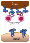
γδT cells are key players in cancer immune surveillance because of their ability to recognize malignant transformed cells, which makes them promising therapeutic tools in the treatment of cancer. However, the biological mechanisms of how γδT-cell receptors (TCRs) interact with their ligands are poorly understood. Within this context, we describe the novel allo-HLA-restricted and CD8α-dependent Vγ5Vδ1TCR. In contrast to the previous assumption of the general allo-HLA reactivity of a minor fraction of γδTCRs, we show that classic anti-HLA-directed, γδTCR-mediated reactivity can selectively act on hematological and solid tumor cells, while not harming healthy tissues in vitro and in vivo. We identified the molecular interface with proximity to the peptide-binding groove of HLA-A*24:02 as the essential determinant for recognition and describe the critical role of CD8 as a coreceptor. We conclude that alloreactive γδT-cell repertoires provide therapeutic opportunities, either within the context of haplotransplantation or as individual γδTCRs for genetic engineering of tumor-reactive T cells.


Conflict of interest statement

Conflict-of-interest disclosure: G.J.J.K., D.X.B., Z.S., and J.K. are inventors on different patents with γδTCR sequences, recognition mechanisms, and isolation strategies. G.J.J.K. is employed by Kiadis Pharma. J.K. is scientific advisor and shareholder of Gadeta. The remaining authors declare no competing financial interests.

Figures
None
Graphical abstract
Figure 1.
Figure 1.
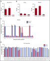
Introduction of FE11 γδTCR in αβT cells can reestablish tumor cell recognition of clone FE11 cells. (A) To assess tumor reactivity, clone FE11 cells were incubated with SW480 or EBV-LCL tumor target cells. IFN-γ secretion was measured by ELISPOT. Healthy PBMCs served as negative control targets. (B) The TCR-γ and -δ chains of clone FE11 cells were sequenced and retrovirally transduced into αβT cells. Transfer of γδTCR-mediated tumor reactivity was tested by coincubating mock- (left) or γδTCR-transduced (right) T cells with indicated target cells in an IFN-γ ELISPOT. (C) The effect of blocking with FE11-like hybridoma supernatant on the recognition of SW480 and LCL-TM cells by FE11 γδTCR–transduced T cells. (D) LABScreen Single Antigen HLA class I beads were incubated with antibodies purified from hybridoma 6 (mAb6) or from hybridoma 12 (mAb12) and secondary α-mIgG-PE measured using Luminex. Error bars represent the standard deviation (SD; n ≥ 1). MFI, mean fluorescence intensity.
Figure 2.
Figure 2.
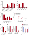
Activation of FE11 γδTCR–transduced T cells is dependent on expression of HLA-A*24:02. (A) Activation of T cells, transduced with FE11 γδTCR by EBV-LCLs with different HLA genotypes. (B) Activation of T cells, transduced with FE11 γδTCR by HLA-A*24:02 or HLA-A*02:01 target cells. (C) Activation of T cells, transduced with FE11 γδTCR by EBV-LCLs with different either homozygous or heterozygous HLA-A*24:02 expression. (D) Total HLA class I expression of HLA-A*24:02-positive and -negative EBV-LCLs compared with TEG011 recognition. (E) The effect of β2m knockout of HLA-A*24:02 target cells on the activation of FE11 γδTCR–transduced T cells. (F) Activation of T cells, transduced with FE11 γδTCR by K562 HLA-A*24:02 cells untreated or overnight monensin incubation. (G) Activation of Jurma cells, transduced with FE11 γδTCR or αβTCR WT1126-134 (control) by LCL-TM or A2-restricted WT1126-134 peptide–loaded T2 cells. CD3 cross-linking by plate-bound α-CD3 mAb clone OKT-3 served as the positive control. Recognition was assessed by measuring IFN-γ secretion using ELISA. Error bars represent the SD (n ≥ 1).
Figure 3.
Figure 3.
Activation of FE11 γδTCR–transduced T cells is limited to HLA-A*24:0+malignant cells. (A) Activation of TEG011 cells by malignant and healthy hematological cells. B cells from multiple HLA-A*24:02+ donors were activated or stressed before the TEG011 cell coculture. (B) TEG011 or NEF134-144 αβTCR–engineered αβ T-cell recognition after coculture with HLA-A*24:02+ healthy tissues. When using NEF134-144 αβTCR–engineered αβ T cells, 10 μM NEF134-144 was added. (C) Healthy donor B cells (HD1) were EBV transformed and cocultured with TEG011. Recognition was assessed by measuring IFN-γ secretion using ELISA. Error bars represent the SD (n ≥ 2). *P < .05. Cyclo, cyclophosphamide; Fluda, fludarabine.
Figure 4.
Figure 4.
Activation of FE11 γδTCR–transduced T cells is dependent on the presence of a specific HLA-A*24:02-restricted peptide. (A) Activation of TEG011 by HLA-A*24:02-positive or -negative target cells. (B) The differences between HLA-A*02:01 and HLA-A*24:02 mapped on the structure of HLA-A*24:02 (Protein Data Bank: 3wl9), the 2 nonhomologous amino acids between HLA-A*24:02 and HLA-A*24:03 are show in the red circle (top). Alignment of HLA-A*24:02, 02:01, 24:03, and 25:01 with the 2 nonhomologous amino acids in red (bottom). (C) Activation of T cells, transduced with γδTCR-FE11, by HLA-A*24:02-transduced, TAP-deficient T2 cells not loaded or loaded with the A*24-restricted viral peptides NEF134-144 or CMV341-349 (pp65 341-349). (D) WT1126-134 tetramer, NY-ESO1157-165 pentamer, and CMV341-349 pentamer binding to WT1126-134-specific TCR, NY-ESO1157-165-specific TCR, and FE11 TCR–transduced T cells. (E) The effect of bortezomib treatment of HLA-A*24:02–transduced target cells COS-7 (left) and K562 (right) on the activation of FE11 γδTCR–transduced T cells. (F) Homodimerization was assessed on HLA-A*24:02+ cells, recognized and not recognized by flow cytometry FRET. (G) Activation of TEGs (left) or T cells transduced with the WT1126-134-specific αβTCR (control) (right), by HLA-A*24:02-transduced COS-7 and K562 cells or HLA-A*02:01 (control). *Out of range. (H) Proximity Ligation Assay (PLA) was performed on HLA-A*24:02+ fibroblasts and the SW480 cell line. Cells were stained for 4′,6-diamidino-2-phenylindole and PLA signal. Where indicated, target cells were fixed before coincubation; target cells were coincubated with WT1126-134. Error bars represent the SD (n ≥ 1).
Figure 5.
Figure 5.
Recognition of LCL-TM cells cannot be outcompeted by peptides. (A) T2 cells were transduced with HLA-A*24:02 and loaded with 10 µM of 15 different transformation-associated peptides (supplemental Table 1), after which they were coincubated with TEG011 cells. Activation of TEG011 cells was assessed by measuring IFN-γ production. (B) The 4 residues of NEF134-144 that are pointed out of the peptide binding groove of HLA-A*24:02 are indicated. (C) Ten micromoles of the generated NEF134-144-derived mutant peptides were loaded on LCL-TM cells, after which they were coincubated with TEG011 cells. (D) HLA-A*24:02-transduced K562 cells were loaded with 10 µM NEF134-144 before loading with increasing concentrations of an HLA-A*24:02-restricted MiHA peptide (K.F., unpublished data). The peptide-loaded loaded cells were coincubated with TEG011 cells, αβT cells engineered with an HLA-A*24:02-restricted NEF134-144-specific TCR or a MiHA-specific αβT cell clone. T-cell activation was assessed by measuring IFN-γ. Error bars represent the SD (n ≥ 2).
Figure 6.
Figure 6.
The FE11 γδTCR critically depends on the CD8 coreceptor for tumor recognition. (A) CD8α or CD8β expression on clone FE11 cells and FE11 γδTCR–transduced αβ T cells. (B) CD4+ and CD8+ αβT cells transduced with the FE11 γδTCR were sorted and cocultured with mock (left) and TEG011 (right) target cells. (C) TEG011 T-cell activation was assessed by IFN-γ ELISPOT. CD4+ and CD8+ αβT cells expressing the FE11 γδTCR were coincubated with SW480 target cells as in panel B, but in the presence of a control antibody or blocking antibodies against CD8α or CD8β. (D) αβT cells were transduced with WT CD8α or a truncated, signaling-deficient CD8α variant (CD8α′), alongside the γδTCR-FE11, after which the CD4+, CD8+, CD4+CD8α+, and CD4+CD8α′+ T-cell populations were sorted. Recognition of healthy PBMCs and SW480 tumor target cells was assessed by measuring IFN-γ secretion using ELISPOT. Error bars represent the standard error of the mean (n ≥ 1). *P < .05; **P < .01; ***P < .001.
Figure 7.
Figure 7.
TEG011 treatment leads to efficient tumor control of K562-HLA*A24 tumors, whereas it shows no toxicity in vivo in NSG-A24 transgenic mice. NOD.Cg-Prkdc Il2rg Tg(HLA-A24)3Dvs/Sz (NSG-A24) mice were injected with 1 × 10⁵ K562 HLA-A*24:02 Luciferase cells on day 0 followed by 1 × 10⁷ TEG011 or LM1 transduced T cells on days 1 and 6 (n = 10 per group). In parallel, non-tumor–bearing mice also received 1 × 10⁷ TEG011 or LM1 transduced T cells on days 1 and 6 (n = 5 per group). Overall survival of treated K562-HLA*A24 luciferase tumor-bearing mice for monitoring efficacy (A) and overall survival of non-tumor–bearing mice for monitoring toxicity (B) was recorded for 57 days. Data represent the mean ± SD of all mice in each group. Statistical significance was calculated by log-rank (Mantel-Cox) test; *P < .05.

References

    1. Girardi M, Oppenheim DE, Steele CR, et al. . Regulation of cutaneous malignancy by gammadelta T cells. Science. 2001;294(5542):605-609. - PubMed
    1. Scheper W, van Dorp S, Kersting S, et al. . γδT cells elicited by CMV reactivation after allo-SCT cross-recognize CMV and leukemia. Leukemia. 2013;27(6):1328-1338. - PubMed
    1. Lalor SJ, McLoughlin RM. Memory γδ T Cells-Newly Appreciated Protagonists in Infection and Immunity. Trends Immunol. 2016;37(10):690-702. - PubMed
    1. Dadi S, Chhangawala S, Whitlock BM, et al. . Cancer Immunosurveillance by Tissue-Resident Innate Lymphoid Cells and Innate-like T Cells. Cell. 2016;164(3):365-377. - PMC - PubMed
    1. Hidalgo JV, Bronsert P, Orlowska-Volk M, et al. . Histological Analysis of γδ T Lymphocytes Infiltrating Human Triple-Negative Breast Carcinomas. Front Immunol. 2014;5:632. - PMC - PubMed

Publication types

Substances