In vivo non-invasive monitoring of dystrophin correction in a new Duchenne muscular dystrophy reporter mouse
- PMID: 31586095
- PMCID: PMC6778191
- DOI: 10.1038/s41467-019-12335-x
In vivo non-invasive monitoring of dystrophin correction in a new Duchenne muscular dystrophy reporter mouse
Abstract
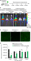
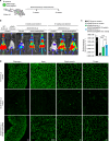
Duchenne muscular dystrophy (DMD) is a fatal genetic disorder caused by mutations in the dystrophin gene. To enable the non-invasive analysis of DMD gene correction strategies in vivo, we introduced a luciferase reporter in-frame with the C-terminus of the dystrophin gene in mice. Expression of this reporter mimics endogenous dystrophin expression and DMD mutations that disrupt the dystrophin open reading frame extinguish luciferase expression. We evaluated the correction of the dystrophin reading frame coupled to luciferase in mice lacking exon 50, a common mutational hotspot, after delivery of CRISPR/Cas9 gene editing machinery with adeno-associated virus. Bioluminescence monitoring revealed efficient and rapid restoration of dystrophin protein expression in affected skeletal muscles and the heart. Our results provide a sensitive non-invasive means of monitoring dystrophin correction in mouse models of DMD and offer a platform for testing different strategies for amelioration of DMD pathogenesis.
Conflict of interest statement
L.A., R.B.D., and E.N.O. are consultants for Exonics Therapeutics. L.A., C.L., R.B.D., and E.N.O. are listed as co-inventors on two filed patents regarding the mouse model (provisional filing application number 62/431,699) and strategy (provisional filing application number 62/442,606) presented in this study. Remaining authors declare no competing interests.
Figures


Similar articles
-
Full-length dystrophin restoration via targeted exon integration by AAV-CRISPR in a humanized mouse model of Duchenne muscular dystrophy.Mol Ther. 2021 Nov 3;29(11):3243-3257. doi: 10.1016/j.ymthe.2021.09.003. Epub 2021 Sep 10. Mol Ther. 2021. PMID: 34509668 Free PMC article.
-
Creation of a Novel Humanized Dystrophic Mouse Model of Duchenne Muscular Dystrophy and Application of a CRISPR/Cas9 Gene Editing Therapy.J Neuromuscul Dis. 2017;4(2):139-145. doi: 10.3233/JND-170218. J Neuromuscul Dis. 2017. PMID: 28505980 Free PMC article.
-
In Vivo Genome Editing Restores Dystrophin Expression and Cardiac Function in Dystrophic Mice.Circ Res. 2017 Sep 29;121(8):923-929. doi: 10.1161/CIRCRESAHA.117.310996. Epub 2017 Aug 8. Circ Res. 2017. PMID: 28790199 Free PMC article.
-
Therapeutic Applications of CRISPR/Cas for Duchenne Muscular Dystrophy.Curr Gene Ther. 2017;17(4):301-308. doi: 10.2174/1566523217666171121165046. Curr Gene Ther. 2017. PMID: 29173172 Review.
-
Molecular correction of Duchenne muscular dystrophy by splice modulation and gene editing.RNA Biol. 2021 Jul;18(7):1048-1062. doi: 10.1080/15476286.2021.1874161. Epub 2021 Jan 20. RNA Biol. 2021. PMID: 33472516 Free PMC article. Review.
Cited by
-
Endogenous bioluminescent reporters reveal a sustained increase in utrophin gene expression upon EZH2 and ERK1/2 inhibition.Commun Biol. 2023 Mar 25;6(1):318. doi: 10.1038/s42003-023-04666-9. Commun Biol. 2023. PMID: 36966198 Free PMC article.
-
CRISPR-Based Therapeutic Gene Editing for Duchenne Muscular Dystrophy: Advances, Challenges and Perspectives.Cells. 2022 Sep 22;11(19):2964. doi: 10.3390/cells11192964. Cells. 2022. PMID: 36230926 Free PMC article. Review.
-
Genome Editing for Rare Diseases.Curr Stem Cell Rep. 2020 Sep;6(3):41-51. doi: 10.1007/s40778-020-00175-1. Epub 2020 Jul 7. Curr Stem Cell Rep. 2020. PMID: 33184603 Free PMC article.
-
Ubiquitous Luciferase Expression in "Firefly Rats" Does Not Alter the Pancreatic Islet Morphology, Metabolism, and Function.Cell Transplant. 2023 Jan-Dec;32:9636897231182497. doi: 10.1177/09636897231182497. Cell Transplant. 2023. PMID: 37345228 Free PMC article.
-
Gene Editing for Duchenne Muscular Dystrophy: From Experimental Models to Emerging Therapies.Degener Neurol Neuromuscul Dis. 2025 Apr 12;15:17-40. doi: 10.2147/DNND.S495536. eCollection 2025. Degener Neurol Neuromuscul Dis. 2025. PMID: 40241992 Free PMC article. Review.
References
Publication types
MeSH terms
Substances
Grants and funding
LinkOut - more resources
Full Text Sources
Other Literature Sources
Medical
Molecular Biology Databases
Research Materials

