Review
. 2019 Oct;11(5):783-793.
doi: 10.1007/s12551-019-00582-7. Epub 2019 Oct 4.

The mechanobiology of tight junctions

Affiliations
Review

The mechanobiology of tight junctions

Sandra Citi. Biophys Rev. 2019 Oct.

Abstract

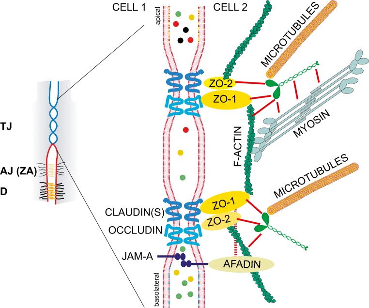
Tight junctions (TJ) play a central role in the homeostasis of epithelial and endothelial tissues, by providing a semipermeable barrier to ions and solutes, by contributing to the maintenance of cell polarity, and by functioning as signaling platforms. TJ are associated with the actomyosin and microtubule cytoskeletons, and the crosstalk with the cytoskeleton is fundamental for junction biogenesis and physiology. TJ are spatially and functionally connected to adherens junctions (AJ), which are essential for the maintenance of tissue integrity. Mechano-sensing and mechano-transduction properties of several AJ proteins have been characterized during the last decade. However, little is known about how mechanical forces act on TJ and their proteins, how TJ control the mechanical properties of cells and tissues, and what are the underlying molecular mechanisms. Here I review recent studies that have advanced our understanding of the relationships between mechanical force and TJ biology.

Keywords: Actin; Mechanobiology; Tight junction; ZO-1.


Figures

Fig. 1
Fig. 1
Scheme of tight junctions (TJ) and their interactions with the actin and microtubule cytoskeletons. The small scheme on the left illustrates the apical position of the TJ (also called zonulae occludentes or ZO) with respect to the more basolateral adherens junctions (AJ) and desmosomes (D). The circumferential AJ of polarized cells is also called zonula adhaerens (ZA). The larger scheme on the right is an expansion of the TJ region of the apical junctional complex of a polarized epithelial cell. During junction biogenesis, several TJ and AJ components are intermingled, and the spatial segregation between TJ and ZA occurs only in mature, fully polarized epithelial cells. The junctional membrane, selected transmembrane proteins of TJ (claudin, occludin, JAM-A), and the major cytoplasmic proteins involved in TJ-cytoskeleton interactions are schematically depicted in the larger scheme. Actin filaments interact with ZO proteins, afadin, and cingulin, and microtubules interact with cingulin. Additional interactions of cingulin with ZO proteins and myosin (Cordenonsi et al. ; D'Atri et al. 2002) are indicated by red lines. The colored circles in the space between cells represent solutes that are selectively blocked or not blocked by the TJ barrier, which forms a selective filter for the paracellular pathway. The barrier function of TJ is controlled by the expression of specific claudin isoforms and by the cytoskeleton (see text)

Similar articles

Cited by

References

    1. Aijaz S, D'Atri F, Citi S, Balda MS, Matter K. Binding of GEF-H1 to the tight junction-associated adaptor cingulin results in inhibition of Rho signaling and G1/S phase transition. Dev Cell. 2005;8:777–786. - PubMed
    1. Anderson JM, Van Itallie CM. Physiology and function of the tight junction. Cold Spring Harb Perspect Biol. 2009;1(a002584):1–16. - PMC - PubMed
    1. Bazellieres E, Conte V, Elosegui-Artola A, Serra-Picamal X, Bintanel-Morcillo M, Roca-Cusachs P, Munoz JJ, Sales-Pardo M, Guimera R, Trepat X. Control of cell-cell forces and collective cell dynamics by the intercellular adhesome. Nat Cell Biol. 2015;17:409–420. - PMC - PubMed
    1. Birukova AA, Smurova K, Birukov KG, Usatyuk P, Liu F, Kaibuchi K, Ricks-Cord A, Natarajan V, Alieva I, Garcia JG, Verin AD. Microtubule disassembly induces cytoskeletal remodeling and lung vascular barrier dysfunction: role of Rho-dependent mechanisms. J Cell Physiol. 2004;201:55–70. - PubMed
    1. Birukova AA, Fu P, Xing J, Yakubov B, Cokic I, Birukov KG. Mechanotransduction by GEF-H1 as a novel mechanism of ventilator-induced vascular endothelial permeability. Am J Physiol Lung Cell Mol Physiol. 2010;298:L837–L848. - PMC - PubMed

LinkOut - more resources