. 2019 Oct;7(19):e14245.
doi: 10.14814/phy2.14245.

Synaptic GABAergic transmission in the central amygdala (CeA) of rats depends on slice preparation and recording conditions

Affiliations

Synaptic GABAergic transmission in the central amygdala (CeA) of rats depends on slice preparation and recording conditions

Elizabeth M Avegno et al. Physiol Rep. 2019 Oct.

Abstract

The central nucleus of the amygdala (CeA) is a primarily GABAergic brain region implicated in stress and addictive disorders. Using in vitro slice electrophysiology, many studies measure GABAergic neurotransmission to evaluate the impact of experimental manipulations on inhibitory tone in the CeA, as a measure of alterations in CeA activity and function. In a recent study, we reported spontaneous inhibitory postsynaptic current (sIPSC) frequencies higher than those typically reported in CeA neurons in the literature, despite utilizing similar recording protocols and internal recording solutions. The purpose of this study was to systematically evaluate two common methods of slice preparation, an NMDG-based aCSF perfusion method and an ice-cold sucrose solution, as well as the use of an in-line heater to control recording temperature, on measures of intrinsic excitability and spontaneous inhibitory neurotransmission in CeA neurons. We report that both slice preparation and recording conditions significantly impact spontaneous GABAergic transmission in CeA neurons, and that recording temperature, but not slicing solution, alters measures of intrinsic excitability in CeA neurons. Bath application of corticotropin-releasing factor (CRF) increased sIPSC frequency under all conditions, but the magnitude of this effect was significantly different across recording conditions that elicited different baseline GABAergic transmission. Furthermore, CRF effects on synaptic transmission differed according to data reporting methods (i.e., raw vs. normalized data), which is important to consider in relation to baseline synaptic transmission values. These studies highlight the impact of experimental conditions and data reporting methods on neuronal excitability and synaptic transmission in the CeA.

Keywords: CRF; NMDG; central amygdala; electrophysiology; neurotransmission.


Conflict of interest statement

NWG owns shares in Glauser Life Sciences, Inc., a start‐up company with interest in development of therapeutics for treatment of mental illness. All other authors declare no competing financial interests.

Figures
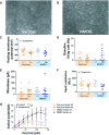
Figure 1
Figure 1
Effects of slice preparation and recording temperature on intrinsic excitability measures on CeA neurons. (A) Representative brightfield image of CeA‐containing tissue section from sucrose preparation, demonstrating reduced visibility of neurons relative to slices derived from NMDG preparation (B). (C), Resting membrane potential of CeA neurons from slices prepared in NMDG (blue) and sucrose (orange), recorded with (dark) and without (light) the heater on. *Indicates main effect of recording temperature; P < 0.05, two‐way ANOVA. (D) Baseline firing rat of each neuron recorded. (E) Rheobase values for each neuron recorded. (F) Input resistance values for each neuron recorded. *Indicates main effect of recording temperature; P < 0.05, two‐way ANOVA. (G) Number of action potentials generated in response to increasing current injections for neurons under each condition. *Indicates main effect of current and recording temperature, as well as an interaction between the two; P < 0.05, three‐way ANOVA. Values in (C‐G) shown as mean ± SEM. The number of cells and rats for each condition were as follows: NMDG‐heater on, eight cells from three rats; NMDG‐heater off, eight cells from four rats; sucrose‐heater on, eight cells from four rats; sucrose‐heater off, 15 cells from 5 rats.
Figure 2
Figure 2
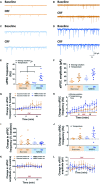
Effects of slice preparation and recording temperature on spontaneous inhibitory postsynaptic currents (sIPSCs) recorded in CeA neurons. Representative sIPSC traces at baseline (top) and in the presence of CRF (bottom) from CeA neurons in the sucrose‐heater off (A), sucrose‐heater on (B), NMDG‐heater off (C), and NMDG‐heater on (D) conditions. Scale bar shown in (D) describes panels A‐D. (E) Baseline sIPSC frequency of CeA neurons recorded under each condition. *Indicates main effect of slicing solution and recording temperature; P < 0.05, two‐way ANOVA; P < 0.05 relative to NMDG‐heater on, Tukey’s multiple comparisons test. (F) Baseline sIPSC amplitude of CeA neurons recorded under each condition. *Indicates main effect of slicing solution and recording temperature; P < 0.05, two‐way ANOVA. (G) Change in sIPSC frequency in the presence of CRF, expressed as a percentage of baseline. No significant effect of time, slicing solution, or recording temperature indicated by three‐way ANOVA. (H) Change in sIPSC frequency in the presence of CRF, expressed as a raw change from baseline. *Indicates main effect of slicing solution, recording temperature, and time, as well as an interaction between the time and recording temperature; P < 0.05, three‐way ANOVA. (I) Average CRF‐induced change in sIPSC frequency, expressed as a percentage of baseline. (J) Average CRF‐induced change in sIPSC frequency, expressed as a raw change from baseline. *Indicates main effect of recording temperature; p < 0.05, two‐way ANOVA. Relatively small changes in sIPSC frequency in (J) appear as large changes in (I) for those neurons with a low baseline sIPSC frequency, and vice versa. (K) Change in sIPSC amplitude in the presence of CRF, expressed as a percentage of baseline. No significant effect of time, slicing solution, or recording temperature indicated by three‐way ANOVA. (L) Change in sIPSC amplitude in the presence of CRF, expressed as a raw change from baseline. No significant effect of time, slicing solution, or recording temperature indicated by three‐way ANOVA. Data in (E‐L) shown as mean ± SEM. The number of cells and rats for each condition were as follows: NMDG‐heater on, eight cells from six rats; NMDG‐heater off, seven cells from four rats; sucrose‐heater on, nine cells from eight rats; sucrose‐heater off, seven cells from six rats.

References

    1. Aghajanian, G. K. , and Rasmussen K.. 1989. Intracellular studies in the facial nucleus illustrating a simple new method for obtaining viable motoneurons in adult rat brain slices. Synapse 3:331–338. - PubMed
    1. Avegno, E. M. , Lobell T. D., Itoga C. A., Baynes B. B., Whitaker A. M., Weera M. M., et al. 2018. Central amygdala circuits mediate hyperalgesia in alcohol‐dependent rats. J. Neurosci. 38:7761–7773. - PMC - PubMed
    1. Baginskas, A. , Palani D., Chiu K., and Raastad M.. 2009. The H‐current secures action potential transmission at high frequencies in rat cerebellar parallel fibers. Eur. J. Neurosci. 29:87–96. - PubMed
    1. Brahma, B. , Forman R. E., Stewart E. E., Nicholson C., and Rice M. E.. 2000. Ascorbate inhibits edema in brain slices. J. Neurochem. 74:1263–1270. - PubMed
    1. Cao, X.‐J. , and Oertel D.. 2005. Temperature Affects voltage‐sensitive conductances differentially in octopus cells of the mammalian cochlear nucleus. J. Neurophysiol. 94:821–832. - PubMed

Publication types