Review
. 2019 Nov;1455(1):34-58.
doi: 10.1111/nyas.14249. Epub 2019 Oct 6.

Chromogranin A and its fragments in cardiovascular, immunometabolic, and cancer regulation

Affiliations
Review

Chromogranin A and its fragments in cardiovascular, immunometabolic, and cancer regulation

Sushil K Mahata et al. Ann N Y Acad Sci. 2019 Nov.

Abstract

Chromogranin A (CgA)-the index member of the chromogranin/secretogranin secretory protein family-is ubiquitously distributed in endocrine, neuroendocrine, and immune cells. Elevated levels of CgA-related polypeptides, consisting of full-length molecules and fragments, are detected in the blood of patients suffering from neuroendocrine tumors, heart failure, renal failure, hypertension, rheumatoid arthritis, and inflammatory bowel disease. Full-length CgA and various CgA-derived peptides, including vasostatin-1, pancreastatin, catestatin, and serpinin, are expressed at different relative levels in normal and pathological conditions and exert diverse, and sometime opposite, biological functions. For example, CgA is overexpressed in genetic hypertension, whereas catestatin is diminished. In rodents, the administration of catestatin decreases hypertension, cardiac contractility, obesity, atherosclerosis, and inflammation, and it improves insulin sensitivity. By contrast, pancreastatin is elevated in diabetic patients, and the administration of this peptide to obese mice decreases insulin sensitivity and increases inflammation. CgA and the N-terminal fragment of vasostatin-1 can enhance the endothelial barrier function, exert antiangiogenic effects, and inhibit tumor growth in animal models, whereas CgA fragments lacking the CgA C-terminal region promote angiogenesis and tumor growth. Overall, the CgA system, consisting of full-length CgA and its fragments, is emerging as an important and complex player in cardiovascular, immunometabolic, and cancer regulation.

Keywords: cancer; cardiovascular diseases; catestatin; immunometabolism; pancreastatin; vasostatin.


Figures

Figure 1
Figure 1
Schematic diagram showing the major peptide domains of CgA protein. Relative locations of vasostatin‐1, pancreastatin (PST), catestatin (CST), and serpinin domains along with the first and last amino acids for each peptide domain are shown except for CST, where an arginine at position 373 is shown in addition to the last amino acid. Major functions of each of the peptide are shown above the peptide domain.
Figure 2
Figure 2
Schematic diagram showing the inhibition of catecholamine secretion and regulation of cardiovascular functions by CST. Acetylcholine released from preganglionic sympathetic endings at the splanchnic–adrenal synapse binds to nicotinic acetylcholine receptors and induces influx of Na+ inside chromaffin cells resulting in the depolarization of the cell membrane and opening of voltage‐gated Ca2+ channels. Increased cytosolic Ca2+ concentration stimulates the transcription of Chga and induces exocytotic release of the secretory cocktail containing catecholamines, CST, ATP, chromogranins, and neuropeptides. Exocytotically released CST inhibits further release of catecholamines by an autocrine/paracrine mechanism; decreases blood pressure and improves BRS and HRV by inhibiting catecholamine secretion; causes vasodilation by stimulating massive release of histamine; and decreases inotropy and lusitropy by activating the β2‐AR‐Gi/o‐protein–PI‐3K–AKT–NO–cGMP signaling pathway. Ach, acetylcholine; BRS, baroreflex sensitivity; CA, catecholamine; CBP, CREB binding protein; CgA, chromogranin A protein; CREB, cAMP‐responsive element–binding protein; DA, dopamine; ERK, extracellular signal–regulated kinase; GA, Golgi apparatus; HRV, heart rate variability; MC, mitochondria; NE, norepinephrine; NTS, nucleus tractus solitarius; PKC, protein kinase C; PSNS, peripheral sympathetic nervous system; RER, rough endoplasmic reticulum; SNS, sympathetic nervous system; Tyr, tyrosine.
Figure 3
Figure 3
Schematic diagram showing the signaling pathways used by CST to provide cardioprotection. CST‐induced cardioprotection begins by the activation of G protein–coupled or cytokine receptors and the consequent recruitment of signaling pathways: (1) the reperfusion injury salvage kinase (RISK) pathway, including PI‐3K–AKT, ERK1/2, and the downstream target glycogen synthase kinase 3 beta (GSK‐3β); and (2) the PKG pathways. These salvage pathways activate downstream mediators, such as endothelial nitric oxide synthase (eNOS), GSK‐3β, hexokinase II (HKII), protein kinase C‐epsilon (PKCε), the mitochondrial ATP–dependent potassium channel (KATP) with consequent inhibition of mitochondrial permeability transition pore (MPTP). PDK1, pyruvate dehydrogenase kinase 1; ROS, reactive oxygen species.
Figure 4
Figure 4
Schematic diagram showing the putative signaling pathways for the anti‐inflammatory actions of CST. Since CST decreases plasma TNF‐α levels, it is expected that less TNF‐α will be available for binding to its receptor (TNFR1), resulting in the attenuation of the IκB–NF‐κB signaling pathway leading to the translocation of fewer NF‐κB to the nucleus. This reduced NF‐κB signaling will affect the formation of NLRP3 inflammasome resulting in less cleavage procaspase 1 to active caspase 1 leading to less conversion of pro–IL‐1β and pro–IL‐18 to IL‐1β and IL‐18 and secretion of IL‐1β and IL‐18.
Figure 5
Figure 5
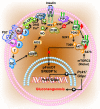
Schematic diagram showing the PST inhibition of gluconeogenesis in hepatocytes. PST initiates a GTP‐binding protein–linked signaling cascade, which results in the activation of diacylglycerol (DAG) and calcium‐dependent conventional PKC (cPKC), thereby attenuating IRS–PI3K–PDK1–AKT signaling pathway. In addition, the stimulation of the cGMP–NOS pathway also dampens this signaling pathway by nitrosylation of IRS. Suppression of this pathway by PST allows FoxO1 and SREBP1c to stimulate the expression of gluconeogenic genes, Pck1 (also known as Pepck) and G6pc (also known as G6Pase), and thus prevents insulin action.
Figure 6
Figure 6
Hypothetical model of the CgA‐dependent angiogenic switch. According to this model, in normal conditions, full‐length CgA1–439 exerts antiangiogenic effects by inducing protease nexin‐1 (PN‐1), an antiangiogenic protein. Since PN‐1 is also a potent inhibitor of plasmin and thrombin, this protein can also prevent CgA cleavage by these enzymes and preserve its antiangiogenic activity. In damaged tissues or tumors, the balance of protease/antiprotease activity is altered leading to CgA cleavage and formation of fragments, for example, CgA1–373, capable of interacting with neuropilin‐1 on endothelial cells (via its PGPQLR sequence) and stimulating angiogenesis (see text).

References

    1. Banks, P. & Helle K.. 1965. The release of protein from the stimulated adrenal medulla. Biochem. J. 97: 40C–41C. - PMC - PubMed
    1. Blaschko, H. , Comline R.S., Schneider F.H., et al 1967. Secretion of a chromaffin granule protein, chromogranin, from the adrenal gland after splanchnic stimulation. Nature 215: 58–59. - PubMed
    1. Schneider, F.H. , Smith A.D. & Winkler H.. 1967. Secretion from the adrenal medulla: biochemical evidence for exocytosis. Br. J. Pharmacol. Chemother. 31: 94–104. - PMC - PubMed
    1. Konecki, D.S. , Benedum U.M., Gerdes H.H., et al 1987. The primary structure of human chromogranin A and pancreastatin. J. Biol. Chem. 262: 17026–17030. - PubMed
    1. Winkler, H. & Fischer‐Colbrie R.. 1992. The chromogranins A and B: the first 25 years and future perspectives. Neuroscience 49: 497–528. - PMC - PubMed

Publication types