Review
. 2019 Nov;9(11):e01440.
doi: 10.1002/brb3.1440. Epub 2019 Oct 6.

Reversible lesion in the splenium of the corpus callosum

Affiliations
Review

Reversible lesion in the splenium of the corpus callosum

Syuichi Tetsuka. Brain Behav. 2019 Nov.

Abstract

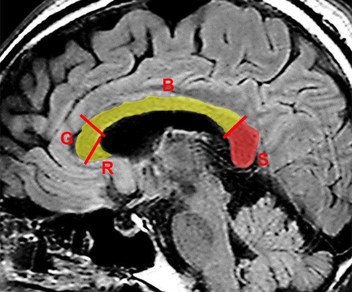
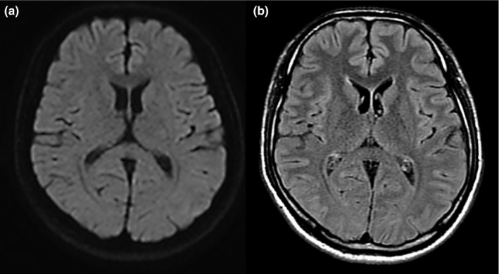
Aim of review: The presence of isolated, reversible lesions in the splenium of the corpus callosum (SCC) is essential to confirm the diagnosis of mild encephalitis/encephalopathy. The lesions usually heal within a month after the onset of neurological symptoms. Magnetic resonance imaging (MRI) has increasingly been used as a diagnostic tool, which has led to the publication of an increasing number of case reports. These have highlighted some inconsistencies about encephalitis/encephalopathy. First, the condition is not always mild and may be severe. Second, reversible lesions in the SCC have been identified in various diseases and conditions other than viral encephalitis/encephalopathy. Third, lesions in SCC are not always completely reversible. On this note, this review describes the specific clinical and radiological features of encephalitis/encephalopathy.

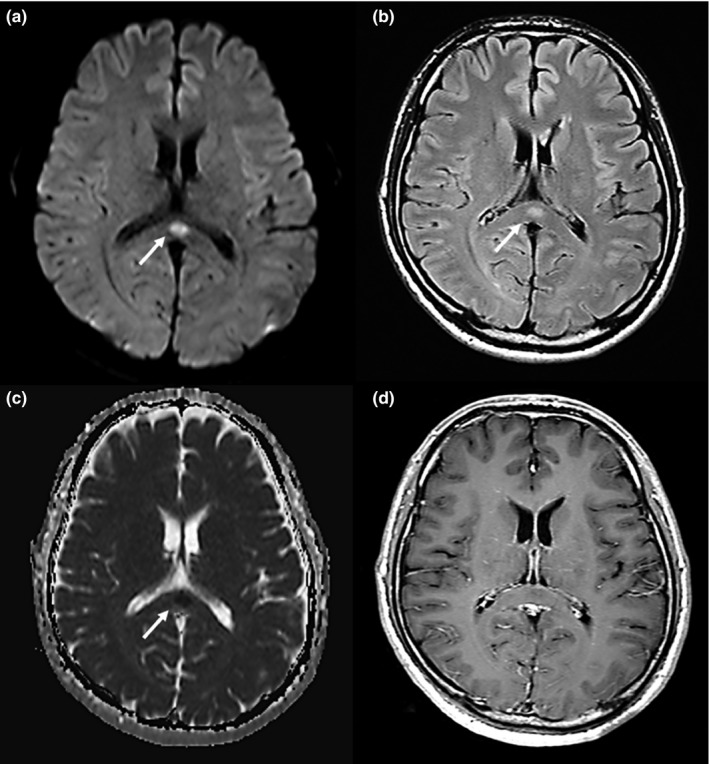
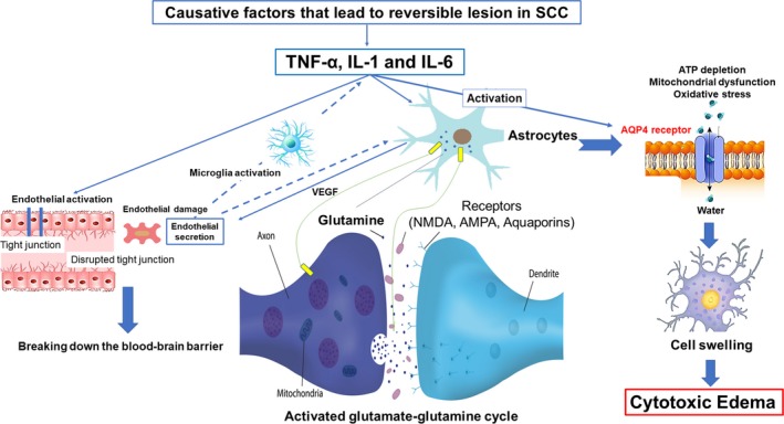
Findings: The reversible lesion in SCC is an MRI finding observable in a wide variety of diseases and conditions. Thus, it should be considered as a secondary change rather than a peculiar feature associated with mild encephalitis/encephalopathy. If reversible lesions are present in the SCC, the symptoms and prognosis are not necessarily favorable, with manifestations of encephalitis/encephalopathy varying from absent to severe. Neuroradiological features that appear as isolated high-intensity signals on diffusion-weighted images and a decreased apparent diffusion coefficient of the lesion might indicate a diagnosis of cytotoxic edema. Findings of previous studies suggest that cytokine-mediated cytotoxic edema of the SCC may be an important pathophysiological manifestation of this condition.

Conclusion: The reversible lesions in the SCC found on MRI are not exclusive to encephalitis/encephalopathy but may be secondary to other disorders.

Keywords: apparent diffusion coefficient; cytokine; cytotoxic edema; reversible; splenium of the corpus callosum.


Conflict of interest statement

The author declares no conflict of interest.

Figures

Figure 1
Figure 1
Midsagittal view of the splenium. Midsagittal fluid‐attenuated inversion recovery—weighted magnetic resonance image shows the corpus callosum and the splenium (S, in red). According to the conventional partitioning scheme, the splenium corresponds to the posterior 20% of the corpus callosum, which is separated by the border line perpendicular to the line linking the most anterior and posterior points of the corpus callosum. B, body; G, genu; R, rostrum
Figure 2
Figure 2
A 41‐year‐old man was admitted with fever, headache, and fatigue. He received a diagnosis of clinically mild encephalitis. Magnetic resonance imaging was performed on day 1 hospitalization. Diffusion‐weighted (a) and fluid‐attenuated inversion recovery (b) imaging showed a marked hyperintense signal (arrows) on the splenium of the corpus callosum (SCC). The apparent diffusion coefficient (c) showed a hypointense signal (arrow). T1‐weighted imaging with contrast material (d) showed an isointense signal that was not enhanced by the contrast material. Arrows indicate the SCC
Figure 3
Figure 3
Magnetic resonance images on day 10 of hospitalization of the patient described in Figure 2. Axial diffusion‐weighted image (a) and fluid‐attenuated inversion recovery image (b) showed either disappearance or remarkable shrinking of the previously observed abnormalities. The patient was discharged without sequelae or medication
Figure 4
Figure 4
Depiction of the pathophysiological hypothesis of the reversible lesion in the splenium of the corpus callosum (SCC) that leads to cytotoxic edema. Cytokines, such as tumor necrosis factor alpha (TNF‐α), interleukin‐1 (IL‐1), and interleukin‐6 (IL‐6), are released. Endothelial damage causes activation of microglia that further accelerates the release of these cytokines. IL‐1 can also induce astrocytes to take up glutamate, which reacts with ammonia to form glutamine, and then glutamine is transported to the extracellular fluid, where it is again taken up by neurons; then the glutamate synthesis process restarts, thus increasing extracellular glutamine. Intracellular ATP depletion, resulting in mitochondrial dysfunction and oxidative stress, is induced by activated glutamate–glutamine cycle. The excitotoxic action of receptors, such as N‐methyl‐d‐aspartate (NMDA), α‐amino‐3‐ hydroxy‐5‐methyl‐4‐isoxazole propionic acid (AMPA), and aquaporin 4 (AQP4) receptors activation, is triggered through a complex cell–cytokine, that results in an influx of water into both astrocytes and neurons, which leads to cytotoxic edema, characterized by intracellular accumulation of fluid and Na+ and resulting in cell swelling. VEGF, vascular endothelial growth factor

References

    1. Al Brashdi, Y. H. , & Albayram, M. S. (2015). Reversible restricted‐diffusion lesion representing transient intramyelinic cytotoxic edema in a patient with traumatic brain injury. The Neuroradiology Journal, 28(4), 409–412. 10.1177/1971400915598071 - DOI - PMC - PubMed
    1. Avcu, G. , Kilinc, M. A. , Eraslan, C. , Karapinar, B. , & Vardar, F. (2017). Mild encephalitis/encephalopathy with reversible splenial lesion (MERS) associated with Streptococcus pneumoniae bacteraemia. Journal of Infection and Public Health, 10(4), 479–482. 10.1016/j.jiph.2016.08.019 - DOI - PMC - PubMed
    1. Badaut, J. , Fukuda, A. M. , Jullienne, A. , & Petry, K. G. (2014). Aquaporin and brain diseases. Biochimica et Biophysica Acta, 1840(5), 1554–1565. 10.1016/j.bbagen.2013.10.032 - DOI - PMC - PubMed
    1. Baehring, J. M. , & Fulbright, R. K. (2012). Diffusion‐weighted MRI in neuro‐oncology. CNS Oncology, 1(2), 155–167. 10.2217/cns.12.28 - DOI - PMC - PubMed
    1. Bin, C. H. , & Lee, S. J. (2011). Teaching neuroImages: Reversible splenial cytotoxic edema in acute mountain sickness. Neurology, 77(16), e94 10.1212/WNL.0b013e318233b326 - DOI - PubMed

MeSH terms