. 2019 Nov 8;5(11):1915-1925.
doi: 10.1021/acsinfecdis.9b00245. Epub 2019 Oct 24.

Ureadepsipeptides as ClpP Activators

Affiliations

Ureadepsipeptides as ClpP Activators

Elizabeth C Griffith et al. ACS Infect Dis. .

Abstract

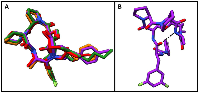
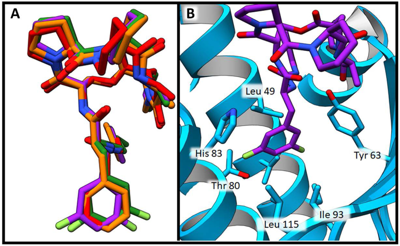
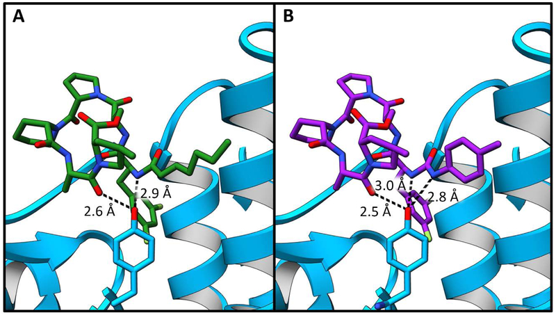
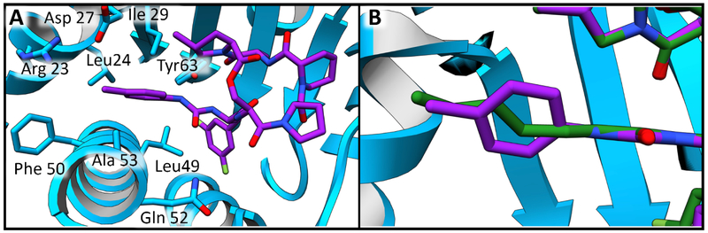
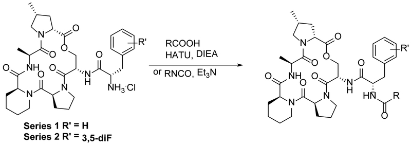
Acyldepsipeptides are a unique class of antibiotics that act via allosterically dysregulated activation of the bacterial caseinolytic protease (ClpP). The ability of ClpP activators to kill nongrowing bacteria represents a new opportunity to combat deep-seated biofilm infections. However, the acyldepsipeptide scaffold is subject to rapid metabolism. Herein, we explore alteration of the potentially metabolically reactive α,β unsaturated acyl chain. Through targeted synthesis, a new class of phenyl urea substituted depsipeptide ClpP activators with improved metabolic stability is described. The ureadepsipeptides are potent activators of Staphylococcus aureus ClpP and show activity against Gram-positive bacteria, including S. aureus biofilms. These studies demonstrate that a phenyl urea motif can successfully mimic the double bond, maintaining potency equivalent to acyldepsipeptides but with decreased metabolic liability. Although removal of the double bond from acyldepsipeptides generally has a significant negative impact on potency, structural studies revealed that the phenyl ureadepsipeptides can retain potency through the formation of a third hydrogen bond between the urea and the key Tyr63 residue in the ClpP activation domain. Ureadepsipeptides represent a new class of ClpP activators with improved drug-like properties, potent antibacterial activity, and the tractability to be further optimized.

Keywords: ClpP; acyldepsipeptide; antibiotic; biofilm; ureadepsipeptide.


Figures

Figure 1.
Figure 1.
Casein-BODIPY ClpP mediated degradation assay, a comparison of matched urea and non-urea pairs. These graphs show the rate of SaClpP casein-BODIPY degradation (normalized to the maximum rate of SaClpP degradation in the presence of ADEP4) as a function of experimental compound concentration. A) Compounds represented in this graph have a non-fluorinated phenylalanine motif. Compounds 5 and 12 (round markers) have urea substitutions while compounds 4 and 11 (square markers) retain the acyl side chains. Compounds 5 and 4 (green) contain phenyl side chains. Compounds 12 and 11 (red) have a para methyl addition to the phenyl side chain. B) Compounds represented in this graph have a 3,5 difluorinated phenylalanine motif. Compounds 9 and 16 (round markers) have urea substitutions while compounds 8 and 15 (square markers) retain the acyl side chains. Compounds 9 and 8 (green) contain phenyl side chains. Compounds 15 and 16 (red) have a para methyl addition to the phenyl side chain.
Figure 2.
Figure 2.
In vitro S. aureus biofilm killing by UDEPs and in combination with rifampin. Assays were performed at 10 times the MIC and statistical analysis completed with 1way ANOVA and Dunnett’s multiple comparisons test.
Figure 3.
Figure 3.
Isothermal titration calorimetry results. Top panels show the change in enthalpy per injection of SaClpP into experimental compounds. Bottom panels show the integrated enthalpies and the results of nonlinear regression fitting of the integrated enthalpies. A) SaClpP injected into compound 8. B) SaClpP injected into compound 9.C) SaClpP injected into compound 15. D) SaClpP injected into compound 16.
Figure 4.
Figure 4.
Surface plasmon resonance sensograms. A) Compound 8. B) Compound 9.C) Compound 15. D) Compound 16
Figure 5.
Figure 5.
Depsipeptide lactone core binding positions A) Structure overlay demonstrating similarity of lactone core binding positions. Compound 2 is shown in red, compound 5 is shown in orange, compound 16 is shown in purple and ADEP4 is shown in green. B) Intramolecular hydrogen bond within the lactone core of compound 16 is modeled with dashed black line.
Figure 6.
Figure 6.
Depsipeptide phenylalanine motif binding pocket. A) Structure overlay demonstrating similarity of phenylalanine binding positions. Compound 2 is shown in red, compound 5 is shown in orange, compound 16 is shown in purple and ADEP4 is shown in green. B) Binding pocket of compound 16 shown in purple. SaClpP is shown in blue ribbon structure. SaClpP side chains within 4 Å of the phenyl ring of this ligand are shown as blue sticks.
Figure 7.
Figure 7.
Tyr 63 interactions. SaClpP is represented in blue ribbons with Tyr 63 shown in blue sticks. A) SaClpP bound to ADEP4 shown here as a green stick figure. Hydrogen bonding interactions between ADEP4 and Tyr 63 are represented with black dashed lines. B) SaClpP bound to compound 16, which is shown here as a purple stick figure. Hydrogen bonding interactions between compound 16 and Tyr 63 are represented with black dashed lines.
Figure 8.
Figure 8.
Hydrophobic side chain binding pocket. A) The binding pocket of compound 16 shown in purple sticks. Residues within 4 Å of the urea side chain are shown in blue sticks. B) An overlay of ADEP4 shown in green sticks and compound 16 shown in purple sticks. Image focused on the acyl and urea side chain of the compounds.
Scheme 1.
Scheme 1.
The chemical synthesis approach for depsipeptide analogs.

References

    1. Kourtis AP; Hatfield K; Baggs J; Mu Y; See I; Epson E; Nadle J; Kainer MA; Dumyati G; Petit S; Ray SM; Emerging Infections Program, M. a. g.; Ham D; Capers C; Ewing H; Coffin N; McDonald LC; Jernigan J; Cardo D, Vital Signs: Epidemiology and Recent Trends in Methicillin-Resistant and in Methicillin-Susceptible Staphylococcus aureus Bloodstream Infections - United States. MMWR Morb Mortal Wkly Rep 2019, 68 (9), 214–219. - PMC - PubMed
    1. Hall-Stoodley L; Costerton JW; Stoodley P, Bacterial biofilms: from the natural environment to infectious diseases. Nat Rev Microbiol 2004, 2 (2), 95–108. - PubMed
    1. Conlon BP; Rowe SE; Lewis K, Persister cells in biofilm associated infections. Adv Exp Med Biol 2015, 831, 1–9. - PubMed
    1. Lewis K, Persister cells: molecular mechanisms related to antibiotic tolerance. Handb Exp Pharmacol 2012, (211), 121–33. - PubMed
    1. Fauvart M; De Groote VN; Michiels J, Role of persister cells in chronic infections: clinical relevance and perspectives on anti-persister therapies. J Med Microbiol 2011, 60 (Pt 6), 699–709. - PubMed

Publication types

MeSH terms

LinkOut - more resources