Review
. 2019 Oct 5;20(19):4929.
doi: 10.3390/ijms20194929.

Pyruvate Substitutions on Glycoconjugates

Affiliations
Review

Pyruvate Substitutions on Glycoconjugates

Fiona F Hager et al. Int J Mol Sci. .

Abstract

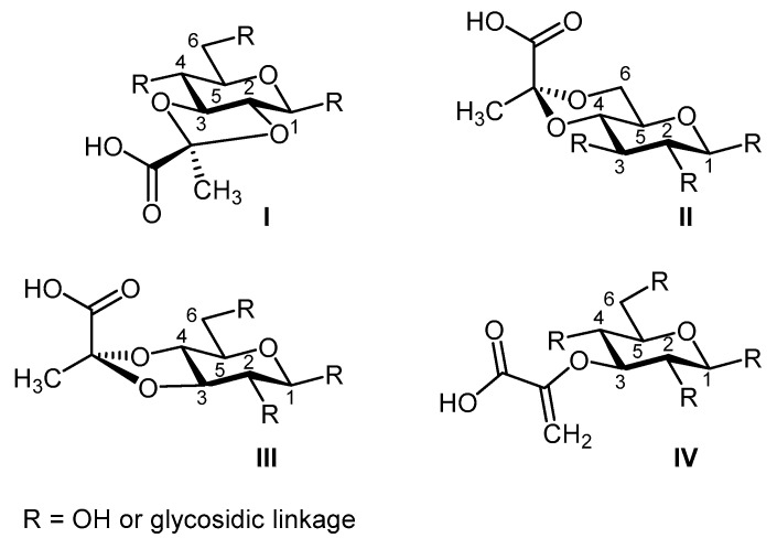
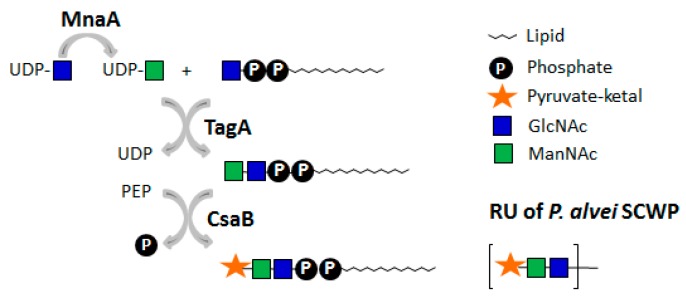
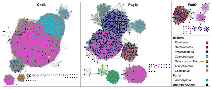
Glycoconjugates are the most diverse biomolecules of life. Mostly located at the cell surface, they translate into cell-specific "barcodes" and offer a vast repertoire of functions, including support of cellular physiology, lifestyle, and pathogenicity. Functions can be fine-tuned by non-carbohydrate modifications on the constituting monosaccharides. Among these modifications is pyruvylation, which is present either in enol or ketal form. The most commonly best-understood example of pyruvylation is enol-pyruvylation of N-acetylglucosamine, which occurs at an early stage in the biosynthesis of the bacterial cell wall component peptidoglycan. Ketal-pyruvylation, in contrast, is present in diverse classes of glycoconjugates, from bacteria to algae to yeast-but not in humans. Mild purification strategies preventing the loss of the acid-labile ketal-pyruvyl group have led to a collection of elucidated pyruvylated glycan structures. However, knowledge of involved pyruvyltransferases creating a ring structure on various monosaccharides is scarce, mainly due to the lack of knowledge of fingerprint motifs of these enzymes and the unavailability of genome sequences of the organisms undergoing pyruvylation. This review compiles the current information on the widespread but under-investigated ketal-pyruvylation of monosaccharides, starting with different classes of pyruvylated glycoconjugates and associated functions, leading to pyruvyltransferases, their specificity and sequence space, and insight into pyruvate analytics.

Keywords: N-glycans; biosynthesis; capsular polysaccharides; cell wall glycopolymers; exopolysaccharides; lipopolysaccharides; pyruvate analytics; pyruvylation; pyruvyltransferase; sequence space.


Conflict of interest statement

The authors declare no conflict of interest. The funders had no role in the design of the study; in the collection, analyses, or interpretation of data; in the writing of the manuscript, or in the decision to publish the results.

Figures

Figure 1
Figure 1
Overview on the most common modes of monosaccharide pyruvylation. Shown is pyruvate ketal at bridging positions 2,3 (I), 4,6 (II), and 3,4 (III), and enol pyruvate (IV). Arabic numbers indicate ring positions.
Figure 2
Figure 2
Scheme of succinoglycan biosynthesis in Shinorhizobium meliloti [43]. The pyruvylation step occurs in the cytoplasm at the stage of the undp-PP-linked RU prior to export and polymerization in the periplasmic space. Pyruvylation (ExoV) is indicated by a star. The order of pyruvylation, acetylation (ExoZ), and succinylation (Exo) is unknown. RU: repeating unit. Monosaccharide symbols are shown according to the Symbol Nomenclature for Glycans (SNFG) [45].
Figure 3
Figure 3
Scheme of capsular polysaccharide (CPS) A biosynthesis in Bacteroides fragilis. The pyruvylation step occurs in the cytoplasm at the stage of the lipid-PP-linked RU precursor. Pyruvylation (WcfO) is indicated by a star. Notably, in contrast to succinoglycan biosynthesis (Figure 2), pyruvylation of the internal Galp of the RU needs to proceed prior to completion of the lipid-PP-linked tetrasaccharide repeat. RU: repeating unit. Monosaccharide symbols are shown according to the Symbol Nomenclature for Glycans (SNFG) [45].
Figure 4
Figure 4
I and II, equatorial-oriented methyl groups of 4,6 galacto- and manno-pyranosyls. III, identification of the attachment site of pyruvylation via hetero multiple bond correlation (HMBC). IV, through-space correlation of the pyruvate methyl group to a ring proton—in this case H3. Pink arrows indicate though-bond interactions of neighbouring protons (HMBC), while blue arrows indicate through-space interactions of neighbouring protons (NOE).
Figure 5
Figure 5
In vitro one-pot reaction involving the Paenibacillus alvei enzymes MnaA, TagA, and CsaB in combination with a synthetic lipid-PP-GlcNAc primer, demonstrating the preference of P. alvei CsaB for a lipid-PP-linked disaccharide substrate [109]. Pyruvylation (CsaB) is indicated by a star. RU: repeating unit. Monosaccharide symbols are shown according to the Symbol Nomenclature for Glycans (SNFG) [45].
Figure 6
Figure 6
Sequence similarity networks illustrating the extant sequence space around Paenibacillus alvei CsaB (K4ZGN3), Schizosaccharomyces pombe Pvg1p (Q9UT27), and Bacteroides fragilis WcfO (Q5LFK7). The three characterized sequences—CsaB, Pvg1p, and WcfO—are highlighted by yellow circles.

References

    1. Bennet L.G., Bishop C.T. The pyruvate ketal as a stereospecific immunodeterminant in the type XXVII Streptococcus pneumoniae (pneumococcal) capsular polysaccharide. Immunochemistry. 1977;14:693–696. doi: 10.1016/0019-2791(77)90143-4. - DOI
    1. Lovering A.L., Safadi S.S., Strynadka N.C. Structural perspective of peptidoglycan biosynthesis and assembly. Annu. Rev. Biochem. 2012;81:451–478. doi: 10.1146/annurev-biochem-061809-112742. - DOI - PubMed
    1. Andreishcheva E.N., Kunkel J.P., Gemmill T.R., Trimble R.B. Five genes involved in biosynthesis of the pyruvylated Galβ1,3-epitope in Schizosaccharomyces pombe N-linked glycans. J. Biol. Chem. 2004;279:35644–35655. doi: 10.1074/jbc.M403574200. - DOI - PubMed
    1. Leone S., Izzo V., Silipo A., Sturiale L., Garozzo D., Lanzetta R., Parrilli M., Molinaro A., Di Donato A. A novel type of highly negatively charged lipooligosaccharide from Pseudomonas stutzeri OX1 possessing two 4,6-O-(1-carboxy)-ethylidene residues in the outer core region. Eur. J. Biochem. 2004;271:2691–2704. doi: 10.1111/j.1432-1033.2004.04197.x. - DOI - PubMed
    1. Toukach P.V., Egorova K.S. Carbohydrate structure database merged from bacterial, archaeal, plant and fungal parts. Nucleic Acids Res. 2016;44:D1229–D1236. doi: 10.1093/nar/gkv840. - DOI - PMC - PubMed

MeSH terms

LinkOut - more resources