. 2019 Oct 6;11(10):2380.
doi: 10.3390/nu11102380.

Leptin and EGF Supplementation Enhance the Immune System Maturation in Preterm Suckling Rats

Affiliations

Leptin and EGF Supplementation Enhance the Immune System Maturation in Preterm Suckling Rats

Blanca Grases-Pintó et al. Nutrients. .

Abstract

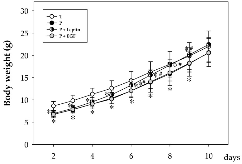
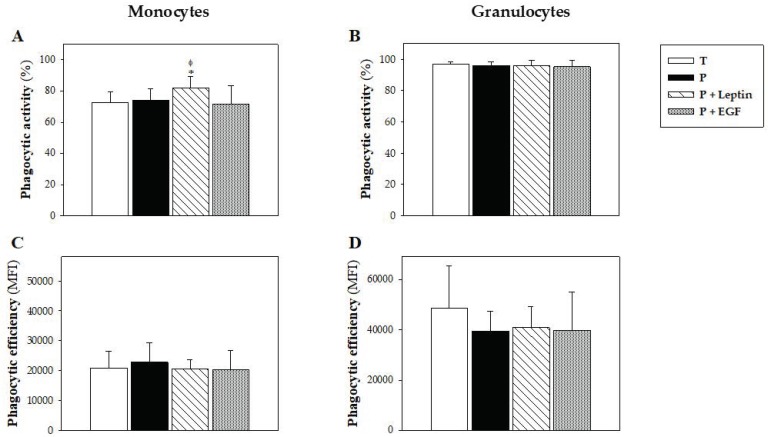
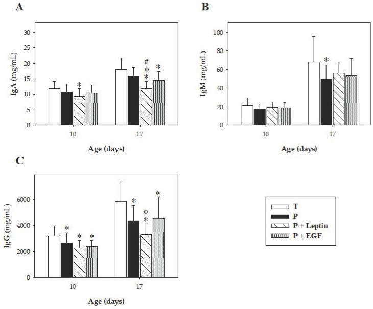
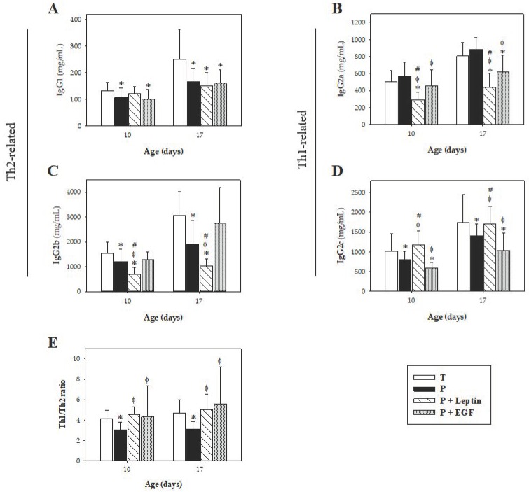
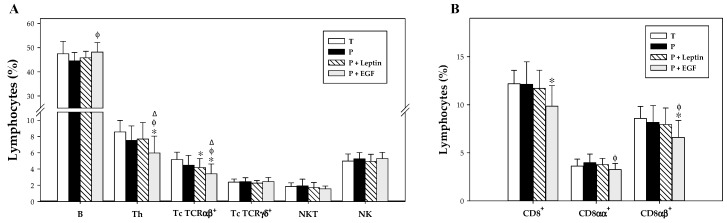
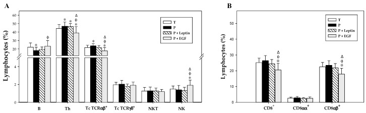
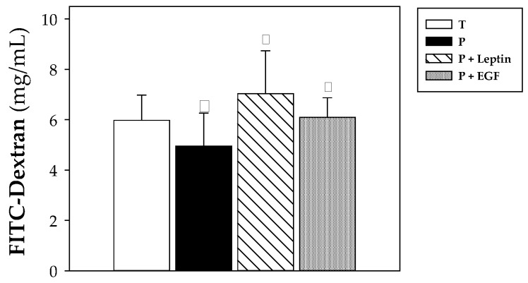
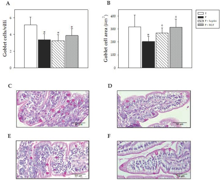
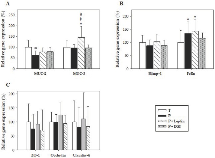
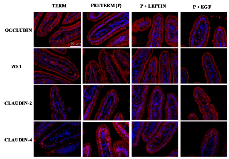
In preterm newborns the immaturity of the immune system is remarkable, with reduced innate and adaptive immune responses. Many bioactive compounds in breast milk, such as growth factors and adipokines, contribute to the immune system's maturation in early life. However, studies on the immunoregulatory activity in preterm neonates are practically nonexistent. The aim of the present study was to determine whether a nutritional supplementation in early life with leptin or epidermal growth factor (EGF) was able to promote the maturation of the systemic and intestinal immune system in preterm conditions. For this purpose, premature rats were daily supplemented by oral gavage with leptin or EGF. Term and Preterm groups receiving vehicle were used as controls. Preterm rats showed deficiencies compared to full-term ones, such as lower body weights, erythrocyte counts, plasma IgG and IgM concentrations and B cell percentages, and higher values of Th and Tc TCRαβ+ cells in mesenteric lymph nodes, and intestinal permeability, among others. However, leptin and EGF supplementation were able to revert some of these deficiencies and to improve the premature immune system's development. These results suggest that leptin and EGF are involved in enhancing the maturation of the systemic and intestinal immune system in preterm conditions.

Keywords: EGF; adaptive immunity; breast milk; innate immunity; leptin; prematurity; suckling rat.


Conflict of interest statement

The authors declare no conflict of interest.

Figures

Figure 1
Figure 1
Body weight from the four groups: Term (T), Preterm (P), P+Leptin, and P+epidermal growth factor (P+EGF) the first 10 days of the study. Results are expressed as mean ± SD (n = 12–30 pups/group). Statistical differences: * p < 0.05 versus T group; φ p < 0.05 versus P group; # p < 0.05 versus P+EGF group.
Figure 2
Figure 2
Phagocytic function of blood leukocytes from the four groups: Term (T), Preterm (P), P+Leptin, and P+epidermal growth factor (P+EGF). Phagocytic activity (A,B) and efficiency (C,D) at day 10 from monocytes and granulocytes, respectively. Results are expressed as mean ± SD (n = 9 pups/group). Statistical differences: * p < 0.01 versus T group; φ p < 0.05 versus P group.
Figure 3
Figure 3
Plasma Ig concentrations at day 10 and 17. Plasma IgA (A), IgM (B), and IgG (C) concentrations from the four groups: Term (T), Preterm (P), P+Leptin, and P+epidermal growth factor (P+EGF), are expressed as mean ± SD (n = 8–12 pups/group). Statistical differences: * p < 0.05 versus T group; φ p < 0.05 versus P group; # p < 0.05 versus P+EGF group.
Figure 4
Figure 4
Plasmatic concentration of IgG subclasses at days 10 and 17. IgG1 (A), IgG2a (B), IgG2b (C), IgG2c (D), and Th1/Th2 ratios (E) from the four groups: Term (T), Preterm (P), P+Leptin, and P+epidermal growth factor (P+EGF) are expressed as mean ± SD (n = 8–12 pups/group). Statistical differences: * p < 0.05 versus T group; φ p < 0.05 versus P group; # p < 0.05 versus P+EGF group.
Figure 5
Figure 5
Main lymphocyte subsets (A) and CD8+ cells and their both forms (CD8αα+ and CD8αβ+) cell percentages (B) in the spleen from the four groups: Term (T), Preterm (P), P+Leptin, and P+epidermal growth factor (P+EGF) at day 17. The results are expressed as mean ± SD (n = 8–12 pups/group). Statistical differences: * p < 0.05 versus T group; φ p < 0.05 versus P group; Δ p < 0.05 versus P+Leptin group.
Figure 6
Figure 6
Main lymphocyte subsets (A) and CD8+cells and their both forms (CD8αα+ and CD8αβ+) cell percentages (B) in the mesenteric lymph nodes (MLNs) from the four groups: Term (T), Preterm (P), P+Leptin, and P+epidermal growth factor (P+EGF) at day 17. The results are expressed as mean ± SD (n = 8–12 pups/group). Statistical differences: * p < 0.05 versus T group; φ p < 0.05 versus P group; Δ p < 0.05 versus P+Leptin group.
Figure 7
Figure 7
Intestinal permeability to 4 kDa-FITC-dextran from the four groups: Term (T), Preterm (P), P+Leptin, and P+epidermal growth factor (P+EGF) at day 10. Results are expressed as mean ± SD (n = 9 pups/group). Statistical differences: * p < 0.05 versus T group; φ p < 0.05 versus P group.
Figure 8
Figure 8
Number of goblet cells/villi (A), goblet cells area (B), and representative images of histological sections of the jejunum with periodic acid−Schiff (PAS) staining from the four groups: Term (C), Preterm (D), P+Leptin (E), and P+epidermal growth factor (P+EGF) (F) at day 10 of the suckling period. Results of Figure 8A,B are expressed as mean ± SD (n = 6 pups/group). Statistical differences: * p < 0.05 versus T group; φ p < 0.05 versus P group. Goblet cells with densely stained granules can be observed along the length of the villi (Figure 8C–F). Scale bar = 50 μm for 400 ×.
Figure 9
Figure 9
Gene expression in small intestine samples from the four groups: Term (T), Preterm (P), P+Leptin, and P+epidermal growth factor (P+EGF) at day 10 of the suckling period. MUC-2 and MUC-3 (A), Blimp-1 and FcRn (B), ZO-1, occludin, and claudin-4 (C). Results are expressed as mean ± SD (n = 9 pups/group). Statistical differences: * p < 0.05 versus T group; φ p < 0.05 versus P group; # p < 0.05 versus P+EGF group.
Figure 10
Figure 10
Immunofluorescent staining of small intestine tissue for occludin, ZO-1, claudin-2, and claudin-4. Representative images from Term (T), Preterm (P), P+Leptin, and P+epidermal growth factor (P+EGF) intestines at day 10. Similar results were obtained in five animals in each group. Nuclei were stained with DAPI (blue). Localization of occludin, ZO-1, claudin-2, and claudin-4 (red) were observed by fluorescence microscopy at 400× magnification. Scale bar: 50 µm.

References

    1. Boquien C.Y. Human milk: An ideal food for nutrition of preterm newborn. Front. Pediatrics. 2018;6:295. doi: 10.3389/fped.2018.00295. - DOI - PMC - PubMed
    1. El Homsi M., Ducroc R., Claustre J., Jourdan G., Gertler A., Estienne M., Bado A., Scoazec J.Y., Plaisancié P. Leptin modulates the expression of secreted and membrane-associated mucins in colonic epithelial cells by targeting PKC, PI3K, and MAPK pathways. Am. J. Physiol. Liver Physiol. 2007;293:G365–G373. doi: 10.1152/ajpgi.00091.2007. - DOI - PubMed
    1. Neu J. Gastrointestinal development and meeting the nutritional needs of premature infants. Am. J. Clin. Nutr. 2007;85:629S–634S. doi: 10.1093/ajcn/85.2.629S. - DOI - PubMed
    1. Gartner L.M., Morton J., Lawrence R.A., Naylor A.J., O’Hare D., Schanler R.J., Eidelman A.I., American Academy of Pediatrics Section on Breastfeeding Breastfeeding and the use of human milk. Pediatrics. 2005;115:496–506. - PubMed
    1. Underwood M.A. Human milk for the premature infant. Pediatrics Clin. North Am. 2013;60:189–207. doi: 10.1016/j.pcl.2012.09.008. - DOI - PMC - PubMed

MeSH terms