Review
. 2019 Oct 7;21(1):61.
doi: 10.1186/s12968-019-0575-y.

Machine learning in cardiovascular magnetic resonance: basic concepts and applications

Affiliations
Review

Machine learning in cardiovascular magnetic resonance: basic concepts and applications

Tim Leiner et al. J Cardiovasc Magn Reson. .

Abstract

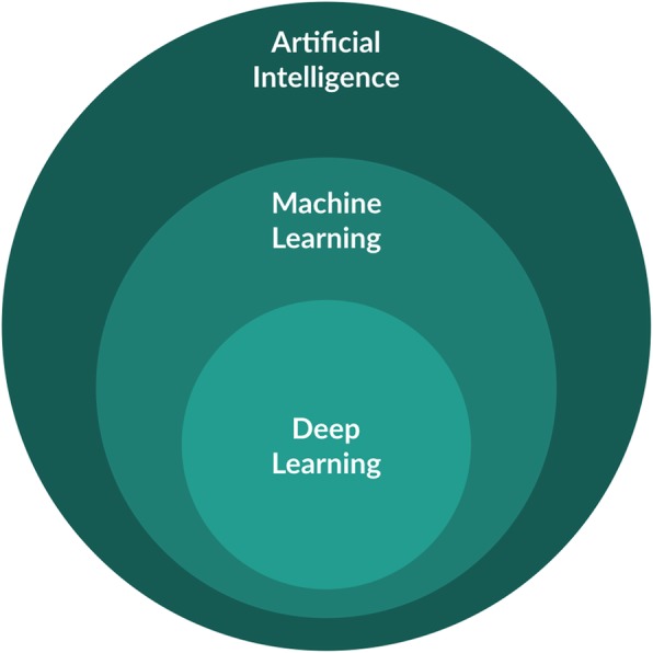
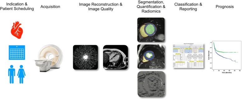
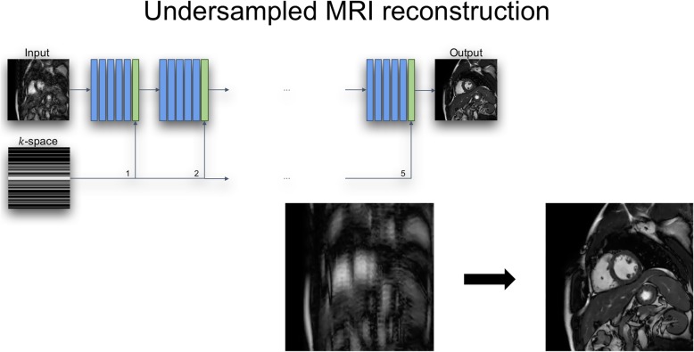
Machine learning (ML) is making a dramatic impact on cardiovascular magnetic resonance (CMR) in many ways. This review seeks to highlight the major areas in CMR where ML, and deep learning in particular, can assist clinicians and engineers in improving imaging efficiency, quality, image analysis and interpretation, as well as patient evaluation. We discuss recent developments in the field of ML relevant to CMR in the areas of image acquisition & reconstruction, image analysis, diagnostic evaluation and derivation of prognostic information. To date, the main impact of ML in CMR has been to significantly reduce the time required for image segmentation and analysis. Accurate and reproducible fully automated quantification of left and right ventricular mass and volume is now available in commercial products. Active research areas include reduction of image acquisition and reconstruction time, improving spatial and temporal resolution, and analysis of perfusion and myocardial mapping. Although large cohort studies are providing valuable data sets for ML training, care must be taken in extending applications to specific patient groups. Since ML algorithms can fail in unpredictable ways, it is important to mitigate this by open source publication of computational processes and datasets. Furthermore, controlled trials are needed to evaluate methods across multiple centers and patient groups.

Keywords: Cardiovascular magnetic resonance; Deep learning; Machine learning; Radiomics.


Conflict of interest statement

Dr. Nezafat has issued and pending patents related to methods for improved cardiovascular MRI, received source code from Philips Healthcare, and receives patent royalties from Phillips Healthcare and Samsung Electronics. Drs. Leiner and disclosed institutional grants received from Pie Medical B.V.

Figures

Fig. 1
Fig. 1
Artificial intelligence (AI) can be seen as any technique that enables computers to perform tasks characteristic of human intelligence. Machine learning (ML) is generally seen as the subdiscipline of AI which uses a statistical model together with training data to learn how to make predictions. Deep learning (DL) is a specific form of ML that uses artificial neural networks with hidden layers to make predictions directly from datasets
Fig. 2
Fig. 2
Machine learning will impact all aspects of cardiovascular magnetic resonance imaging from patient scheduling to image analysis and prognosis
Fig. 3
Fig. 3
Deep learning network for reconstruction of undersampled CMR images
Fig. 4
Fig. 4
Late gadolinium enhancement (LGE; red arrows) images with isotropic spatial resolution of 1.4 mm3 reconstructed using deep learning from a prospectively five-fold randomly undersampled 3D LGE dataset in a patient with hypertrophic cardiomyopathy
Fig. 5
Fig. 5
Some examples of deep learning based myocardial segmentation on long-axis CMR images, trained from almost 5000 cases. A U-Net network architecture was used in this case to classify myocardium (red) and cavity (blue)
Fig. 6
Fig. 6
Scar and myocardium segmentation results for slices from four different patients. Contours resulting from manual (top row) and automatic (lower row) segmentations for the epicardium (blue), endocardium (red), and scar (yellow) boundaries are overlaid on late gadolinium enhancement (LGE) images
Fig. 7
Fig. 7
Myocardial T1 mapping at five short axial slices (apex to base from left to right respectively) of the left ventricle of one patient. a Automatically reconstructed map (after automatic removal of myocardial boundary pixels) overlaid on a T1 weighted image with shortest inversion time; (a) Manually reconstructed T1 map. The contours in (b) represent the myocardium region of interest manually selected by the reader. In Fig. c scatter plots are shown of the automatic versus manual myocardium T1 values averaged over the patient volume (left) and each imaging slice (right). Solid lines represent the unity slope line
Fig. 8
Fig. 8
Radiomics in CMR. Radiomic feature extraction can be performed on all types of CMR images, e.g. cine images or T1 / T2 maps. The myocardium is segmented either manually or automatically using DL algorithms and feature extraction is performed. Whereas shape features are of high interest in oncologic imaging, radiomics in CMR mostly rely on intensity based / histogram, texture features and filter methods such as wavelet transform. After extracting a high number of quantitative features from CMR images, high-level statistical modelling involving ML and DL methods is applied in order to perform classification tasks or make predictions in a given dataset

References

    1. Krittanawong C, Zhang H, Wang Z, Aydar M, Kitai T. Artificial Intelligence in Precision Cardiovascular Medicine. J Am Coll Cardiol. 2017;69:2657–2664. doi: 10.1016/j.jacc.2017.03.571. - DOI - PubMed
    1. Johnson KW, Torres Soto J, Glicksberg BS, Shameer K, Miotto R, Ali M, et al. Artificial Intelligence in Cardiology. J Am Coll Cardiol. 2018;71:2668–2679. doi: 10.1016/j.jacc.2018.03.521. - DOI - PubMed
    1. Miller DD, Brown EW. Artificial intelligence in medical practice: the question to the answer? Am J Med. 2018;131:129–133. doi: 10.1016/j.amjmed.2017.10.035. - DOI - PubMed
    1. Krittanawong C. The rise of artificial intelligence and the uncertain future for physicians. Eur J Inter Med. 2018;48:e13–e14. doi: 10.1016/j.ejim.2017.06.017. - DOI - PubMed
    1. Deo RC. Machine learning in medicine. Circulation. 2015;132:1920–1930. doi: 10.1161/CIRCULATIONAHA.115.001593. - DOI - PMC - PubMed

Publication types

MeSH terms