. 2019 Oct 22;116(43):21739-21747.
doi: 10.1073/pnas.1911108116. Epub 2019 Oct 7.

Screening Legionella effectors for antiviral effects reveals Rab1 GTPase as a proviral factor coopted for tombusvirus replication

Affiliations

Screening Legionella effectors for antiviral effects reveals Rab1 GTPase as a proviral factor coopted for tombusvirus replication

Jun-Ichi Inaba et al. Proc Natl Acad Sci U S A. .

Abstract

Bacterial virulence factors or effectors are proteins targeted into host cells to coopt or interfere with cellular proteins and pathways. Viruses often coopt the same cellular proteins and pathways to support their replication in infected cells. Therefore, we screened the Legionella pneumophila effectors to probe virus-host interactions and identify factors that modulate tomato bushy stunt virus (TBSV) replication in yeast surrogate host. Among 302 Legionella effectors tested, 28 effectors affected TBSV replication. To unravel a coopted cellular pathway in TBSV replication, the identified DrrA effector from Legionella was further exploited. We find that expression of DrrA in yeast or plants blocks TBSV replication through inhibiting the recruitment of Rab1 small GTPase and endoplasmic reticulum-derived COPII vesicles into the viral replication compartment. TBSV hijacks Rab1 and COPII vesicles to create enlarged membrane surfaces and optimal lipid composition within the viral replication compartment. To further validate our Legionella effector screen, we used the Legionella effector LepB lipid kinase to confirm the critical proviral function of PI(3)P phosphoinositide and the early endosomal compartment in TBSV replication. We demonstrate the direct inhibitory activity of LegC8 effector on TBSV replication using a cell-free replicase reconstitution assay. LegC8 inhibits the function of eEF1A, a coopted proviral host factor. Altogether, the identified bacterial effectors with anti-TBSV activity could be powerful reagents in cell biology and virus-host interaction studies. This study provides important proof of concept that bacterial effector proteins can be a useful toolbox to identify host factors and cellular pathways coopted by (+)RNA viruses.

Keywords: effector; host factor; tomato bushy stunt virus; viral replication; yeast.


Conflict of interest statement

The authors declare no competing interest.

Figures
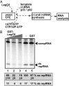
Fig. 1.
Fig. 1.
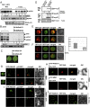
Cell-free TBSV replication assay supports an inhibitory effect for the Legionella LegC8 effector on TBSV replication. (A) Scheme of the CFE-based TBSV replication assay. (B) Nondenaturing PAGE analysis of the 32P-labeled TBSV repRNA products obtained in the CFE-based assay programmed with in vitro transcribed TBSV DI-72 (+)repRNA and purified recombinant MBP-p33 and MBP-p92pol replication proteins of TBSV. The affinity-purified GST-LegC8 from Escherichia coli was added 15 min before the TBSV replicase reconstitution. The CFEs were prepared from the BY4741 yeast strain. Each experiment was repeated 3 times and the data were used to calculate SD.
Fig. 2.
Fig. 2.
Expression of the Legionella LepB effector inhibits TBSV replication in yeast and plants. (A) The effect of Legionella LepB effector on TBSV repRNA accumulation was measured by Northern blot 16 h after initiation of TBSV replication in BY4741 yeast. The accumulation level of repRNA was normalized based on the ribosomal RNA (rRNA). (Bottom) Ethidium-bromide stained gel with ribosomal RNA, as a loading control. (B) Expression of LepB inhibits TBSV replication in N. benthamiana. Total RNAs were extracted from the TBSV sap-inoculated leaves 1.5 d after the inoculation, followed by Northern blot analysis. There was a lack of phenotype on the agroinfiltrated leaves at the time of sampling. Each experiment was performed 3 times. (C) Reduced TBSV replicase activity in CFE in the presence of affinity-purified GST-LepB. Denaturing PAGE analysis was used to detect the 32P-labeled TBSV repRNA products. CFEs were prepared from BY4741 yeast. The CFEs were programmed with TBSV (+)repRNA transcripts and comparable amounts of replication proteins and added purified proteins, shown in a SDS/PAGE. Purified GST-GUS was used as a negative control in the CFE assay. (D and E) Expression of LepB prevents the enrichment of PI(3)P in the viral replication compartment in N. benthamiana protoplasts infected with TBSV. Distribution of PI(3)P was detected via expression of GFP-FYVE, which specifically binds to PI(3)P. DIC, differential interference contrast. (F and G) Enrichment of PE phospholipid in the replication compartment is reduced by the expression of LepB in plant cells. PE localization was monitored with duramycin staining of N. benthamiana protoplasts infected with TBSV.
Fig. 3.
Fig. 3.
Expression of the Legionella DrrA effector inhibits TBSV replication in yeast and plants. (A) The effect of Legionella DrrA effector on TBSV repRNA accumulation was tested in BY4741 yeast. Replication of the TBSV repRNA was measured by Northern blot 16 h after initiation of TBSV replication in yeast. The accumulation level of repRNA was normalized based on the rRNA. (Middle) Ethidium-bromide stained gel with ribosomal RNA, as a loading control. The accumulation levels of His6-p33 and His6-p92pol replication proteins were tested using Western blot and anti-His antibody. (B) Reduced TBSV replicase activity in CFE in the presence of affinity-purified GST-DrrA. Denaturing PAGE analysis was used to detect the 32P-labeled TBSV repRNA products. CFEs were prepared from BY4741 yeast. The CFEs were programmed with TBSV (+)repRNA transcripts and comparable amounts of replication proteins and purified proteins. (C) Inhibition of TBSV replication in N. benthamiana by DrrA expression. Total RNAs were extracted from the TBSV sap-inoculated leaves 1 d after inoculation, followed by Northern blot analysis. DrrA and its mutants were detected via Western blotting using anti-His antibody. Each experiment was performed 3 times. (D) Lack of phenotype on the agroinfiltrated leaves at the time of sampling. Note that TBSV does not induce visible symptoms on the inoculated leaves at the early time point shown.
Fig. 4.
Fig. 4.
Rab1 small GTPase is coopted to enhance TBSV replication in yeast and plants. (A) Depletion of Ypt1 (Rab1) level in TET::YPT1 yeast inhibits TBSV replication. Doxycycline (Dox) was used to down-regulate the expression of Ypt1 from the TET promoter. Replication of the TBSV repRNA in TET::YPT1 yeast coexpressing the tombusvirus p33 and p92pol replication proteins was measured by Northern blotting 24 h after initiation of TBSV replication. The accumulation level of His6-p33 replication protein was tested by Western blot and anti-His antibody. (B) Coexpression of DN mutant of AtRab-D1 and DN AtRab-D2 by agroinfiltration suppressed TBSV replication in N. benthamiana, based on Northern blot analysis of TBSV RNA accumulation. Total RNA was extracted from the inoculated leaves 1 d after the inoculation. Empty vector was used as a negative control in this experiment. (C) Lack of phenotype on the agroinfiltrated leaves at the time of sampling. (D) Copurification of GFP-Ypt1 with Flag-p33 and Flag-p92 replication proteins from the membranous fraction of yeast. (E and F) Confocal microscopy-based partial colocalization of the coopted Ypt1 with p33 replication protein in yeast not expressing or expressing DrrA. Manders’ coefficient for the quantitative analysis of colocalization of p33-RFP and GFP-Ypt1 is shown. (Scale bars, 5 μm.) (G) Partial colocalization of TBSV p33 replication protein and AtRab-D1. The RFP-tagged p33 and GFP-AtRab-D1 were expressed via agroinfiltration in plant epidermal cells, which were also infected with TBSV, followed by confocal microscope imaging. (H) Interaction between the TBSV p33 replication protein and AtRab-D1 in plant was detected via BiFC. The signal between TBSV p33-cYFP and the nYFP-AtRab-D1 protein overlaps with RFP-SKL (peroxisomal matrix marker), which suggests the p33 and AtRab-D1 interaction occurs in the TBSV replication compartment. The fluorescence-tagged proteins were expressed by agroinfiltration in N. benthamiana leaves. The infiltrated leaves were also inoculated with TBSV to induce the formation of the large replication compartment consisting of aggregated peroxisomes. The combination of p33-cYFP and nYFP-MBP was used as negative control for BiFC. (I) Inhibition of colocalization of TBSV p33 replication protein and AtRab-D1 by expression of DrrA. The p33-RFP and GFP-AtRab-D1 and DrrA were expressed via agroinfiltration in plant epidermal cells, which were also infected with TBSV, followed by confocal microscope imaging. (J) Different subcellular localization of GFP-AtRab-D1 and the peroxisomal RFP-SKL in N. benthamiana. These proteins were expressed via agroinfiltration in plant epidermal cells, which were mock infected.
Fig. 5.
Fig. 5.
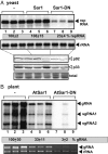
Expression of dominant-negative mutant of Sar1 inhibits TBSV replication in yeast and plants. (A) The effect of Sar1-DN on TBSV repRNA accumulation was tested in BY4741 yeast by Northern blot 24 h after initiation of TBSV replication. The accumulation level of repRNA was normalized based on the rRNA. (Middle) Northern blot analysis of ribosomal RNA level, as a loading control. The accumulation levels of His6-p33 and His6-p92pol replication proteins were tested using Western blot and anti-His antibody. (B) Inhibition of TBSV replication in N. benthamiana by AtSar1-DN expression. Total RNAs were extracted from the TBSV sap-inoculated leaves 1 d after the inoculation, followed by Northern blot analysis. Each experiment was performed 3 times.
Fig. 6.
Fig. 6.
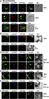
Recruitment of COPII cargo to the large replication compartment in plants infected with TBSV. (A) Partial colocalization of TBSV p33 replication protein and GFP-HDEL, in TBSV-infected protoplasts. RFP-SKL marks the aggregated peroxisomes, the site of TBSV replication. The Bottom shows the lack of colocalization of GFP-HDEL with the peroxisomal RFP-SKL in the absence of TBSV. (Scale bars, 5 μm.) (B) Partial colocalization of TBSV p33 replication protein and GFP-HDEL, in TBSV-infected N. benthamiana plants. The p33-BFP and GFP-HDEL were expressed via agroinfiltration in plant epidermal cells, which were also infected with TBSV, followed by confocal microscope imaging. The Bottom shows the lack of colocalization of GFP-HDEL with the peroxisomal RFP-SKL in the absence of TBSV. (C and D) Expression of AtSar1-DN or DrrA effector interferes with the recruitment of GFP-HDEL to the large replication compartment in plants infected with TBSV. See further details in B. (E and F) Expression of DrrA-ma and DrrA-mm mutants only partially interferes with the recruitment of GFP-HDEL to the large replication compartment in plants infected with TBSV. See further details in B.
Fig. 7.
Fig. 7.
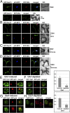
DrrA blocks the recruitment of COPII vesicles to the replication compartment in plants infected with TBSV. (A) Partial colocalization of TBSV p33 replication protein and GFP-Sec13 coat protein of COPII vesicles, in TBSV-infected N. benthamiana plants. The p33-BFP and GFP-Sec13 were coexpressed via agroinfiltration in plant epidermal cells, which were also infected with TBSV, followed by confocal microscope imaging. The Bottom shows the lack of colocalization of GFP-Sec13 with the peroxisomal RFP-SKL in the absence of TBSV. (B) Expression of DrrA blocks the recruitment of GFP-Sec13 to the replication compartment in plants infected with TBSV. See further details in A. (C and D) Expression of DrrA-ma does not block, whereas DrrA-mm only partially interferes with the recruitment of GFP-Sec13 to the replication compartment in plants infected with TBSV. See further details in A. (E and F) The effect of induction or repression of the expression of syntaxin-18 like Ufe1 SNARE protein on the colocalization of GFP-Ypt1 and p33-RFP in GALS::UFE1 yeast strain, documented via confocal microscopy. Manders’ coefficient for the quantitative analysis of colocalization of p33-RFP and GFP-Ypt1 is shown. (G and H) The effect of induction or repression of the expression of the ER-resident Use1 SNARE protein on the colocalization of GFP-Ypt1 and p33-RFP in the GAL1::USE1 yeast strain, documented via confocal microscopy. Each experiment was performed 2 times.

Similar articles

Cited by

References

    1. Wang A., Dissecting the molecular network of virus-plant interactions: The complex roles of host factors. Annu. Rev. Phytopathol. 53, 45–66 (2015). - PubMed
    1. Altan-Bonnet N., Lipid tales of viral replication and transmission. Trends Cell Biol. 27, 201–213 (2017). - PMC - PubMed
    1. den Boon J. A., Ahlquist P., Organelle-like membrane compartmentalization of positive-strand RNA virus replication factories. Annu. Rev. Microbiol. 64, 241–256 (2010). - PubMed
    1. Nagy P. D., Pogany J., The dependence of viral RNA replication on co-opted host factors. Nat. Rev. Microbiol. 10, 137–149 (2011). - PMC - PubMed
    1. Diaz A., Wang X., Bromovirus-induced remodeling of host membranes during viral RNA replication. Curr. Opin. Virol. 9, 104–110 (2014). - PubMed

Publication types

MeSH terms