. 2019 Nov 15;294(46):17301-17313.
doi: 10.1074/jbc.RA119.010518. Epub 2019 Oct 7.

Regulation of human trophoblast syncytialization by histone demethylase LSD1

Affiliations

Regulation of human trophoblast syncytialization by histone demethylase LSD1

Jessica Milano-Foster et al. J Biol Chem. .

Abstract

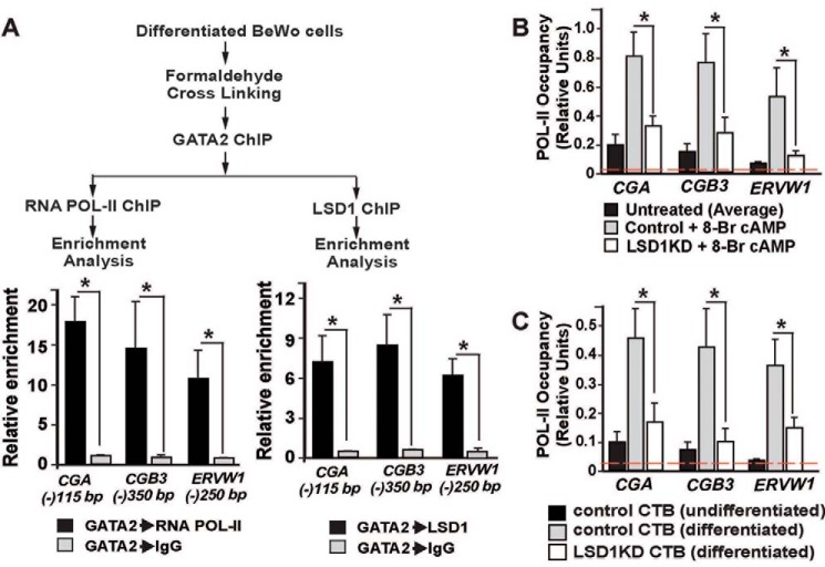
A successful pregnancy is critically dependent upon proper placental development and function. During human placentation, villous cytotrophoblast (CTB) progenitors differentiate to form syncytiotrophoblasts (SynTBs), which provide the exchange surface between the mother and fetus and secrete hormones to ensure proper progression of pregnancy. However, epigenetic mechanisms that regulate SynTB differentiation from CTB progenitors are incompletely understood. Here, we show that lysine-specific demethylase 1 (LSD1; also known as KDM1A), a histone demethylase, is essential to this process. LSD1 is expressed both in CTB progenitors and differentiated SynTBs in first-trimester placental villi; accordingly, expression in SynTBs is maintained throughout gestation. Impairment of LSD1 function in trophoblast progenitors inhibits induction of endogenous retrovirally encoded genes SYNCYTIN1/endogenous retrovirus group W member 1, envelope (ERVW1) and SYNCYTIN2/endogenous retrovirus group FRD member 1, envelope (ERVFRD1), encoding fusogenic proteins critical to human trophoblast syncytialization. Loss of LSD1 also impairs induction of chorionic gonadotropin α (CGA) and chorionic gonadotropin β (CGB) genes, which encode α and β subunits of human chorionic gonadotrophin (hCG), a hormone essential to modulate maternal physiology during pregnancy. Mechanistic analyses at the endogenous ERVW1, CGA, and CGB loci revealed a regulatory axis in which LSD1 induces demethylation of repressive histone H3 lysine 9 dimethylation (H3K9Me2) and interacts with transcription factor GATA2 to promote RNA polymerase II (RNA-POL-II) recruitment and activate gene transcription. Our study reveals a novel LSD1-GATA2 axis, which regulates human trophoblast syncytialization.

Keywords: LSD1; cytotrophoblast; histone demethylase; human; placenta; syncytiotrophoblast; transcription regulation; trophoblast.


Conflict of interest statement

The authors declare that they have no conflicts of interest with the contents of this article

Figures

Figure 1.
Figure 1.
LSD1 is abundantly expressed in human SynTBs. A, immunohistochemistry showing LSD1 is highly expressed within trophoblast cells (both CTBs (red arrows) and SynTBs (green arrows)) of a first-trimester (9-week) human placenta. IgG was used as a negative control for the immunostaining experiment. B, immunohistochemistry showing LSD1 is highly expressed in SynTBs (green arrows) of a term human placenta. C, micrographs showing altered morphology of BeWo cells when they were treated with 8-Br-cAMP for 72 h to induce SynTB differentiation. D, analysis of mRNA expression showing significant induction of SynTB-specific genes in 8-Br-cAMP–treated BeWo cells (expression level of a gene in untreated BeWo cells was considered as 1; mean ± S.E.; n = 3; *, p ≤ 0.001; Error bars represent S.E.). E, immunofluorescence images showing a high level of LSD1 expression at the nuclei of BeWo cells. F, Western blots showing LSD1 protein expression is maintained in 8-Br-cAMP–treated, differentiated BeWo cells.
Figure 2.
Figure 2.
Loss of LSD1 impairs SynTB differentiation. A and B, quantitative RT-PCR (mean ± S.E.; n = 3; *, p ≤ 0.001) (A) and Western blot (B) analyses showing depletion of LSD1 mRNA and protein expressions in BeWo cells upon shRNA-mediated RNAi (LSD1KD BeWo). BeWo cells, which were transduced with a nonfunctional shRNA, were considered as a control. C, analysis of mRNA expression showing impaired induction of SynTB-specific genes in 8-Br-cAMP–treated LSD1KD BeWo cells (mean ± S.E.; n = 3; *, p ≤ 0.001). D, immunofluorescence images showing E-Cadherin (E-CAD) expression and nuclei of control and LSD1-depleted BeWo cells in undifferentiated and differentiating (with 8-Br-cAMP) culture conditions. Efficient syncytialization of 8-Br-cAMP–treated control BeWo cells is evident from loss of E-Cadherin and cell–cell fusion (red boundaries). Impaired syncytialization in 8-Br-cAMP–treated LSD1KD BeWo cells is evident from maintenance of E-Cadherin expression and lack of cell–cell fusion. Only a few regions (red boundary, right panel) show partial loss of E-Cadherin expression. E, Western blot analyses showing depletion of LSD1 protein expression in human term CTBs upon shRNA-mediated RNAi (LSD1KD CTBs). F, micrographs showing impaired syncytialization (monitored by cell–cell fusion; white boundaries in left control panel) in LSD1KD CTBs. The syncytialization was not evident with LSD1KD CTBs (right panel). G, quantitative RT-PCR (mean ± S.E.; n = 3; *, p ≤ 0.001) analyses showing impaired induction of ERVW1, CGA, and CGB transcription in LSD1KD CTBs. Note that the SynTB differentiation of term CTBs was not associated with an induction in ERVFRD1 transcription. Error bars in panels A, C, and G represent S.E.
Figure 3.
Figure 3.
LSD1 demethylates H3K9Me2 mark at key gene promoters during SynTB differentiation. A, cartoon showing two modes of LSD1 function. LSD1 demethylates H3K4 methylation when it interacts with the CoREST or NuRD repressive complexes at transcriptionally active gene loci. In contrast, LSD1 demethylates H3K9 methylation and promotes gene transcription when it interacts with certain transcription factors like AR, ER, and cMYB. B, Western blot analyses showing similar levels of global H3K4Me2 and H3K9Me2 modifications in control and LSD1KD BeWo cells. C and D, plots showing quantitative ChIP assessment of H3K4Me2 (C) and H3K9Me2 (D) deposition at the promoter regions of SynTB-associated genes in control and LSD1KD BeWo cells. Signal from 0.2% of total input chromatin before immunoprecipitation (for details, see Ref. 94) was considered as 1 to generate the relative enrichment plot. *, p < 0.01; three independent experiments. The red dotted line indicates the average nonspecific enrichment using IgG as the negative control antibody for ChIP analyses. E, plot showing quantitative assessment of H3K9Me2 deposition at the promoter regions of SynTB-associated genes in control and LSD1KD CTBs (*, p < 0.01; three independent experiments). HDAC, histone deacetylase. Error bars in panels C, D, and E represent S.E.
Figure 4.
Figure 4.
LSD1 facilitates recruitment of GATA2 at the CGA, CGB, and ERVW1 gene loci during SynTB differentiation. A, immunohistochemistry showing GATA2 is highly expressed in SynTBs (green arrows) of a term human placenta. B, Western blot analyses showing GATA2 protein expressions in control and LSD1KD BeWo cells. C, sequence alignments of human and rhesus monkey CGA and CGB3 loci showing the presence of conserved WGATAR motifs around the transcription start site. The red vertical bars on top indicate positions of the conserved WGATAR motifs. D, positions and sequences of conserved WGATAR motifs at human CGA and CGB3 loci. E, sequence around −250 bp region of the human ERVW1 locus showing GATA motifs, which were implicated earlier in transcriptional regulation of ERVW1. F, plot showing quantitative ChIP assessment of GATA2 occupancy at the CGA, CGB3, and ERVW1 loci in control and LSD1KD BeWo cells (signal from 0.2% of total input chromatin before immunoprecipitation was considered as 1 to generate the relative enrichment plot; *, p < 0.001; three independent experiments). The red dotted line indicates the average nonspecific enrichment using IgG as the negative control antibody for ChIP analyses. Note that GATA2 occupancy was not significantly enriched at the −335 bp region of the CGA locus, which contains a WGATA(T) motif, instead of a WGATAR(A/G) motif. G, plot showing quantitative ChIP assessment of GATA2 occupancy at the CGA, CGB3, and ERVW1 loci in control and LSD1KD CTBs (*, p < 0.01; three independent experiments). Error bars in panels F and G represent S.E.
Figure 5.
Figure 5.
LSD1, GATA2, and RNA-POL-II physically interact at the SynTB-associated gene loci during SynTB differentiation, and loss of LSD1 impairs RNA-POL-II recruitment. A, sequential ChIP showing co-occupancy of LSD1, GATA2, and RNA-POL-II at human CGA, CGB3, and ERVW1 gene loci. The signal from GATA2 ChIP samples that were further subjected to sequential ChIP with control IgG was considered as 1 to determine the relative enrichment (*, p < 0.001; three independent experiments). B and C, plots showing quantitative ChIP assessment of RNA-POL-II occupancy at the promoter region of human CGA, CGB3, and ERVW1 loci in control and LSD1KD BeWo cells (B) and control and LSD1KD CTBs (C) (signal from 0.2% of total input chromatin before immunoprecipitation was considered as 1 to generate the relative enrichment plot; *, p < 0.01; three independent experiments). The red dotted line indicates the average nonspecific enrichment using IgG as the negative control antibody for ChIP analyses. Error bars in all panels represent S.E.
Figure 6.
Figure 6.
The LSD1–GATA2 gene regulatory axis during SynTB differentiation. The model illustrates an LSD1–GATA2 gene regulatory axis that directly controls RNA-POL-II recruitment at GATA2 target genes to promote transcriptional activation during SynTB differentiation. From our experimental data, we propose that in the undifferentiated CTBs, SynTB-associated genes such as CGA harbor both H3K4Me2 and H3K9Me2 modifications, leading to a relatively compact chromatin inaccessible to GATA2 binding. The lack of GATA2 binding in turn results in impaired RNA-POL-II recruitment and suppression of transcription. In response to cell signaling like PKA signaling, a LSD1–GATA2 complex forms at the SynTB-associated genes, which leads to H3K9Me2 demethylation by LSD1 and instigates RNA-POL-II recruitment and gene transcription.

References

    1. Soares M. J., Varberg K. M., and Iqbal K. (2018) Hemochorial placentation: development, function, and adaptations. Biol. Reprod. 99, 196–211 10.1093/biolre/ioy049 - DOI - PMC - PubMed
    1. Knöfler M., Haider S., Saleh L., Pollheimer J., Gamage T. K. J. B., and James J. (2019) Human placenta and trophoblast development: key molecular mechanisms and model systems. Cell. Mol. Life Sci. 76, 3479–3496 10.1007/s00018-019-03104-6 - DOI - PMC - PubMed
    1. James J. L., Carter A. M., and Chamley L. W. (2012) Human placentation from nidation to 5 weeks of gestation. Part I: what do we know about formative placental development following implantation? Placenta 33, 327–334 10.1016/j.placenta.2012.01.020 - DOI - PubMed
    1. Boss A. L., Chamley L. W., and James J. L. (2018) Placental formation in early pregnancy: how is the centre of the placenta made? Hum. Reprod. Update 24, 750–760 10.1093/humupd/dmy030 - DOI - PubMed
    1. Baird D. D., Weinberg C. R., McConnaughey D. R., and Wilcox A. J. (2003) Rescue of the corpus luteum in human pregnancy. Biol. Reprod. 68, 448–456 10.1095/biolreprod.102.008425 - DOI - PubMed

Publication types

MeSH terms