. 2019 Oct 7;10(10):761.
doi: 10.1038/s41419-019-1992-4.

Intermittent compressive force promotes osteogenic differentiation in human periodontal ligament cells by regulating the transforming growth factor-β pathway

Affiliations

Intermittent compressive force promotes osteogenic differentiation in human periodontal ligament cells by regulating the transforming growth factor-β pathway

Jeeranan Manokawinchoke et al. Cell Death Dis. .

Abstract

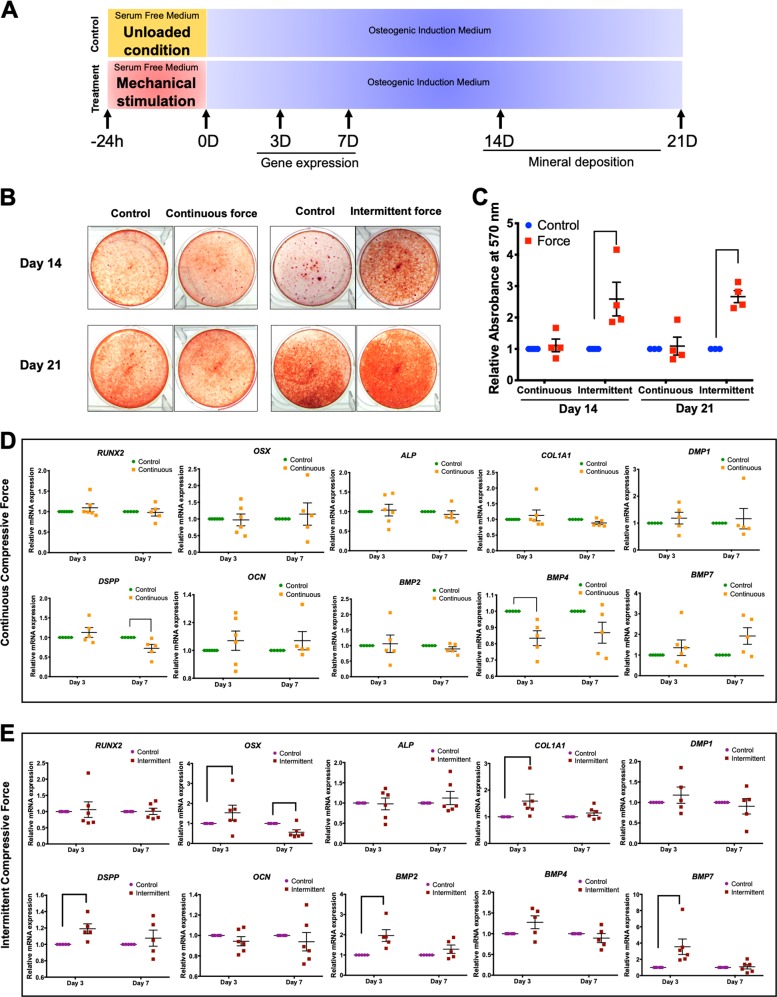
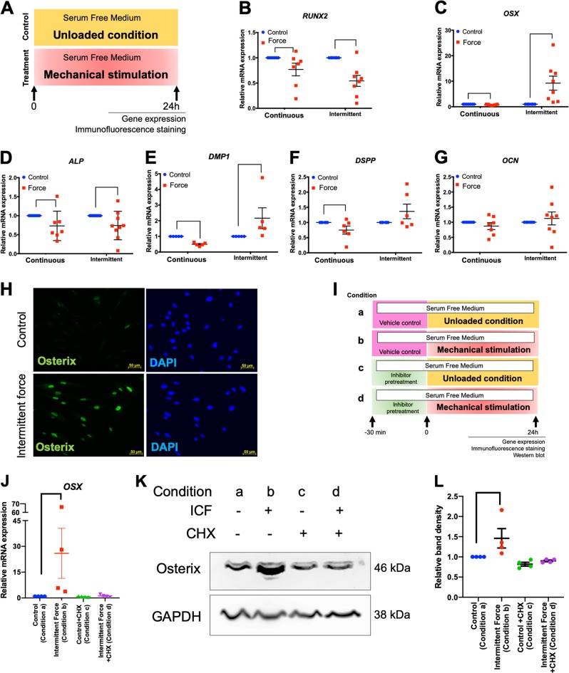
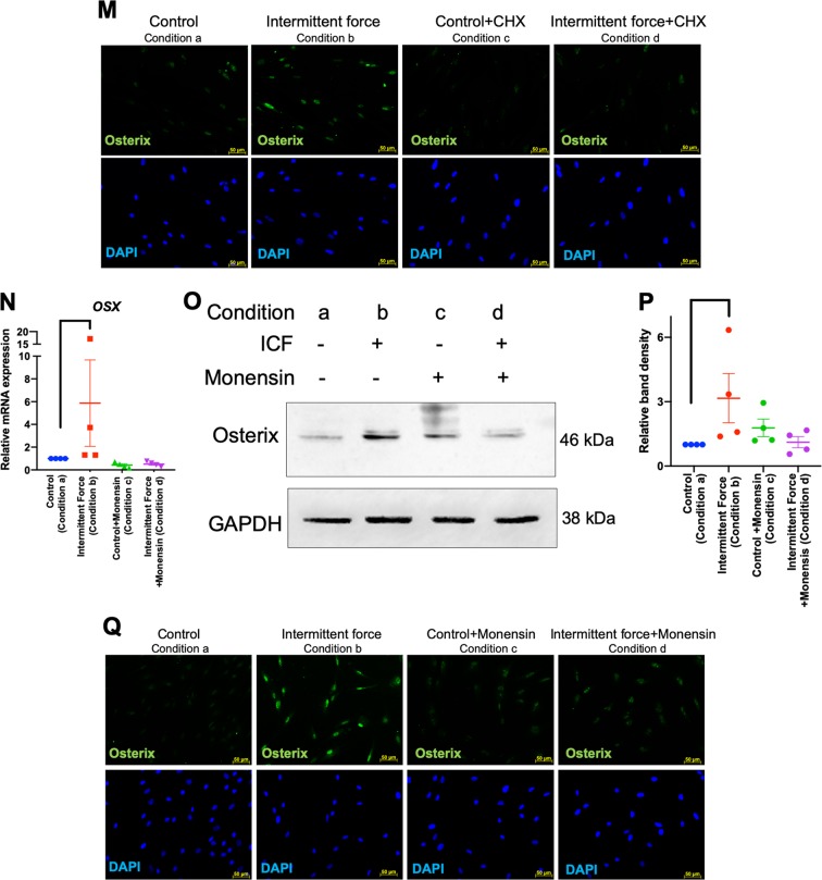
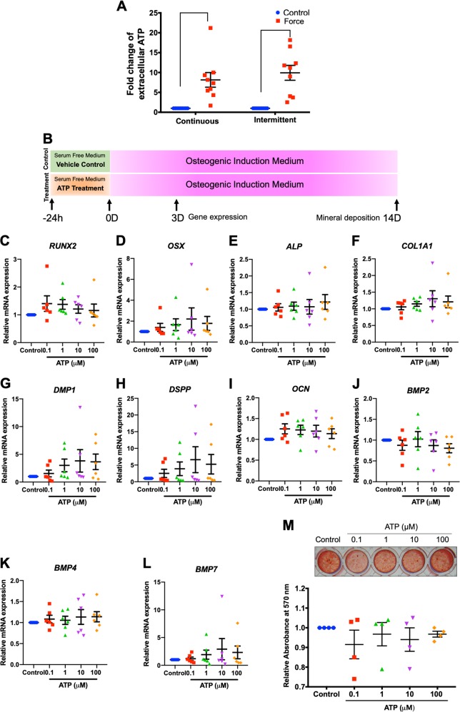
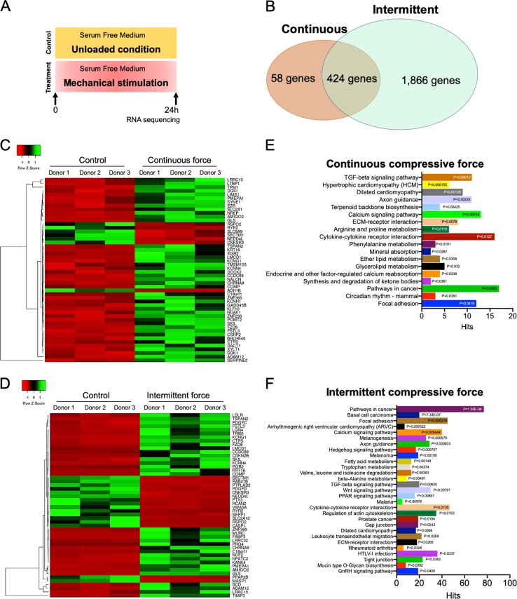
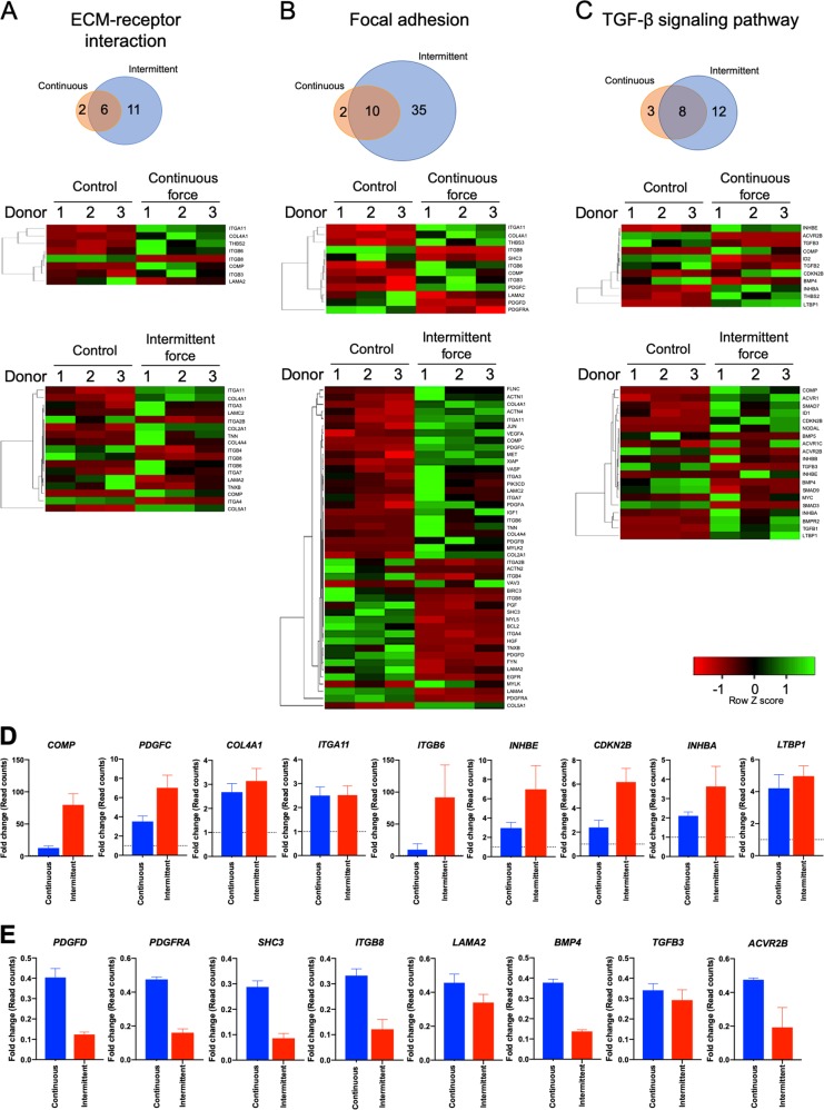
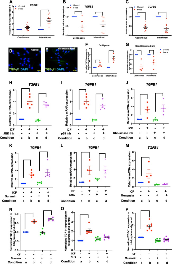
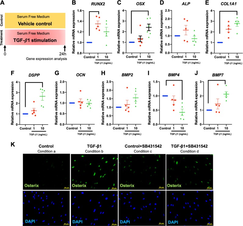
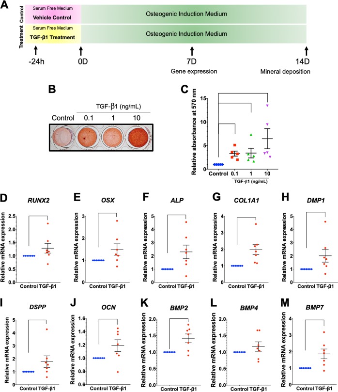
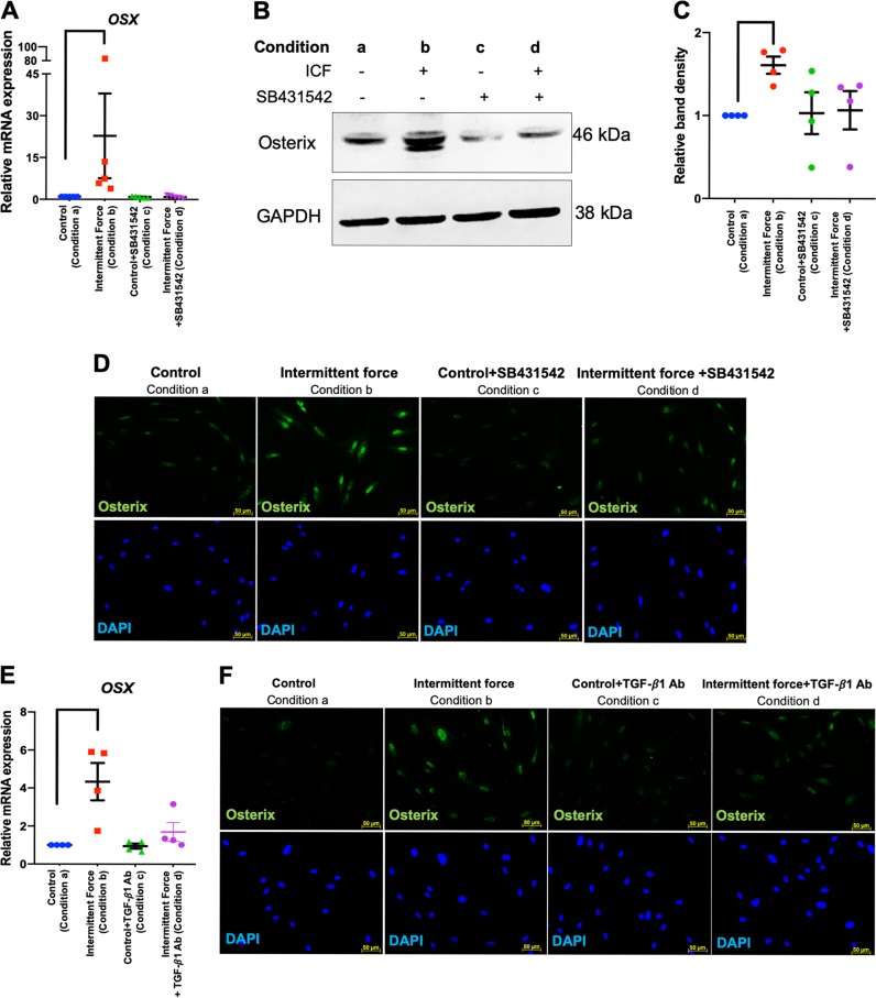
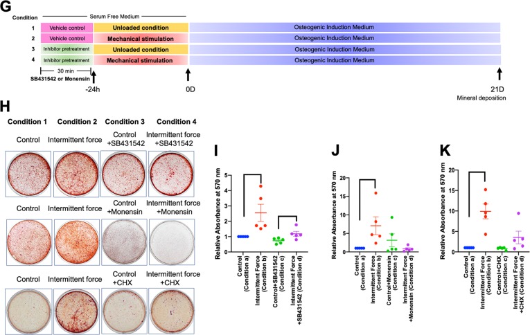
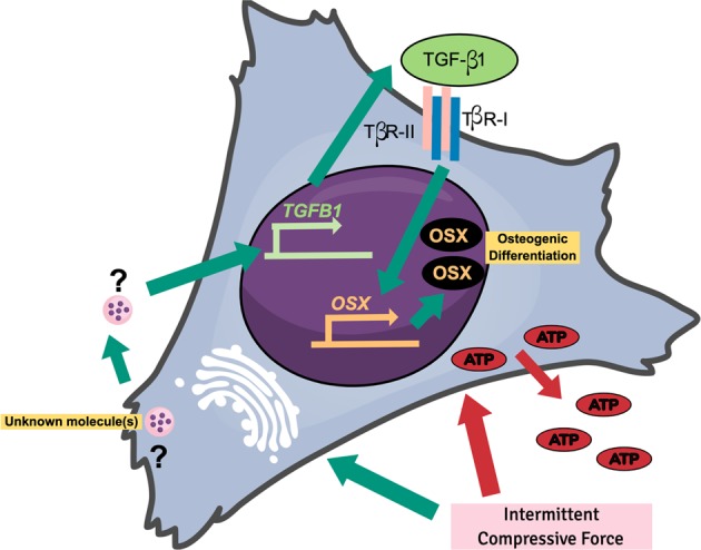
Mechanical force regulates periodontal ligament cell (PDL) behavior. However, different force types lead to distinct PDL responses. Here, we report that pretreatment with an intermittent compressive force (ICF), but not a continuous compressive force (CCF), promoted human PDL (hPDL) osteogenic differentiation as determined by osteogenic marker gene expression and mineral deposition in vitro. ICF-induced osterix (OSX) expression was inhibited by cycloheximide and monensin. Although CCF and ICF significantly increased extracellular adenosine triphosphate (ATP) levels, pretreatment with exogenous ATP did not affect hPDL osteogenic differentiation. Gene-expression profiling of hPDLs subjected to CCF or ICF revealed that extracellular matrix (ECM)-receptor interaction, focal adhesion, and transforming growth factor beta (TGF-β) signaling pathway genes were commonly upregulated, while calcium signaling pathway genes were downregulated in both CCF- and ICF-treated hPDLs. The TGFB1 mRNA level was significantly increased, while those of TGFB2 and TGFB3 were decreased by ICF treatment. In contrast, CCF did not modify TGFB1 expression. Inhibiting TGF-β receptor type I or adding a TGF-β1 neutralizing antibody attenuated the ICF-induced OSX expression. Exogenous TGF-β1 pretreatment promoted hPDL osteogenic marker gene expression and mineral deposition. Additionally, pretreatment with ICF in the presence of TGF-β receptor type I inhibitor attenuated the ICF-induced mineralization. In conclusion, this study reveals the effects of ICF on osteogenic differentiation in hPDLs and implicates TGF-β signaling as one of its regulatory mechanisms.


Conflict of interest statement

The authors declare that they have no conflict of interest.

Figures

Fig. 1
Fig. 1. ICF stimulated osteogenic differentiation in hPDLs.
Cells were exposed to CCF or ICF in serum-free media for 24 h and subsequently maintained in osteogenic medium (a). In the control condition, cells were cultured in serum-free culture medium for 24 h without mechanical loading and further maintained in osteogenic medium (a). Mineral deposition shown by alizarin red s staining at 14 and 21 days after osteogenic induction (b). The relative absorbance of the solubilized alizarin red dye was demonstrated (c). After pretreating cells with the CCF (d) or ICF (e), osteogenic marker gene expression was evaluated compared with the unloaded control using real-time polymerase chain reaction at 3 and 7 days after osteogenic induction. Bars indicate a significant difference between conditions
Fig. 2
Fig. 2. ICF induced OSX expression in hPDLs.
Cells were treated with CCF or ICF in serum-free media for 24 h and cells cultured in the same condition without mechanical force application were used as the control (a). The mRNA expression of osteogenic marker genes was examined using real-time polymerase chain reaction (b–g). OSX protein expression was evaluated using immunofluorescence staining (h). In the inhibition experiments, cells were pretreated with inhibitor 30 min prior to ICF stimulation. Experimental conditions were illustrated (i). The OSX mRNA (j, n) were determined using real-time polymerase chain reaction. Osterix protein expression was evaluated using western blot (k, o) and the normalized band density was demonstrated (l, p). In addition, osterix protein expression was also determined by immunofluorescence staining (m, q). Bars indicate a significant difference between conditions. Scale bars indicate 50 μm
Fig. 3
Fig. 3. ATP priming did not influence osteogenic differentiation in hPDLs.
Cells were treated with CCF or ICF in serum-free media for 24 h. Extracellular ATP was evaluated using an ATP assay in culture medium (a). Schematic diagram of the experimental plan of the ATP priming was illustrated (b). Cells were exposed to ATP for 24 h in serum-free medium. Thereafter, the culture medium was changed to osteogenic medium. Osteogenic marker gene expression was determined using real-time polymerase chain reaction at day 3 after osteogenic induction (cl). Mineral deposition was examined using alizarin red s staining at day 14 (m). The normalized absorbance of the solubilized dye. Bars indicate a significant difference between conditions
Fig. 4
Fig. 4. Gene expression profiling of mechanical force treated hPDLs.
Cells were treated with CCF or ICF in serum-free medium for 24 h. Cells maintained in serum-free medium without mechanical loading were employed as the control (a). The gene expression profile compared with the unloaded control was determined using RNA sequencing and bioinformatic analyses. Common differentially expressed genes in the CCF- or ICF-treated hPDLs are shown (b). Heat maps demonstrating the top 50 differentially expressed genes in the CCF- (c) or ICF- (d) treated cells compared with the unloaded control. Pathway enrichment of all differentially expressed genes using KEGG database revealed the differentially regulated pathways after CCF (e) and ICF (f) treatment
Fig. 5
Fig. 5. Mechanical force regulated ECM-receptor interaction, focal adhesion, and TGF-β signaling pathways in hPDLs.
Bioinformatic analysis demonstrated the common differentially expressed genes and heat maps in ECM-receptor interaction (a), focal adhesion (b), and TGF-β signaling (c) pathways. The significantly upregulated (d) and downregulated (e) genes were selected. The fold-change of the raw read counts from the RNA sequencing data were presented. Dotted lines indicate the normalized expression value of the unloaded control
Fig. 6
Fig. 6. ICF induced TGF-β1 expression.
Cells were treated with CCF or ICF in serum-free media for 24 h. Cells cultured in the same condition without mechanical force application were used as the control. Experimental conditions were illustrated in Fig. 2a. The mRNA expression of TGFB1 (a), TGFB2 (b), and TGFB3 (c) was evaluated using real-time polymerase chain reaction. TGF-β1 protein expression was examined using immunofluorescence staining (d, e) and enzyme linked immunosorbent assay (f, g). To investigate the regulatory pathways, cells were pretreated chemical inhibitor 30 min prior to ICF treatment. Experimental conditions were illustrated in Fig. 2i. Chemical inhibitors were JNK inhibitor (h), p38 inhibitor (i), Rho-kinase inhibitor (j), suramin (k, n), cycloheximide (l, o), or monensin (m, p). TGFB1 mRNA and protein levels were examined using real-time polymerase chain reaction and enzyme linked immunosorbent, respectively. Bars indicate a significant difference between conditions. ICF intermittent compressive force treatment, CHX cycloheximide. Scale bars indicate 50 μm
Fig. 7
Fig. 7. Recombinant human TGF-β1 promoted osteogenic marker gene expression.
Cells were treated with 1 and 10 ng/mL of recombinant human TGF-β1 in serum-free culture medium for 24 h. In the control condition, cells were cultured in serum-free condition and the vehicle control was added in the medium. Schematic diagram of the experimental plan was illustrated (a). Osteogenic marker gene expression was determined using real-time polymerase chain reaction (bj). OSX protein expression was evaluated using immunofluorescence staining (k). In some conditions, cells were pretreated with SB431542 30 min prior to TGF-β1 exposure. Experimental conditions were illustrated in Fig. 2i. Bars indicate a significant difference between conditions. Scale bars indicate 50 μm
Fig. 8
Fig. 8. Priming with recombinant human TGF-β1 promoted osteogenic differentiation in hPDLs.
Schematic diagram of the experimental plan of TGF-β1 priming and subsequent osteogenic induction (a). Mineralization was examined using alizarin red s staining at day 14 after osteogenic induction (b). The normalized absorbance of alizarin red dye (c). Osteogenic marker gene expression was determined using real-time polymerase chain reaction at day 7 after osteogenic induction (dm). Bars indicate a statistically significant difference between conditions
Fig. 9
Fig. 9. TGF-β1 participated in the ICF-induced osteogenic differentiation in hPDLs.
Cells were pretreated with SB431542 or TGF-β1 neutralizing antibody for 30 min prior to ICF stimulation for 24 h in serum-free culture medium. Experimental conditions were illustrated in Fig. 2i. OSX mRNA expression was evaluated using real-time polymerase chain reaction (a, e). OSX protein expression was demonstrated using western blot (b) and immunofluorescence staining (d, f). The normalized band density was illustrated (c). Schematic diagram of the experimental plan for evaluating the influence of TGF-β1 on mineralization in hPDLs was demonstrated (g). Cells were exposed with SB431542 or monensin or cycloheximide for 30 min prior to ICF stimulation in serum-free culture medium for 24 h and subsequently maintained in osteogenic medium. Mineral deposition was examined using alizarin red s staining at day 21 after osteogenic induction (h). The relative absorbance of the solubilized alizarin red dye was demonstrated (i–k). Bars indicate a significant difference between conditions. Scale bars indicated 50 μm
Fig. 10
Fig. 10. Schematic diagram of the potential regulation of OSX expression under ICF in hPDLs.
ICF induces the release of molecule(s) from the golgi apparatus and subsequently enhances TGFB1 expression. Further, TGF-β1 is released and binds to TGF-β receptors, leading to the induction of OSX expression

References

    1. Tomokiyo, A. et. al. Detection, characterization, and clinical application of mesenchymsl stem cells in periodontal ligament tissue. Stem. Cell. Int.2018, 5450768 (2018). - PMC - PubMed
    1. Iwata Takanori, Yamato Masayuki, Ishikawa Isao, Ando Tomohiro, Okano Teruo. Tissue Engineering in Periodontal Tissue. The Anatomical Record. 2013;297(1):16–25. doi: 10.1002/ar.22812. - DOI - PubMed
    1. Smith PC, Martinez C, Martinez J, McCulloch CA. Role of fibroblast populations in periodontal wound healing and tissue remodeling. Front. Physiol. 2019;10:270. doi: 10.3389/fphys.2019.00270. - DOI - PMC - PubMed
    1. Mason AG, Scott BJ, van der Glas HW, Linden RW, Cadden SW. Remote noxious stimuli modulate jaw reflexes evoked by activation of periodontal ligament mechanoreceptors in man. Exp. Physiol. 2002;87:699–706. doi: 10.1113/eph8702452. - DOI - PubMed
    1. Turker KS, Sowman PF, Tuncer M, Tucker KJ, Brinkworth RS. The role of periodontal mechanoreceptors in mastication. Arch. Oral. Biol. 2007;52:361–364. doi: 10.1016/j.archoralbio.2006.11.014. - DOI - PubMed

Publication types

MeSH terms