. 2019 Oct 7;10(10):755.
doi: 10.1038/s41419-019-1984-4.

The oncometabolite 2-hydroxyglutarate produced by mutant IDH1 sensitizes cells to ferroptosis

Affiliations

The oncometabolite 2-hydroxyglutarate produced by mutant IDH1 sensitizes cells to ferroptosis

Tian-Xiang Wang et al. Cell Death Dis. .

Abstract

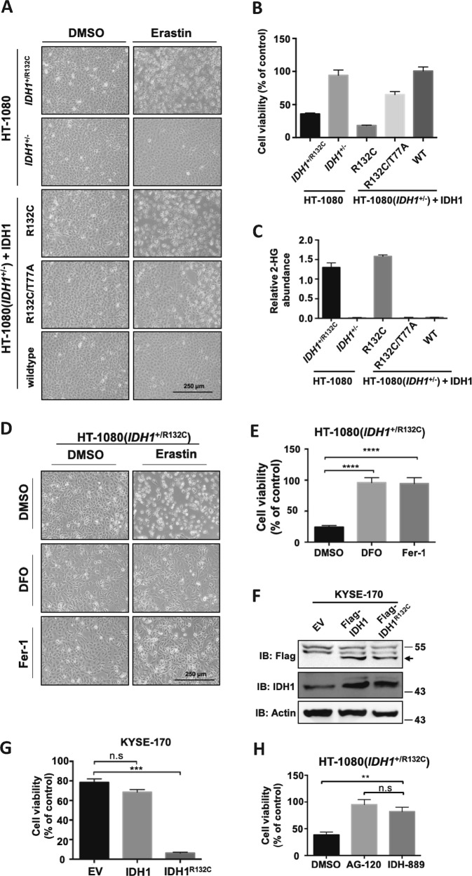
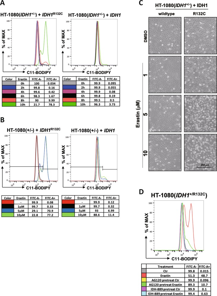
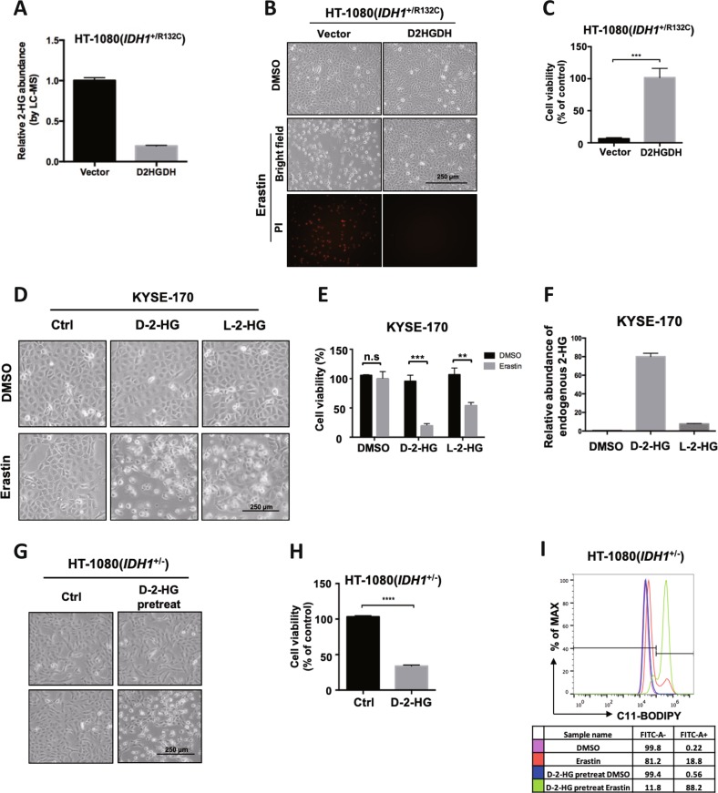
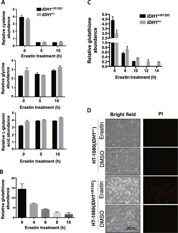
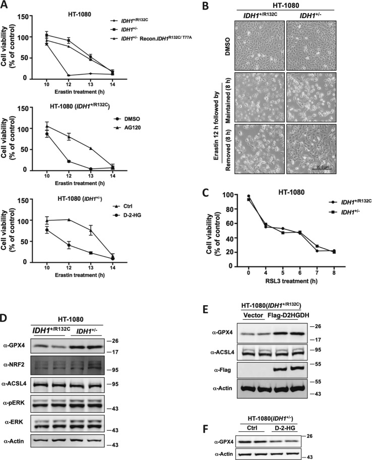
Ferroptosis is a non-apoptotic form of cell death characterized by the iron-dependent lipid peroxidation and is implicated in several human pathologies, such as tissue ischemia, neurodegeneration, and cancer. Ferroptosis appears to be high cell-context dependent and the regulation of ferroptosis by physiological or pathological conditions are unclear. Here, we report that tumor-derived IDH1 mutation sensitizes cells to ferroptosis. Deletion of the mutant IDH1 allele in IDH1 heterozygous tumor cells or pharmacological inhibition of mutant IDH1 to produce the oncometabolite D-2-hydroxyglutarate (D-2-HG) confers resistance to erastin-induced ferroptosis. Conversely, ectopic expression of mutant IDH1 or treatment of cells with cell-permeable D-2-HG promotes the accumulation of lipid reactive oxygen species (ROS) and subsequently ferroptosis. Mechanistically, mutant IDH1 reduces the protein level of the glutathione peroxidase 4 (GPX4), a key enzyme in removing lipid ROS and ferroptosis, and promotes depletion of glutathione. Our results uncover a new role of mutant IDH1 and 2-HG in ferroptosis.


Conflict of interest statement

Kun-Liang Guan is a co-founder of Vivace Therapeutics. Yue Xiong is a co-founder of Cullgen Inc. The remaining authors declare that they have no conflict of interest.

Figures

Fig. 1
Fig. 1. Mutant IDH1 promotes erastin-induced ferroptosis.
a IDH1R132C mutation promotes erastin-induced ferroptosis. HT-1080 cells with indicated IDH1 genotypes were treated with erastin (10 μM) for 12 h, and microscopically photographed. b Cell viability in (a) were assayed by trypan blue staining. c Endogenous 2-HG levels of HT-1080 cells with different IDH1 genotypes in (a) were determined by GC-MS. d Ferroptosis induction in HT-1080 cells. HT-1080 cells were treated with erastin, DFO, and Fer-1 as indicated for 12 h, and microscopically photographed. e Cell viability in (d) were assayed by trypan blue staining (n = 3; ****p < 0.0001 vs. DMSO group, Dunnett’s multiple comparisons test). f Expression of Flag-IDH1 or Flag-IDH1R132C in KYSE-170 cells were detected by Western blot. g IDH1R132C mutation promotes erastin-induced ferroptosis in KYSE-170 cells. KYSE-170 cells expressing Flag-IDH1 or Flag-IDH1R132C were treated with erastin for 16 h and cell viability was assayed by trypan blue staining (n = 3; ***p < 0.001 vs. EV group, Dunnett’s multiple comparisons test). h Pharmacological inhibition of mutant IDH1 reduces cells’ sensitivity to erastin. HT-1080 cells were treated with AG-120 or IDH-889 for 12 h, and then with erastin for additional 12 h. Cell viability were assayed by trypan blue staining (n = 3; **p < 0.01 and ***p < 0.001, Bonferroni’s multiple comparisons test)
Fig. 2
Fig. 2. Mutant IDH1 enhances erastin-induced lipid ROS accumulation.
a IDH1R132C mutation enhances erastin-induced lipid ROS accumulation in a time-dependent manner. HT-1080(IDH1+/−) cells expressing ectopic IDH1 or IDH1R132C were treated with erastin (10 μM) for indicated time and the accumulation of lipid ROS was assessed by C11 BODIPY 581/591 staining coupled with flow cytometry analysis. b Mutant IDH1 enhances erastin-induced lipid ROS accumulation in a concentration-dependent manner. HT-1080(IDH1+/−) cells expressing ectopic IDH1 or IDH1R132C were treated with different concentrations of erastin for 10 h and the accumulation of lipid ROS within cells was assessed by C11 BODIPY 581/591 staining coupled with flow cytometry analysis. c Cell state in (b) was captured by microscope. d Mutant IDH1 inhibitors inhibit erastin-induced lipid ROS accumulation. HT-1080(IDH1+/R132C) cells were treated with mutant IDH1 inhibitors AG-120 or IDH-889 for 12 h, and then with erastin for additional 10 h. The accumulation of lipid ROS within cells was assessed by C11 BODIPY 581/591 staining coupled with flow cytometry analysis
Fig. 3
Fig. 3. D-2-HG promotes erastin-induced ferroptosis.
a Overexpression of D2HGDH inhibits 2-HG accumulation. Cellular 2-HG level in HT-1080 cells with empty vector or D2HGDH overexpression were determined by LC-MS. b Clearance of D-2-HG by D2HGDH overexpression inhibits erastin-induced ferroptosis. HT-1080 cells with empty vector or D2HGDH overexpression were treated with erastin for 12 h and cell state was captured by microscope. Dead cells were stained by propidium iodide (PI). c Cell viability in (b) were assayed by trypan blue staining g (n = 3; **p < 0.01, two-tailed unpaired t-test). d 2-HG promotes erastin-induced ferroptosis. KYSE-170 cells were treated with D-2-HG or L-2-HG for 24 h, and then cells were treated with erastin for additional 16 h. Cell states were captured by microscope. e Cell viability in (d) were assayed by trypan blue staining (n = 3; **p < 0.01 and ***p < 0.001, two-tailed unpaired t-test). f Endogenous 2-HG level of KYSE-170 cells in (d) were determined by GC-MS. g 2-HG promotes erastin-induced ferroptosis. HT-1080 (IDH1+/−) cells were treated with D-2-HG for 24 h and then with erastin for subsequent 12 h, followed by microscopic photograph. h Cell viability in (g) were assayed by trypan blue staining (n = 3; ****p < 0.0001, two-tailed unpaired t-test). i D-2-HG treatment enhances erastin-induced lipid ROS accumulation. HT-1080 (IDH1+/−) cells were first treated with D-2-HG for 24 h and then erastin for 10 h. Lipid ROS accumulation was assessed by C11 BODIPY 581/591 staining coupled with flow cytometry analysis
Fig. 4
Fig. 4. Mutant IDH1 exacerbates erastin-induced glutathione depletion.
a Measurement of cellular levels of glycine, cysteine and L-glutamic acid. HT-1080(IDH1+/R132C) or HT-1080(IDH1+/−) cells were treated with erastin for indicated time and cellular glycine, cysteine and L-glutamic acid levels were determined by GC-MS. b Erastin treatment reduces cellular glutathione level over time. HT-1080(IDH1+/R132C) cells were treated with erastin for indicated time and cellular glutathione levels were measured using Total Glutathione Assay Kit. c Mutant IDH1 exacerbates erastin-induced glutathione depletion. HT-1080(IDH1+/R132C) or HT-1080(IDH1+/−) cells were treated with erastin (10 μM) for indicated time and cellular glutathione levels were measured using Total Glutathione Assay Kit. d HT-1080(IDH1+/R132C) or HT-1080(IDH1+/−) cells were treated with erastin for 12 h and cells were microscopically photographed
Fig. 5
Fig. 5. IDH1 mutation reduces GPX4 protein level.
a IDH1R132C mutation and 2-HG treatment accelerate the onset of erastin-induced ferroptosis. HT-1080 cells with indicated genotypes or treatment were treated with erastin (10 μM) for indicated time, and cell viability were assayed by trypan blue staining. b Ferroptosis is a reversible process before reaching a point of no-return. HT-1080(IDH1+/R132C) or HT-1080(IDH1+/−) cells were treated with erastin for 12 h and then erastin was either removed from or kept in the culture medium for additional 8 h, followed by microscopic photograph. c Mutant IDH1 does not enhance ferroptosis in cells treated with GPX4 inhibitor RSL3. HT-1080(IDH1+/R132C) or HT-1080(IDH1+/−) cells were treated with RSL3 (2 μM) for indicated time, and cell viability were assayed by trypan blue staining. d Knocking out of IDH1R132C allele up-regulates the protein level of GPX4. The protein levels of Nrf2, Acsl4, Erk, p-Erk, GPX4, and Actin in HT-1080(IDH1+/R132C) or HT-1080(IDH1+/−) cells were detected by western blot. e D2HGDH overexpression upregulates the protein level of GPX4. The protein level of Acsl4, Flag-D2HGDH, GPX4 and Actin in HT-1080(IDH1+/R132C) cells expressing vector or Flag-D2HGDH were detected by western blot. f D-2-HG treatment down-regulates the protein level of GPX4. The protein level of GPX4 and Actin in HT-1080(IDH1+/−) cells with or without D-2-HG treatment were detected by western blot

Similar articles

Cited by

References

    1. Dolma S, Lessnick SL, Hahn WC, Stockwell BR. Identification of genotype-selective antitumor agents using synthetic lethal chemical screening in engineered human tumor cells. Cancer Cell. 2003;3:285–296. doi: 10.1016/S1535-6108(03)00050-3. - DOI - PubMed
    1. Soulinna EM, Winberg AM. 7-Ethoxycoumarin O-deethylation and methylumbelliferone conjugation in isolated fetal and adult bovine hepatocytes. Drug Metab. Disposition: Biol. Fate Chem. 1985;13:722–724. - PubMed
    1. Dixon SJ, et al. Ferroptosis: an iron-dependent form of nonapoptotic cell death. Cell. 2012;149:1060–1072. doi: 10.1016/j.cell.2012.03.042. - DOI - PMC - PubMed
    1. Angeli JPF, Shah R, Pratt DA, Conrad M. Ferroptosis inhibition: mechanisms and opportunities. Trends Pharmacol. Sci. 2017;38:489–498. doi: 10.1016/j.tips.2017.02.005. - DOI - PubMed
    1. Shah R, Shchepinov MS, Pratt DA. Resolving the role of lipoxygenases in the initiation and execution of ferroptosis. ACS Cent. Sci. 2018;4:387–396. doi: 10.1021/acscentsci.7b00589. - DOI - PMC - PubMed

Publication types