. 2019 Oct;25(10):1512-1518.
doi: 10.1038/s41591-019-0598-9. Epub 2019 Oct 7.

A luminal unfolding microneedle injector for oral delivery of macromolecules

Affiliations

A luminal unfolding microneedle injector for oral delivery of macromolecules

Alex Abramson et al. Nat Med. 2019 Oct.

Abstract

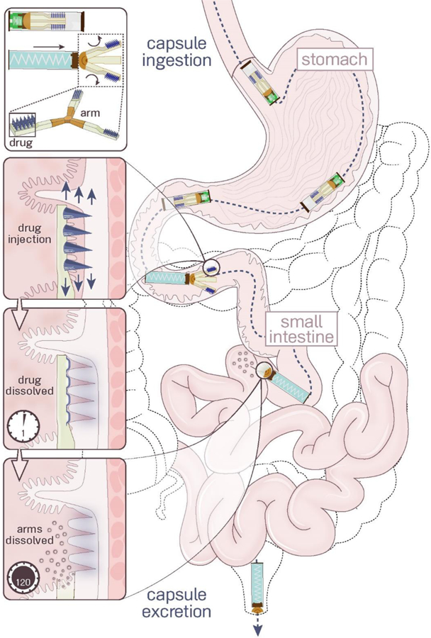
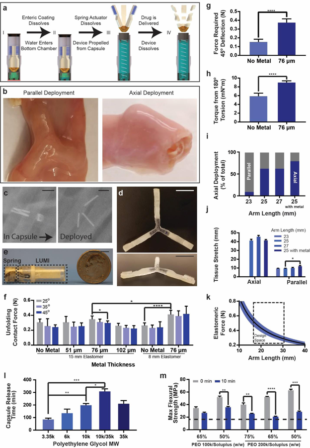
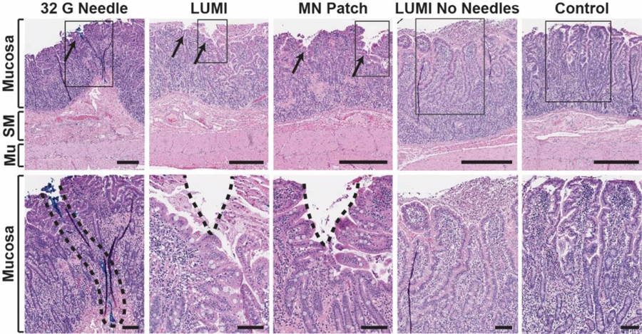
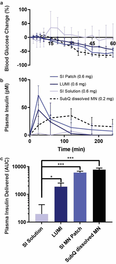
Insulin and other injectable biologic drugs have transformed the treatment of patients suffering from diabetes1,2, yet patients and healthcare providers often prefer to use and prescribe less effective orally dosed medications3-5. Compared with subcutaneously administered drugs, oral formulations create less patient discomfort4, show greater chemical stability at high temperatures6, and do not generate biohazardous needle waste7. An oral dosage form for biologic medications is ideal; however, macromolecule drugs are not readily absorbed into the bloodstream through the gastrointestinal tract8. We developed an ingestible capsule, termed the luminal unfolding microneedle injector, which allows for the oral delivery of biologic drugs by rapidly propelling dissolvable drug-loaded microneedles into intestinal tissue using a set of unfolding arms. During ex vivo human and in vivo swine studies, the device consistently delivered the microneedles to the tissue without causing complete thickness perforations. Using insulin as a model drug, we showed that, when actuated, the luminal unfolding microneedle injector provided a faster pharmacokinetic uptake profile and a systemic uptake >10% of that of a subcutaneous injection over a 4-h sampling period. With the ability to load a multitude of microneedle formulations, the device can serve as a platform to orally deliver therapeutic doses of macromolecule drugs.


Conflict of interest statement

Competing Interests: A.A., E.C-S., D.D., N.R., M.R.F., Y.G., R.L. and G.T. are co-inventors on multiple patent applications describing oral biologic drug delivery. J.F., M.R.F. and U.R. work for Novo Nordisk. R.L. and G.T. report receiving consulting fees from Novo Nordisk.

Figures

Fig. 1
Fig. 1
Luminal Unfolding Microneedle Injector (LUMI) design. LUMI devices were ingested in waterproof enteric capsules. They actuated and unfolded in the small intestine, injecting drug loaded microneedles into the tissue wall. The microneedle patches and arms dissolved within several hours. The non-degradable parts of the device passed through the GI tract and were excreted.
Fig. 2
Fig. 2
LUMI fabrication and design specifications. (a) The LUMI device was housed inside of a waterproof chamber until it reached the small intestine. After delivering the LUMI, the capsule broke apart into small pieces and passed through the GI tract. (b) LUMI devices opened up in multiple orientations in the small intestine, including in the parallel or axial directions shown in this figure. (c) X-rays confirmed that the capsule actuated and released the LUMI device within 2 hours. The metal rods were used for imaging purposes and were not part of the final design. (d) Photo of an unfolded and (e) encapsulated LUMI. (f) Unfolding contact force applied by the arm (n=9). (g) Forces required for arm deflection and (h) torsion (n=9). (i) Percent of devices deployed axially in vivo (n=15). (j) Tissue stretch from unfolding (n=9). (k) LUMI design space based on arm length and elastomeric force beneficial for administration. (l) Capsule Release time is dependent on molecular weight of PEG coating (n=3). (m) Arm flexural strength before and after dissolution in simulated intestinal fluid at 37°C. The dotted line represented the calculated flexural stress required to break the LUMI arm (n=3). (Error Bars=SD; *P<0.05, **P<0.01, ***P<0.001, ****P<0.0001) Scale Bars are 1 cm.
Fig. 3
Fig. 3
Polyvinylpyrrolidone microneedle (MN) characterization in the small intestine. (a) Microneedles were fabricated using solid API powder to increase their drug loading. A single patch 1 cm2 held up to 0.6 mg in the tips alone. The microneedle patch pictured contained Texas red dye. (b) LUMI arms contained an indentation to house insulin loaded microneedles during encapsulation. (c) MicroCT image of a barium sulfate loaded microneedle patch applied to a section of human small intestine using the LUMI. The tissue is outlined in pink. (d) Texas red microneedle dissolution in human tissue. In the control experiment, patches were not penetrated into tissue but were left on the tissue for 30 s. (e) Histology confirmed that needles applied to the small intestine ex vivo using the LUMI penetrated but did not perforate the tissue. Surgical dye used to coat the needle reached 800 μm below the surface of the tissue (f) Relative dye transfer over time (seconds) of microneedles to small intestine tissue (n=3 over 3 samples). (g) Force and (h) displacement required for needle perforation in the small intestine (n=15 over 3 samples). (i) Optical Coherence Tomography imaging confirmed that microneedles penetrated into the small intestine tissue. (Error Bars=SD; *P<0.05, **P<0.01, ***P<0.001, ****P<0.0001).
Fig 4
Fig 4
In vivo histology. Hematoxylin and eosin stained swine small intestine tissue taken at the site of actuation. The top row represents a full tissue slice and the bottom row shows a zoomed in portion marked by the rectangular outline. From left to right: a 32 G hypodermic needle coated in blue dye was manually inserted into the tissue (dye is a surrogate for needle insertion depth, as no hole in the tissue was seen); a LUMI with microneedles unfolded and made contact with the tissue; a microneedle patch was manually applied to the tissue; a LUMI without microneedles unfolded and made contact with the tissue; a control piece of tissue where no device was applied. SM = Submucosa. Mu = Muscularis Externa. Scale Bars = 0.5 mm (top) 0.1 mm (bottom).
Fig 5
Fig 5
In vivo insulin delivery via LUMI in swine. (a) Blood glucose and (b) plasma insulin levels are determined. Two LUMI devices were deployed in each swine jejunum and delivered a total of 0.6 mg of insulin in polyvinylpyrrolidone microneedle patches. Delivery was compared to an equivalent insulin dose from a microneedle patch dissolved in 10 mL water delivered to the lumen of the jejunum (SI Solution), a microneedle patch applied directly to the jejunum (SI MN Patch), or a microneedle patch dissolved in 0.5 mL of sterile saline, filtered and subcutaneously injected (SubQ dissolved MN) (n=3). After filtration some insulin was lost, and the subcutaneous insulin dose was calculated to be 0.2 mg. (c) The total plasma insulin (calculated using the area under the curve, AUC) delivered by each method after extrapolating the data out to infinite time. (Error Bars=SD; *P<0.05, ***P<0.001).

References

    1. Korytkowski M When oral agents fail: practical barriers to starting insulin. Int. J. Obes 26, S18–S24 (2002). - PubMed
    1. Inzucchi SE et al. Management of hyperglycemia in type 2 diabetes, 2015: a patient-centered approach: update to a position statement of the American Diabetes Association and the European Association for the Study of Diabetes. Diabetes Care 38, 140–9 (2015). - PubMed
    1. Matza LS et al. Utilities and disutilities for type 2 diabetes treatment-related attributes. Qual. Life Res 16, 1251–1265 (2007). - PubMed
    1. Boye KS et al. Utilities and disutilities for attributes of injectable treatments for type 2 diabetes. Eur. J. Heal. Econ 12, 219–230 (2011). - PubMed
    1. Abramson A et al. An ingestible self-orienting system for oral delivery of macromolecules. Science (80-. ) 363, 611–615 (2019). - PMC - PubMed

Publication types