. 2019 Nov 1;142(11):3398-3410.
doi: 10.1093/brain/awz297.

Glycine receptor autoantibodies disrupt inhibitory neurotransmission

Affiliations

Glycine receptor autoantibodies disrupt inhibitory neurotransmission

Sarah J Crisp et al. Brain. .

Abstract

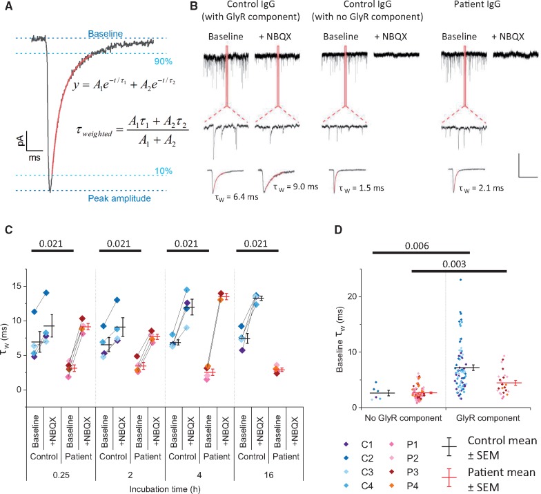
Chloride-permeable glycine receptors have an important role in fast inhibitory neurotransmission in the spinal cord and brainstem. Human immunoglobulin G (IgG) autoantibodies to glycine receptors are found in a substantial proportion of patients with progressive encephalomyelitis with rigidity and myoclonus, and less frequently in other variants of stiff person syndrome. Demonstrating a pathogenic role of glycine receptor autoantibodies would help justify the use of immunomodulatory therapies and provide insight into the mechanisms involved. Here, purified IgGs from four patients with progressive encephalomyelitis with rigidity and myoclonus or stiff person syndrome, and glycine receptor autoantibodies, were observed to disrupt profoundly glycinergic neurotransmission. In whole-cell patch clamp recordings from cultured rat spinal motor neurons, glycinergic synaptic currents were almost completely abolished following incubation in patient IgGs. Most human autoantibodies targeting other CNS neurotransmitter receptors, such as N-methyl-d-aspartate (NMDA) receptors, affect whole cell currents only after several hours incubation and this effect has been shown to be the result of antibody-mediated crosslinking and internalization of receptors. By contrast, we observed substantial reductions in glycinergic currents with all four patient IgG preparations with 15 min of exposure to patient IgGs. Moreover, monovalent Fab fragments generated from the purified IgG of three of four patients also profoundly reduced glycinergic currents compared with control Fab-IgG. We conclude that human glycine receptor autoantibodies disrupt glycinergic neurotransmission, and also suggest that the pathogenic mechanisms include direct antagonistic actions on glycine receptors.

Keywords: autoantibody; glycine receptor; progressive encephalomyelitis with rigidity and myoclonus (PERM); stiff person syndrome (SPS).


Figures

Figure 1
Figure 1
Whole cell patch clamp recordings of glycinergic mIPSCs. (A) Representative images of purified IgG from patient (Patient 1) or control subjects binding to the surface of cultured motor neurons. Scale bar = 10 µm. (B) Representative images of purified IgG from patient (Patient 1) binding to the surface of cultured motor neurons (green) co-stained for alpha1 subunits of glycine receptors with a commercial antibody (red). Lower panels show z-projections of individual processes. Scale bar = 10 µm. (C) Representative traces of miniature postsynaptic currents from whole cell patch clamp recordings of motor neurons following treatment with 0.5 mg/ml IgG purified from patient or control subjects for 2 h. Baseline recordings were made in the presence of TTX, AP5 and bicuculline (upper traces). NBQX was added to block AMPAR-mediated glutamatergic currents and thereby reveal the glycinergic mIPSCs (lower traces). Insets show the average miniature currents recorded in each condition. Vertical scale bar = 50 pA, horizontal scale bar = 1 s (traces) or 10 ms (insets).
Figure 2
Figure 2
IgGs from patients with glycine receptor autoantibodies disrupt glycinergic neurotransmission. (A) Box plots show combined mEPSC/mIPSC frequency before NBQX (left) and glycinergic mIPSC frequency after the addition of NBQX (right) at each time point (mean ± SEM). Each data point represents a single IgG sample (control samples C1–4 and patient samples P1–4). (B) Left box plot shows mean combined mEPSC/mIPSC amplitude before NBQX for patient and control IgG samples. Right: mIPSC amplitude after NBQX is shown for cells where two or more mIPSCs were recorded. (C) Box plots show calculated AMPAR-mediated charge transfer per unit time (calculated by subtracting the glycinergic charge transfer from the total charge transfer before the addition of NBQX, left) and GlyR charge transfer (right). Charge transfer was calculated from the area of all miniature events divided by time (upper panel, vertical scale bar = 50 pA, horizontal scale bar = 100 ms). (D) Box plot showing mean GlyR:AMPAR mediated charge transfer (individual data points = IgG samples, horizontal bar = mean, whiskers = SEM). (AD) Each data point shows the IgG sample mean (calculated from the mean of at least five cells measured following incubation in that particular IgG), horizontal bar indicates the mean of the IgG samples and whiskers indicate SEM (except for mIPSC amplitude after NBQX where n = number of cells where two of more mIPSCs were recorded and is indicated in parentheses; no cells met this criterion for 16 h incubation in patient IgGs). Horizontal bars indicate statistical significance P < 0.05 by Kruskal-Wallis with Dunn’s post hoc comparison (actual P-values shown).
Figure 3
Figure 3
IgGs from patients with glycine receptor autoantibodies alter the shape of average mIPSCs/mEPSCs. (A) Calculation of weighted tau from the fit of the 10–90% of the decay phase with a double exponential function (shown in red). (B) Top row shows example traces from three cells recorded before the addition of NBQX (left panel for each cell) or after NBQX (right panel for each cell) and expanded inset to show difference in individual mIPSC/mEPSC time course. Bottom row shows amplitude scaled average miniature events, with the 10-90% decay phase fitted with a double exponential function (red) to calculate the weighted tau (shown). Scale bar = 50 pA, 1 s (top panel), 50 ms (middle panel) and 10 ms (bottom panel). (C) Weighted tau for patient and control IgGs before and after addition of NBQX. Bars = mean values, error bars = SEM. Horizontal bars indicate statistical significance P < 0.05 by Kruskal-Wallis with Dunn’s post hoc comparison. (D) Weighted tau for cells separated by the presence or absence of glycinergic mIPSCs after the addition of NBQX. Horizontal bars indicate statistical significance P < 0.05 by Kruskal-Wallis with Dunn’s post hoc comparison.
Figure 4
Figure 4
Fab fragments from some patients disrupt glycinergic neurotransmission. (A) Example electrophoresis gel stained with Ponceau S showing a protein ladder (left), the undigested IgG, digest (containing Fc and Fab fragments) and purified Fab fragments. (B) Upper panel shows example traces and example amplitude scaled average miniature events from cells incubated in Fab fragments generated from IgGs from Control Subject 2 and Patients 1–4 (patients). Box plot shows relative (GlyR:AMPAR) charge transfer. (C) Example traces, example scaled average miniature events and box plot of relative charge transfer for neurons previously incubated in Fab fragments from Control Subject 2 or Patient 4 for 15 min. (D) Example traces, example scaled average miniature events and box plot of relative charge transfer for neurons incubated in Fab fragments generated from Control Subject 2 or Patient 3 for 16 h. (BD) Vertical scale bar = 50 pA, horizontal scale bar = 1 s for example traces and 10 ms for example scaled miniature events. Horizontal bar = mean, whiskers = SEM, numbers in parentheses = number of cells. Horizontal bars indicate statistical significance P < 0.05 by two-tailed Mann-Whitney U-tests.

References

    1. Barker RA, Revesz T, Thom M, Marsden CD, Brown P. Review of 23 patients affected by the stiff man syndrome: clinical subdivision into stiff trunk (man) syndrome, stiff limb syndrome, and progressive encephalomyelitis with rigidity. J Neurol Neurosurg Psychiatry 1998; 65: 633–40. - PMC - PubMed
    1. Bode A, Lynch JW. The impact of human hyperekplexia mutations on glycine receptor structure and function. Mol Brain 2014; 7: 2. - PMC - PubMed
    1. Brenner T, Sills GJ, Hart Y, Howell S, Waters P, Brodie MJ, et al.Prevalence of neurologic autoantibodies in cohorts of patients with new and established epilepsy. Epilepsia 2013; 54: 1028–35. - PubMed
    1. Carta E, Chung SK, James VM, Robinson A, Gill JL, Remy N, et al.Mutations in the GlyT2 gene (SLC6A5) are a second major cause of startle disease. J Biol Chem 2012; 287: 28975–85. - PMC - PubMed
    1. Carvajal-Gonzalez A, Leite MI, Waters P, Woodhall M, Coutinho E, Balint B, et al.Glycine receptor antibodies in PERM and related syndromes: characteristics, clinical features and outcomes. Brain 2014; 137: 2178–92. - PMC - PubMed

Publication types

MeSH terms

LinkOut - more resources