. 2019 Nov;9(9):692-702.
doi: 10.1089/brain.2019.0692.

Intrinsic Functional Connectivity of Dentate Nuclei in Autism Spectrum Disorder

Affiliations

Intrinsic Functional Connectivity of Dentate Nuclei in Autism Spectrum Disorder

Sheeba Arnold Anteraper et al. Brain Connect. 2019 Nov.

Abstract

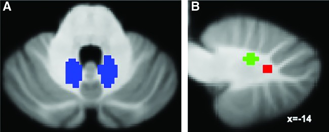
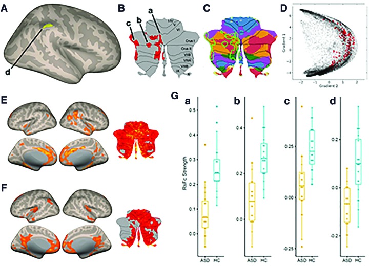
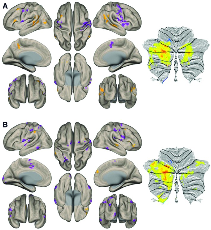
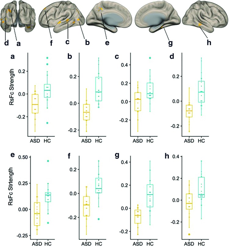
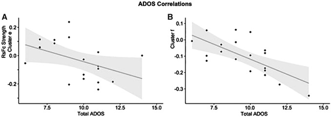
Cerebellar abnormalities are commonly reported in autism spectrum disorder (ASD). Dentate nuclei (DNs) are key structures in the anatomical circuits linking the cerebellum to the extracerebellum. Previous resting-state functional connectivity (RsFc) analyses reported DN abnormalities in high-functioning ASD (HF-ASD). This study examined the RsFc of the DN in young adults with HF-ASD compared with healthy controls (HCs) with the aim to expand upon previous findings of DNs in a dataset using advanced, imaging acquisition methods that optimize spatiotemporal resolution and statistical power. Additional seed-to-voxel analyses were carried out using motor and nonmotor DN coordinates reported in previous studies as seeds. We report abnormal dentato-cerebral and dentato-cerebellar functional connectivity in ASD. Our results expand and, in part, replicate previous descriptions of DN RsFc abnormalities in this disorder and reveal correlations between DN-cerebral RsFc and ASD symptom severity.

Keywords: autism spectrum disorder; cerebellum; dentate nucleus; functional connectivity; resting state networks; social cognition.


Conflict of interest statement

No competing financial interests exist.

Figures

<b>FIG. 1.</b>
FIG. 1.
(A) Quantitative susceptibility mapping-based seed location (He et al., 2017) for left and right DNs. (B) Motor (green) and nonmotor (red) seed locations based on the study by Bernard and colleagues (2014). DN, dentate nucleus. Color images are available online.
<b>FIG. 2.</b>
FIG. 2.
(A) Cerebral cortical results from between-group seed-to-voxel RsFc analysis for HC > ASD contrast (two-sided) for right DN at a height threshold of whole brain, p < 0.001 (T = 3.57), and FDR-corrected cluster threshold of p < 0.05. Age was added as a regressor of no interest. (B) Cerebellar results of the same contrast presented in a cerebellar flat map (Diedrichsen and Zotow, 2015). Cluster labels in (A) and (B) correspond to Table 2. (C) Cerebellar cluster shown in (B) overlaid on cerebellar representations of cerebral cortical networks (Buckner et al., 2011): dark purple, visual; blue, somatomotor; green, dorsal attention; violet, ventral attention; cream, limbic; orange, frontoparietal; and red, default network. (D) Cerebellar cluster shown in (B) represented in the functional gradient space as developed by Guell and colleagues (2018c). Each dot represents one cerebellar voxel; dots shown in color correspond to the voxels that are included in the cerebellar cluster shown in (B). (E) HC within-group connectivity from the right DN seed at a height threshold of whole brain, p < 0.001, and FDR-corrected cluster threshold of p < 0.05. (F) ASD within-group connectivity from the right DN seed using the same thresholds as in (E). (G) Boxplots of ASD and HC connectivity data extracted from clusters shown in (A) and (B). HCs, healthy controls; ASD, autism spectrum disorder; FDR, false discovery rate; RsFc, resting-state functional connectivity. Color images are available online.
<b>FIG. 3.</b>
FIG. 3.
HC (A) and ASD (B) within-group connectivity for the unique effect (nonmotor > motor DN seeds) at a height threshold of whole brain, p < 0.005, and FDR-corrected cluster threshold of p < 0.05. Cerebellar results are presented on a flat map (Diedrichsen and Zotow, 2015). Color images are available online.
<b>FIG. 4.</b>
FIG. 4.
Cerebral cortical results from second-level seed-to-voxel RsFc analysis for HC versus ASD contrast for the unique effect (nonmotor > motor DN seeds) (height threshold = p < 0.005; cluster threshold = p < 0.05 FDR corrected). Age was added as a regressor of no interest. Boxplots (ah) correspond to RsFc strength extracted from clusters a through h (cluster labels correspond to Table 3). Color images are available online.
<b>FIG. 5.</b>
FIG. 5.
Group differences in dentate connectivity for the unique effect (nonmotor > motor DN) correlate with total Autism Diagnostic Observation Schedule scores in the posterior cingulate cortex (A) and superior frontal gyrus (B).

References

    1. Alahmadi AA, Pardini M, Samson RS, Friston KJ, Toosy AT, D'Angelo E, Gandini Wheeler-Kingshott CA. 2017. Cerebellar lobules and dentate nuclei mirror cortical force-related-BOLD responses: beyond all (linear) expectations. Hum Brain Mapp 38:2566–2579 - PMC - PubMed
    1. Allen G, McColl R, Barnard H, Ringe WK, Fleckenstein J, Cullum CM. 2005. Magnetic resonance imaging of cerebellar-prefrontal and cerebellar-parietal functional connectivity. Neuroimage 28:39–48 - PubMed
    1. APA. 2013. Diagnostic and Statistical Manual of Mental Disorders, 5th ed. Arlington, VA: American Psychiatric Association
    1. Arnold Anteraper S, Guell X, D'Mello A, Joshi N, Whitfield-Gabrieli S, Joshi G. 2018. Disrupted cerebrocerebellar intrinsic functional connectivity in young adults with high-functioning autism spectrum disorder: a data-driven, whole-brain, high-temporal resolution functional magnetic resonance imaging study. Brain Connect 9:48–59 - PubMed
    1. Baron-Cohen S. 1989. The autistic child's theory of mind: a case of specific developmental delay. J Child Psychol Psychiatry 30:285–297 - PubMed

Publication types