. 2019 Aug 5;3(10):1373-1387.
doi: 10.1002/hep4.1415. eCollection 2019 Oct.

Liver X Receptor α-Induced Cannabinoid Receptor 2 Inhibits Ubiquitin-Specific Peptidase 4 Through miR-27b, Protecting Hepatocytes From TGF-β

Affiliations

Liver X Receptor α-Induced Cannabinoid Receptor 2 Inhibits Ubiquitin-Specific Peptidase 4 Through miR-27b, Protecting Hepatocytes From TGF-β

Hong Min Wu et al. Hepatol Commun. .

Abstract

Liver X receptor-alpha (LXRα) acts as a double-edged sword in different biological situations. Given the elusive role of LXRα in hepatocyte viability, this study investigated whether LXRα protects hepatocytes from injurious stimuli and the underlying basis. LXRα activation prevented hepatocyte apoptosis from CCl4 challenges in mice. Consistently, LXRα protected hepatocytes specifically from transforming growth factor-beta (TGF-β), whereas LXRα deficiency aggravated TGF-β-induced hepatocyte injury. In the Gene Expression Omnibus database analysis for LXR-/- mice, TGF-β receptors were placed in the core network. Hierarchical clustering and correlation analyses enabled us to find cannabinoid receptor 2 (CB2) as a gene relevant to LXRα. In human fibrotic liver samples, both LXRα and CB2 were lower in patients with septal fibrosis and cirrhosis than those with portal fibrosis. LXRα transcriptionally induced CB2; CB2 then defended hepatocytes from TGF-β. In a macrophage depletion model, JWH133 (a CB2 agonist) treatment prevented toxicant-induced liver injury. MicroRNA 27b (miR-27b) was identified as an inhibitor of ubiquitin-specific peptidase 4 (USP4), deubiquitylating TGF-β receptor 1 (TβRI), downstream from CB2. Liver-specific overexpression of LXRα protected hepatocytes from injurious stimuli and attenuated hepatic inflammation and fibrosis. Conclusion: LXRα exerts a cytoprotective effect against TGF-β by transcriptionally regulating the CB2 gene in hepatocytes, and CB2 then inhibits USP4-stabilizing TβRI through miR-27b. Our data provide targets for the treatment of acute liver injury.


Figures

Figure 1
Figure 1
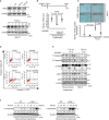
Protection of liver cells by LXRα from injurious stimuli. (A) Assays for hepatocyte apoptosis. TUNEL and hematoxylin and eosin staining were carried out for the liver sections (scale bar = 100 μm) from C57BL/6 mice (n = 4‐7 per group, male). Immunoblottings were done on liver homogenates. (B) Serum ALT and AST activities and quantitative RT‐PCR assays for inflammatory cytokines. (C) Assays for hepatocyte apoptosis. WT or LXRα−/− mice were injected with CCl4 for indicated times (n = 3 or 5 per group, female) and sacrificed 24 hours after last CCl4 injection. TUNEL‐positive cells were indicated with arrowheads. Scale bar = 100 μm. (D) Serum ALT and AST activities in mice treated as in (C). (E) Serum ALT and AST activities at 72 hours after last CCl4 injection (n = 5 or 6 per group, female). For (A) and (C), values represent fold changes relative to control. Quantification was normalized to β‐actin. For (B), (D), and (E), data represent the mean ± SEM (statistical difference was determined by two‐tailed Student t test). Abbreviations: h, hours; H&E, hematoxylin and eosin; iNOS, inducible nitric oxide synthase; and NS, not significant.
Figure 2
Figure 2
Inhibition of TGF‐β1‐induced hepatocyte death by LXRα. (A) Immunoblottings for LXRα in cells treated with 50 ng/mL IL‐6, 60 ng/mL TNF‐α, or 5 ng/mL TGF‐β1 for 24 hours or with 5 ng/mL TGF‐β1 for indicated times. (B) Quantitative RT‐PCR assays for Nr1h3 (LXRα). Mouse primary hepatocytes were treated with 3 μM GW3965 for 1 hour and continuously exposed to 5 ng/mL TGF‐β1 for 24 hours (n = 3 isolations, male). (C) TUNEL staining. The cells were treated as in (B) (n = 3 replicates). (D) Flow cytometric analyses for fluorescein isothiocyanate–annexin V and propidium iodide in cells treated as in (B). Values represent the mean ± SEM of three independent experiments. (E) Immunoblottings in cells treated as in (B). (F) Immunoblottings for C‐caspase3. Hepatocytes prepared from WT or LXRα−/− mice were treated with TGF‐β1 for 24 hours (left panel) or 5 ng/mL TGF‐β1 for the indicated times (right panel). For (A), (E), and (F), multiple assays were done (n = 3), and representative blots were shown. Values represent fold changes relative to control. For (B) and (C), data represent the mean ± SEM (statistical difference was determined by two‐tailed Student t test). Abbreviations: FITC, fluorescein isothiocyanate; and h, hours.
Figure 3
Figure 3
LXRα and CB2 repression under the conditions of liver injury. (A) Heat map and hierarchical correlation analyses of the genes in the complementary DNA microarray obtained from GSE25583. Genes with expressional relevance to LXRα are indicated by the red (the cluster comprising 1% genes) corresponding region in the dendrogram. (B) Quantitative RT‐PCR assays for Nr1h3 (LXRα) and relevant genes in hepatocytes isolated from mice 24 hours after treatment with vehicle or CCl4 (n = 4 per group, male, 2 replicates for each mouse) and correlation of Nr1h3 (LXRα) and Cnr2 (CB2). (C) Correlation between LXRα and CB2 in patients with hepatitis (GSE20140, n = 307) and in the liver of mice with hepatitis (GSE17184). (D) LXRα protein levels and quantitative RT‐PCR assays for CNR2 in patient liver specimens. For (B)‐(D), coefficients were obtained using the Pearson analysis; for (B) and (D), data represent the mean ± SEM (statistical difference was determined by two‐tailed Student t test. Abbreviation: h, hours.
Figure 4
Figure 4
The role of CB2 in hepatocytes in the protection of liver. (A) The hepatoprotective effect of JWH133 in C57BL/6 mice treated with CCl4 and clodronate. Mice were treated with JWH133 (10 mg/kg body weight per day), clodronate (100 μL/mice), and CCl4, as shown in the scheme (n = 5 per group, male). Relative TUNEL positivity was assessed from two or three samples per condition (two samples for JWH133 alone and three samples for the others, randomly selected from five samples), and three random fields per sample were evaluated. Scale bar = 100 μm. (B) Serum ALT and AST activities in mice treated as previously. For (A) and (B), data represent the mean ± SEM (statistical difference was determined by two‐tailed Student t test). For (A), values represent fold changes relative to control. Abbreviations: h, hours; and NS, not significant.
Figure 5
Figure 5
Transcriptional induction of CB2 by LXRα. (A) Quantitative RT‐PCR assays for Cnr2 in the liver of mice (n = 4 per group, male) treated with GW3965 (3 mg/kg body weight per day) for 3 days (left) or in mouse primary hepatocytes (middle, n = 4 isolations, male) and AML12 cells (right) treated with GW3965 (3 μM, 24 hours). (B) Chromatin immunoprecipitation assays for LXRα binding to the promoter region of CB2 gene. Quantitative PCR assays were done for quantification. (C) Promoter reporter assays using pGL3‐CB2 or its LXRE2 deletion mutant construct. AML12 cells were transfected with each construct (1 μg, 24 hours) and then treated with vehicle or GW3965 (3 μM, 12 hours). (D) Immunoblotting for C‐caspase3. AML12 cells were treated with JWH133 (1, 3, and 10 μM for 1 hour) and continuously exposed to 5 ng/mL TGF‐β1 for 24 hours (left). The cells were also treated with 3 μM GW3965 for 1 hour after 4 μM SR144528 treatment for 30 minutes and continuously exposed to TGF‐β1 (middle). Mouse primary hepatocytes were similarly treated with GW3965 and TGF‐β1 after small interfering RNA (siRNA) knockdown for 48 hours (right). For (A)‐(C), data represent the mean ± SEM of three separate experiments (statistical difference was determined by two‐tailed Student t test). For (D), multiple assays were done (n = 3), and representative blots are shown. Values represent fold changes relative to control. Abbreviations: bp, base pair; IgG, immunoglobulin G; IP, immunoprecipitation; and NS, not significant.
Figure 6
Figure 6
USP4 repression by miR‐27b downstream of LXRα and CB2. (A) TβRI stability in AML12 cells treated with GW3965 (3 µM, 24 hours) after transfection with His‐tagged ubiquitin (His‐Ubi). MG132 (10 µM) was added 6 hours before cell lysis (left). The cells were treated with GW3965, SR144528 (4 µM), and cycloheximide (CHX, 20 μg/mL) as indicated (middle and right). (B) Immunoblottings for USP4 and C‐caspase3. Primary hepatocytes were treated with GW3965 or JWH133 (left); AML12 cells were treated with SR144528 (30 minutes) and continuously exposed to GW3965 (24 hours) (middle) or treated with GW3965 and TGF‐β1 after transfection with FLAG‐tagged USP4 (1 µg, 24 hours) (right). (C) miRNA candidates targeting USP4. A Venn diagram and table show an overlapping of decreased miRNAs in the liver of mice treated with CCl4 (GSE66278) and those predicted to target mouse USP4 (microRNA.org). Quantitative RT‐PCR assays for the miRNAs in AML12 cells treated with JWH133 (10 μM, 24 hours). (D) Quantitative RT‐PCR assays for miR‐27b in human specimens and correlation analysis. (E) Quantitative RT‐PCR assays for miR‐27b in AML12 cells treated with SR144528 (30 minutes) and continuously exposed to GW3965 (24 hours) or in mouse primary hepatocytes treated with GW3965 after transfection of CB2 siRNA (or control siRNA) for 48 hours. (F) USP4 3′‐UTR reporter and USP4 immunoblotting assays in AML12 cells transfected with mock or miR‐27b mimic (or ASO). (G) USP4 immunoblots in AML12 cells treated with JWH133 (24 hours) after miR‐27b ASO transfection (48 hours). For (A), (B), and (G), multiple assays were done (n = 3), and representative blots are shown. Values represent fold changes relative to control. For (C), (E), and (F), data represent the mean ± SEM of three separate experiments (statistical difference was determined by two‐tailed Student t test, *P < 0.05, **P < 0.01). Abbreviations: CHX, cycloheximide; h, hours; IB, immunoblotting; IP, immunoprecipitation; min, minutes; NS, not significant; Ub, ubiquitin; and WCL, whole cell lysates.
Figure 7
Figure 7
Amelioration of hepatocyte injury by liver‐specific overexpression of LXRα. (A) Assays for hepatocyte apoptosis. WT or LXRα−/− mice (n = 5 per group, female) were injected with lentiviruses that express control (LV‐Con) or LXRα (LV‐LXRαalb) for 7 days and subjected to CCl4 treatments for 4 weeks. TUNEL‐positive cells were indicated with arrowheads. Scale bar = 100 μm. (B) Quantitative RT‐PCR assays for Cnr2 and miR‐27b and immunoblottings for USP4 and TβRI. (C) Quantitative RT‐PCR assays for inflammatory cytokines. (D) Masson’s trichrome stainings and immunoblottings for fibrosis markers. Scale bar = 100 μm. (E) A schematic illustrating the proposed mechanism by which LXRα protects hepatocytes from TGF‐β‐induced injury. For (A), (B), and (D), values represent fold changes relative to control. For (B) and (C), data represent the mean ± SEM (statistical difference was determined by two‐tailed Student t test). Abbreviations: alb, albumin promoter; Con, control; h, hours; and LV, lentiviral.

References

    1. Racanelli V, Rehermann B. The liver as an immunological organ. Hepatology 2006;43:S54‐S62. - PubMed
    1. Malhi H, Guicciardi ME, Gores GJ. Hepatocyte death: a clear and present danger. Physiol Rev 2010;90:1165‐1194. - PMC - PubMed
    1. Takehara T, Tatsumi T, Suzuki T, Rucker EB, Hennighausen L, Jinushi M, et al. Hepatocyte‐specific disruption of Bcl‐xL leads to continuous hepatocyte apoptosis and liver fibrotic responses. Gastroenterology 2004;127:1189‐1197. - PubMed
    1. Inokuchi S, Aoyama T, Miura K, Osterreicher CH, Kodama Y, Miyai K, et al. Disruption of TAK1 in hepatocytes causes hepatic injury, inflammation, fibrosis, and carcinogenesis. Proc Natl Acad Sci U S A 2010;107:844‐849. - PMC - PubMed
    1. Kohro T, Nakajima T, Wada Y, Sugiyama A, Ishii M, Tsutsumi S, et al. Genomic structure and mapping of human orphan receptor LXR alpha: upregulation of LXRa mRNA during monocyte to macrophage differentiation. J Atheroscl Thromb 2000;7:145‐151. - PubMed