. 2019 Oct 18;8(10):2359-2371.
doi: 10.1021/acssynbio.9b00175. Epub 2019 Oct 8.

An AND-Gated Drug and Photoactivatable Cre- loxP System for Spatiotemporal Control in Cell-Based Therapeutics

Affiliations

An AND-Gated Drug and Photoactivatable Cre- loxP System for Spatiotemporal Control in Cell-Based Therapeutics

Molly E Allen et al. ACS Synth Biol. .

Abstract

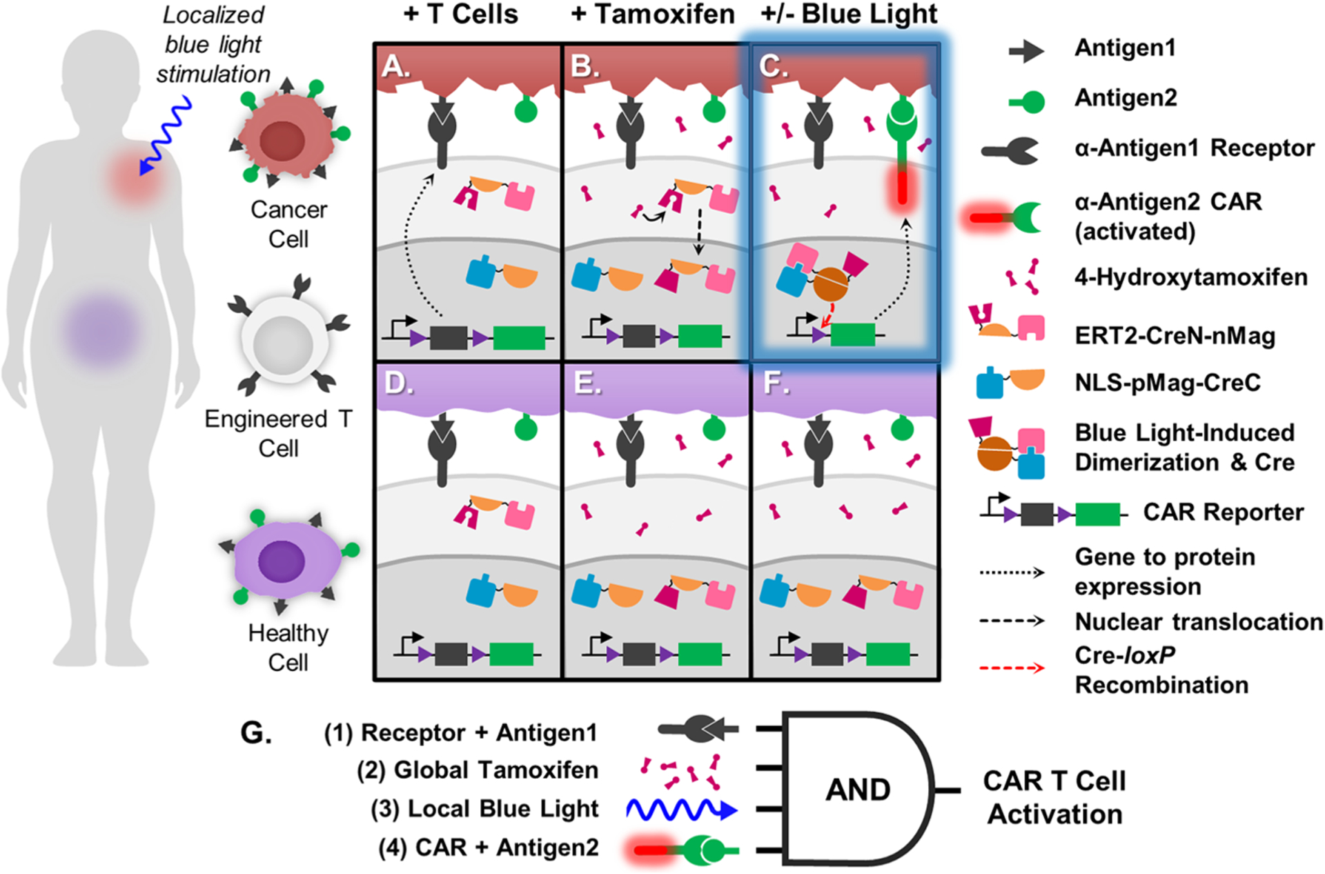
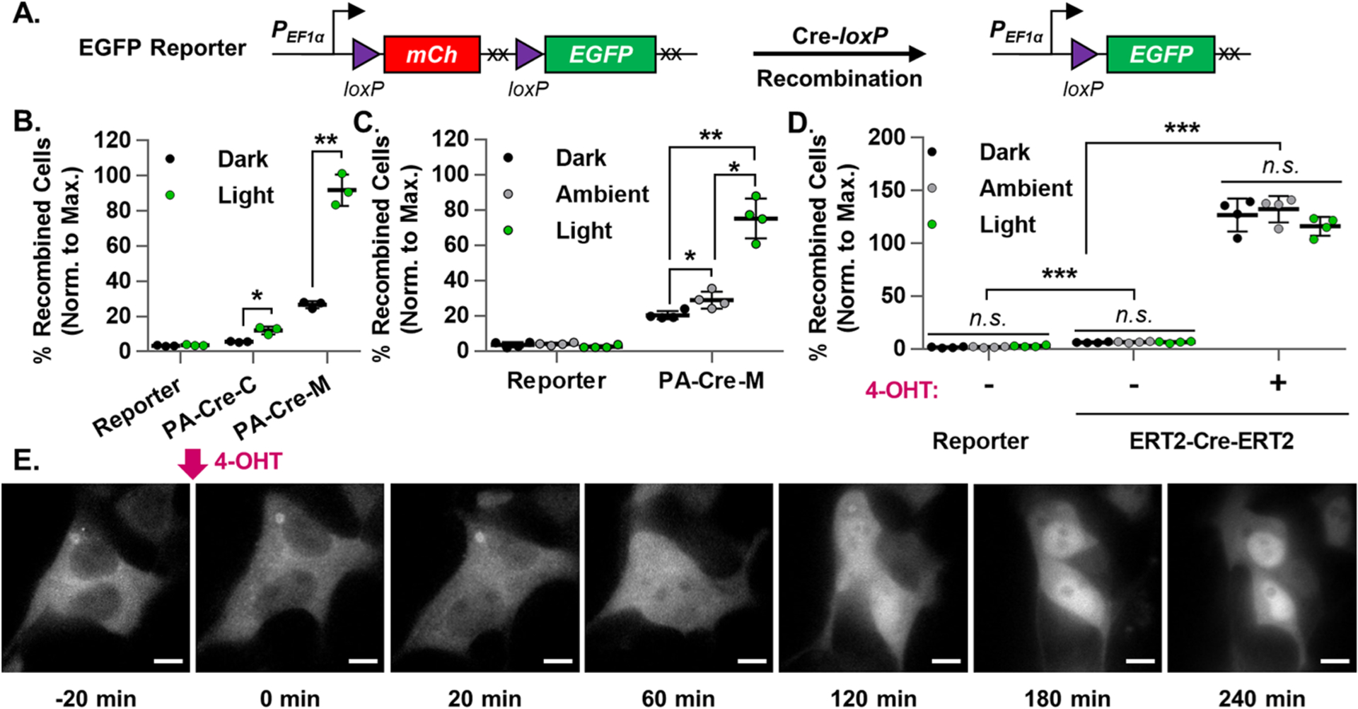
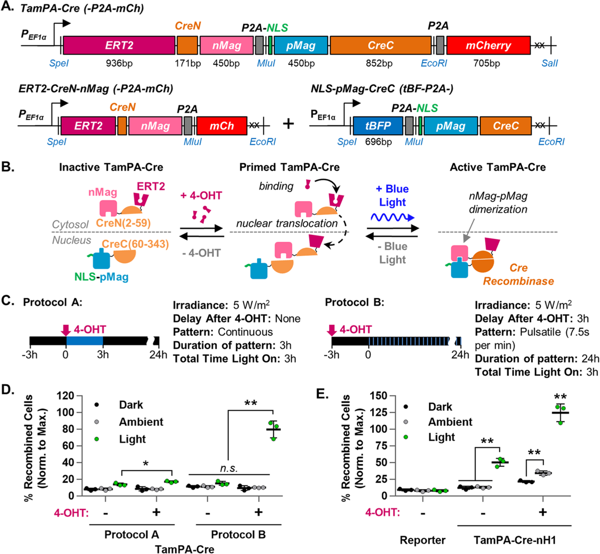
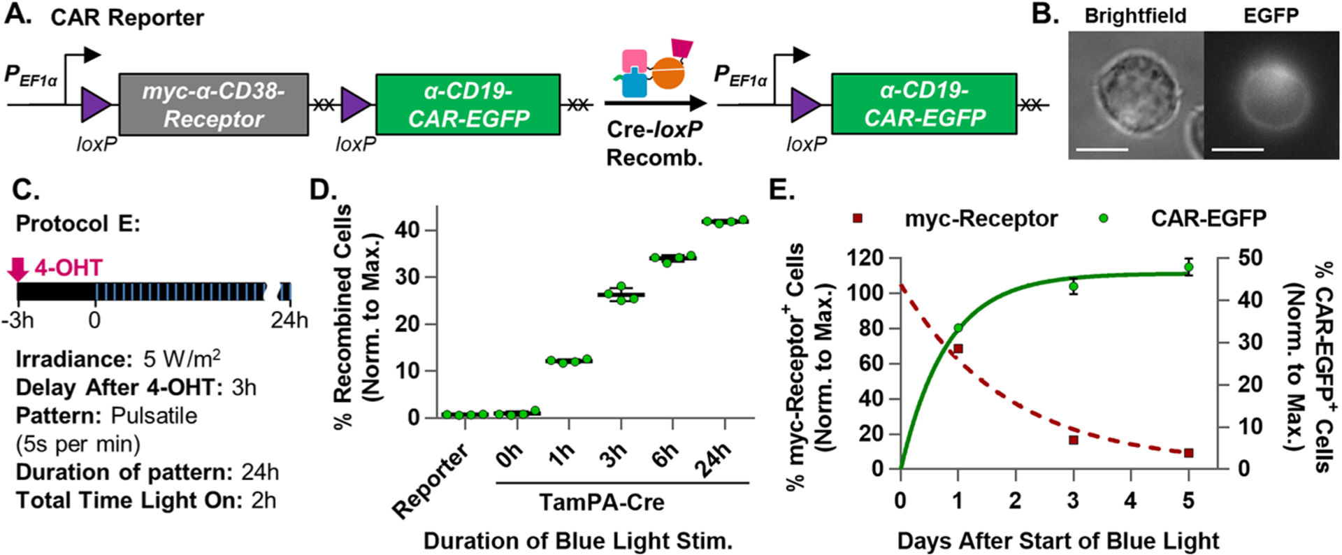
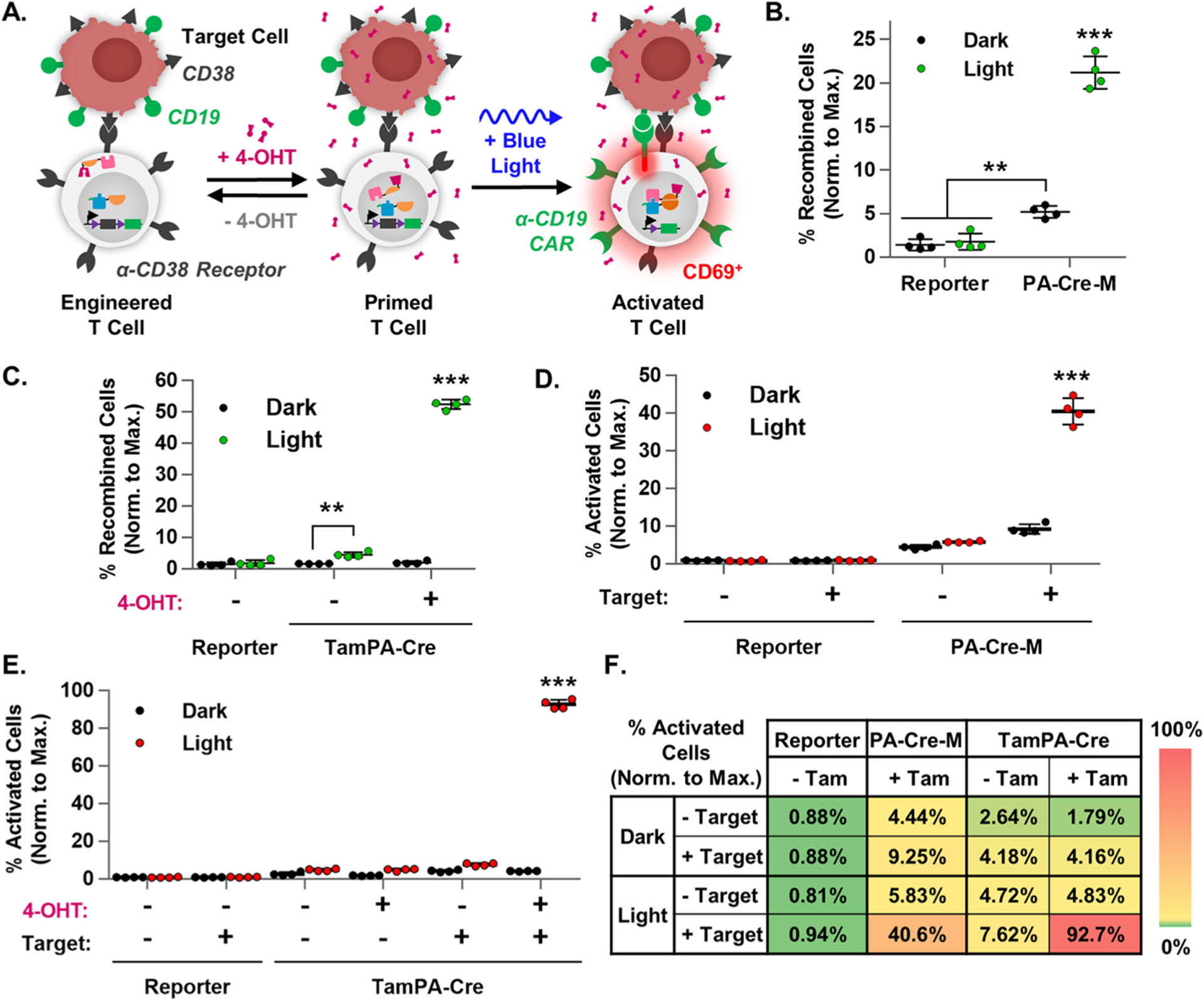
While engineered chimeric antigen receptor (CAR) T cells have shown promise in detecting and eradicating cancer cells within patients, it remains difficult to identify a set of truly cancer-specific CAR-targeting cell surface antigens to prevent potentially fatal on-target off-tumor toxicity against other healthy tissues within the body. To help address this issue, we present a novel tamoxifen-gated photoactivatable split-Cre recombinase optogenetic system, called TamPA-Cre, that features high spatiotemporal control to limit CAR T cell activity to the tumor site. We created and optimized a novel genetic AND gate switch by integrating the features of tamoxifen-dependent nuclear localization and blue-light-inducible heterodimerization of Magnet protein domains (nMag, pMag) into split Cre recombinase. By fusing the cytosol-localizing mutant estrogen receptor ligand binding domain (ERT2) to the N-terminal half of split Cre(2-59aa)-nMag, the TamPA-Cre protein ERT2-CreN-nMag is physically separated from its nuclear-localized binding partner, NLS-pMag-CreC(60-343aa). Without tamoxifen to drive nuclear localization of ERT2-CreN-nMag, the typically high background of the photoactivation system was significantly suppressed. Upon blue light stimulation following tamoxifen treatment, the TamPA-Cre system exhibits sensitivity to low intensity, short durations of blue light exposure to induce robust Cre-loxP recombination efficiency. We finally demonstrate that this TamPA-Cre system can be applied to specifically control localized CAR expression and subsequently T cell activation. As such, we posit that CAR T cell activity can be confined to a solid tumor site by applying an external stimulus, with high precision of control in both space and time, such as light.

Keywords: CAR T cell; Cre-loxP recombination; ERT2; immunotherapy; nMag and pMag; optogenetics.


Conflict of interest statement

The authors declare the following competing financial interest(s): Y.W. is a scientific co-founder of Cell E&G Inc and Acoustic Cell Therapy LLC. However, these financial interests do not affect the design, conduct, or reporting of this research.

Figures

Figure 1.
Figure 1.
Schematic of TamPA-Cre application and molecular mechanism. (Left) Person with Antigen1+ Antigen2+ cancerous (red) and healthy (purple) tissues in separate regions of the body. Engineered T cells express TamPA-Cre (ERT2-CreN-nMag and NLS-pMag-CreC) and the CAR Reporter genetic construct, consisting of a constitutive promoter driving expression of the floxed (purple) α-Antigen1 Receptor CDS with stop codons (black), followed by α-Antigen2 CAR (green). Upon intravenous introduction, the engineered T cells bind and localize to both cancerous (A) and healthy (D) Antigen1+ cells. TamPA-Cre is inactive as its NLS-pMag-CreC and ERT2-CreN-nMag protein halves nuclear and cytosolically localized, respectively. After administration of tamoxifen, metabolite 4-OHT binds with ERT2-CreN-nMag to drive nuclear localization (B,E), priming TamPA-Cre. Next, blue light is applied to the cancerous tissue region only (C), inducing nMag-pMag heterodimerization which restores active TamPA-Cre recombinase activity within the nucleus. The floxed α-Antigen1 Receptor CDS in the CAR Reporter is excised through Cre-loxP recombination along with its stop codons, thus allowing for α-Antigen2 CAR expression. The T cell is finally activated upon CAR-mediated binding to Antigen2. T cells localized to the healthy tissue region (F) are not exposed to blue light and thus do not express α-Antigen2 CAR, effectively protecting healthy cells that express both Antigen1 and Antigen2. (G) Boolean logic representation of the AND-gated TamPA-Cre system. (1) T cells first bind to cells expressing Antigen1 via the Receptor. (2) Then, TamPA-Cre is be primed with tamoxifen before receiving localized (3) blue light stimulation in the cancerous tissue region, driving Cre-loxP recombination and CAR expression. (4) T cell activation is triggered when Antigen2 is recognized by the CAR.
Figure 2.
Figure 2.
Evaluation of photoactivatable and tamoxifen-dependent Cre recombinase systems. (A) Schematic representation of the EGFP Reporter genetic construct before and after Cre-loxP recombination. The hEF1α promoter drives mCherry expression. During Cre-loxP recombination, the floxed mCherry is irreversibly excised along with its stop codons (XX), thus allowing for EGFP expression. The normalized percentage of recombined cells was calculated for EGFP Reporter HEK293T transiently transfected with the following constructs which did (Light) or did not (Ambient, Dark) receive the indicated blue light stimulation (473 ± 29 nm): (B) PA-Cre-C, PA-Cre-M, or Cre PAconstructs, (30 W/m2, 30s) (n = 3), or (C) PA-Cre-M or Cre constructs, (15 W/m2, 1s per min, 24 h) (n = 4), or (D) ERT2-Cre-ERT2 or Cre constructs, (15 W/m2, 1s per min, 24 h) (n = 4). Reporter = untransfected EGFP Reporter HEK293T cell line. Blue light stimulation started, and flow cytometry measurements were taken 24 and 72 h post-transfection, respectively. Percentage of recombined HEK293T cells (normalized to maximal recombination) = 100%*(% of EGFP+ cells)/(mean % of EGFP+ cells in corresponding Cre groups). (E) Representative time-lapse fluorescence microscopy images of HEK293T cells transiently expressing ERT2-mCherry before and after the addition of nuclear-localizing 4-OHT (500 nM) (imaged every 2 min, 100× magnification, scale bar = 10 μm, n = 6 independently measured cells).
Figure 3.
Figure 3.
Design and optimization of the TamPA-Cre system in HEK293T cells. (A) Schematic of TamPA-Cre as a single genetic construct with codon-diversified pMag and marker P2A-mCh (top), or as two genetic constructs: ERT2-CreN-nMag with marker P2A-mCh (bottom left) and NLS-pMag-CreC with marker tBFP-P2A (bottom right). (B) Schematic depicting the mechanism of the tamoxifen- AND blue-light-gated TamPA-Cre system. (C) Schematic illustrating two different tamoxifen and blue light stimulation protocols, each providing the same total amount of blue light energy (473 ± 29 nm, not to scale). Protocol A: 3 h of continuous blue light stimulation (5 W/m2) started immediately after the addition of 4-OHT (500 nM). Protocol B: 24 h of pulsatile blue light stimulation (5 W/m2, 7.5s per min) started 3 h after 4-OHT addition. (D) The percentage of recombined TamPA-Cre+ EGFP Reporter HEK293T cells (normalized to maximal recombination) which were (Light) or were not (Ambient, Dark) subjected to protocol A or B (n = 3). (E) Recombination in TamPA-Cre-nH1+ EGFP Reporter HEK293T cells subjected (Light) or not (Ambient, Dark) to Protocol B (n = 3). Reporter = untransfected EGFP Reporter HEK293T cell line (n = 3). Blue light stimulation started and flow cytometry measurements taken 24 and 72 h post-transfection, respectively. Percentage of recombined HEK293T cells (normalized to maximal recombination) = 100%*(% of EGFP+ cells)/(mean % of EGFP+ cells in corresponding Cre groups).
Figure 4.
Figure 4.
Optimization and characterization of the TamPA-Cre system in Jurkat T cells. (A) Schematic of the CAR Reporter construct before and after TamPA-Cre-mediated Cre-loxP recombination. The hEF1α promoter initially drives myc-α-CD38-Receptor expression. During Cre-loxP recombination, the floxed myc-α-CD38Receptor (with its stop codons, XX) is irreversibly excised allowing for (B) α-CD19CAR-EGFP expression (Jurkat T cell, 100×, scale bar = 10 μm). (C) Schematic illustrating tamoxifen (500 nM) and blue light stimulation (473 ± 29 nm) Protocol E: (5 W/m2, 5 s per min, 24 h) started 3 h after 4-OHT addition. (D) The percentage of recombined TamPA-Cre+ CAR Reporter Jurkat T cells (normalized to maximal recombination) exposed to Protocol E over the course of 0, 1, 3, 6, or 24 h (n = 4). (E) Normalized percentage of myc-Receptor+ and CAR-EGFP+ TamPA-Cre+ CAR Reporter Jurkat T cells stimulated by Protocol E, measured 1, 3, and 5 days after start of blue light stimulation, fitted with exponential decay and association trendlines (GraphPad, Table S2) (n = 4). Reporter = CAR Reporter Jurkat T cell line. CAR-EGFP flow cytometry measurements taken 72 h after the start of blue light stimulation. Percentage of recombined (% CAR-EGFP+) Jurkat T cells (normalized to maximal recombination) = 100%*(% of CAR-EGFP+ cells)/(initial % of CAR Reporter+ cells, measured via myc).
Figure 5.
Figure 5.
TamPA-Cre drives CAR-mediated T cell activation. (A) Schematic of the tamoxifen- and blue light-induced TamPA-Cre system in a CAR Reporter T cells driving recombination and CAR-mediated T cell activation upon binding to a TAA+ Target cells. The percentage of recombined cells (normalized to maximal recombination) in (B) PA-Cre-M+ and (C) TamPA-Cre+ CAR Reporter Jurkat T cells that did (+ 4-OHT) or did not (− 4-OHT) receive tamoxifen (500 nM) stimulation, and did (Light) or did not (Dark) receive blue light stimulation (473 ± 29 nm) as outlined in Protocol E (n = 4). The percentage of activated cells (normalized to maximal recombination) of samples from (B) and (C) that were (+ Target) or were not (− Target) coincubated with CD19+ Target cells (1:1), as reported in (D) and (E) respectively. (n = 4). (F) A heat map summary of T cell activation in Reporter, PA-Cre-M, and TamPA-Cre groups, with higher and lower efficiencies shown in red and green, respectively. Reporter = CAR Reporter Jurkat T cell line. Coincubation started and flow cytometry measurements taken 48 and 72 h after the start of blue light stimulation, respectively. Percentage of recombined Jurkat T cells (normalized to maximal recombination) = 100%*(% of CAR-EGFP+ cells)/(initial% of CAR Reporter+ cells, measured via myc). Percentage of activated Jurkat T cells (normalized to maximal recombination) = 100%*(% of CD69+ cells)/(initial % of CAR Reporter+ cells, measured via myc).

References

    1. Porter DL, Levine BL, Kalos M, Bagg A, and June CH (2011) Chimeric antigen receptor-modified T cells in chronic lymphoid leukemia. N. Engl. J. Med 365 (8), 725–33. - PMC - PubMed
    1. Grupp SA, Kalos M, Barrett D, Aplenc R, Porter DL, Rheingold SR, Teachey DT, Chew A, Hauck B, Wright JF, Milone MC, Levine BL, and June CH (2013) Chimeric antigen receptor-modified T cells for acute lymphoid leukemia. N. Engl. J. Med 368 (16), 1509–1518. - PMC - PubMed
    1. Maude SL, Shpall EJ, and Grupp SA (2014) Chimeric antigen receptor T-cell therapy for ALL. Hematology Am. Soc. Hematol Educ Program 2014 (1), 559–64. - PubMed
    1. Maude SL, Teachey DT, Porter DL, and Grupp SA (2015) CD19-targeted chimeric antigen receptor T-cell therapy for acute lymphoblastic leukemia. Blood 125 (26), 4017–23. - PMC - PubMed
    1. Davila ML, Riviere I, Wang X, Bartido S, Park J, Curran K, Chung SS, Stefanski J, Borquez-Ojeda O, Olszewska M, Qu J, Wasielewska T, He Q, Fink M, Shinglot H, Youssif M, Satter M, Wang Y, Hosey J, Quintanilla H, Halton E, Bernal Y, Bouhassira DC, Arcila ME, Gonen M, Roboz GJ, Maslak P, Douer D, Frattini MG, Giralt S, Sadelain M, and Brentjens R (2014) Efficacy and toxicity management of 19–28z CAR T cell therapy in B cell acute lymphoblastic leukemia. Sci. Transl. Med 6 (224), 224ra25. - PMC - PubMed

Publication types