Review
. 2020 Jan;93(1105):20190677.
doi: 10.1259/bjr.20190677. Epub 2019 Oct 8.

Imaging anatomy of the retrotympanum: variants and their surgical implications

Affiliations
Review

Imaging anatomy of the retrotympanum: variants and their surgical implications

Christian Burd et al. Br J Radiol. 2020 Jan.

Abstract

The retrotympanic anatomy is complex and variable but has received little attention in the radiological literature. With advances in CT technology and the application of cone beam CT to temporal bone imaging, there is now a detailed depiction of the retrotympanic bony structures.With the increasing use of endoscopes in middle ear surgery, it is important for the radiologist to appreciate the nomenclature of the retrotympanic compartments in order to aid communication with the surgeon. For instance, in the context of cholesteatoma, clear imaging descriptions of retrotympanic variability and pathological involvement are valuable in pre-operative planning.The endoscopic anatomy has recently been described and the variants classified. The retrotympanum is divided into medial and lateral compartments with multiple described potential sinuses separated by bony crests.This pictorial review will describe the complex anatomy and variants of the retrotympanum. We will describe optimum reformatting techniques to demonstrate the structures of the retrotympanum and illustrate the associated anatomical landmarks and variants with CT. The implications of anatomical variants with regards to otologic surgery will be discussed.


Figures
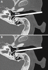
Figure 1.
Figure 1.
(A) Axial CT through the left temporal bone demonstrating the transcanal position of a 30–45° angled endoscope and its field of view, enabling visualization of the the medial but not the lateral retrotympanum. (B) Axial CT through the left temporal bone demonstrating the transcanal position of a 70° angled endoscope and its field of view, enabling visualization of the lateral retrotympanum whilst sacrificing some of the medial view.
Figure 2.
Figure 2.
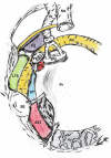
Diagrammatic representation of the retrotympanum from a right ear transcanal view. Key: C, chordiculus; CT, chorda tympani; F, funiculus; FN, facial nerve; FS, facial sinus; Hy, hypotympanum; In, incus; LTS, lateral tympanic sinus; Ma, malleus; P, ponticulus; PE, pyramidal eminence; Pr, cochlear promontory; PS, posterior sinus; RWN, round window niche; S, subiculum; SE, styloid eminence; SFp, stapes footplate; SST, sinus subtympanicum; ST, sinus tympani.
Figure 3.
Figure 3.
Right ear intraoperative middle ear transcanal endoscopic image (a) with comparative labelled endoscopic middle ear image (b) Key: f, funiculus; fn, facial nerve; hy, hypotympanum; in, incus; p, ponticulus; pe, pyramidal eminence; pr, cochlear promontory; ps, posterior sinus; rw, round window; sst, sinus subtympanicus; st, sinus tympani; su, subiculum; t, subcochlear tunnel.
Figure 4.
Figure 4.
Axial CT images demonstrating pars tensa cholesteatoma. (A) Right ear with cholesteatoma filling the epitympanum (white arrow) and displacing the the incus and malleal head laterally (black arrow). (B) Left ear with cholesteatoma opacification tracking down along the retrotympanum surface (white arrow)
Figure 5.
Figure 5.
(A) Left temporal bone axial CT reformat line (dashed white line) through the LSC. (B) Poschl (dashed white squares), parallel to the SSC and Stenvers’ (dashed white rectangles), perpendicular to the SSC, reformat planes on left temporal bone axial CT. (C) Poschl (dashed squares) and Stenvers’ (dashed rectangles) projection planes through the left temporal bone medial and lateral retrotympanic spaces respectively. LSC, lateral semicircular canal; SCC, superiorsemicircular canal.
Figure 6.
Figure 6.
CBCT images (left ear) demonstrating key axial and sagittal retrotympanic anatomy. (A) Sagittal oblique (Poschl) image through the medial retrotympanum. (B) Midline image through the plane of the styloid and pyramidal eminences. (C) Coronal oblique (Stenvers’) image through the lateral retrotympanum. (D) Axial images at the level of the stapes demonstrating the pyramidal eminence, (E) at the superior aspect of the cochlear promontory demonstrating the pyramidal eminence, (F) and at the inferior aspect of cochlear promontory demonstrating the styloid eminence. Key: CBCT, cone beam CT; CE, chordal eminence; CT, chorda tympani; FN, facial nerve; JB, jugular bulb; OW, oval window; Pr, cochlear promontory; PE, pyramidal eminence; PP, posterior pillar of the round window; RW, round window; St, stapes; SE, styloid eminence; TA, tympanic annulus.
Figure 7.
Figure 7.
(AD) Sagittal oblique (Poschl) reformats through the right temporal bone medial retrotympanum with axial slices corresponding to images EH (images B, D). (EH) Axial images superior to the ponticulus (E), at the ponticulus bridge (D), at the subiculum ridge (G), and at the funiculus (H). Key: F, funiculus; FN, facial nerve; Hy, hypotympanum; LSC, lateral semicircular canal; CMT, central mastoid tract; P, ponticulus; PS, posterior sinus; RW, round window; RWN; round window niche; S, subiculum; SST, sinus subtympanicus; ST, sinus tympani; TA, tympanic annulus.
Figure 8.
Figure 8.
(AD) Sagittal oblique (Poschl) reformats through the right temporal bone medial retrotympanum with axial slices corresponding to images EG (images B, D). Axial images at the ponticulus ridge (E), at the subiculum ridge (F) and at the funiculus (G). Key: F, funiculus; Hy, hypotympanum; LSC, lateral semicircular canal; P, ponticulus; PS, posterior sinus; RW, round window; RWN, round window niche; S, subiculum; SST, sinus subtympanicus; ST, sinus tympani; TA, tympanic annulus.
Figure 9.
Figure 9.
(AD) Sagittal oblique (Poschl) reformats through the right temporal bone medial retrotympanum with axial slices corresponding to images EH (images B, D). (EH) Axial images at the oval window (E), at the pyramidal eminence with an absent ponticulus (F), at the subiculum ridge (G) and at the funiculus (H). Key: F, funiculus; FN, facial nerve; Hy, hypotympanum; LSC, lateral semicircular canal; OW, oval window; PE, pyramidal eminence; PS, posterior sinus; RW, round window; RWN, round window niche; S, subiculum; SST, sinus subtympanicus; ST, sinus tympani; TA, tympanic annulus.
Figure 10.
Figure 10.
(AD) Coronal oblique (Stenvers’) reformats through the right temporal bone lateral retrotympanum with axial slices corresponding to images EH (images B, D). (EH) Axial images at the chordiculus insertion into the pyramidal eminence (E), at the proximal chordiculus ridge (F), at the styloid ridge (G) and at the hypotympanum (H). Key: C, chordiculus; CE, chordal eminence; CT, chorda tympani; FN, facial nerve; FS, facial sinus; Hy, hypotympanum; LSC, lateral semicircular canal; LTS, lateral tympanic sinus; PE, pyramidal eminence; PR, pyramidal ridge; S, subiculum; SR, styloid ridge.
Figure 11.
Figure 11.
(AD) Coronal oblique (Stenvers’) reformats through the left temporal bone lateral retrotympanum with axial slices corresponding to images EH (images B, D). (EH) Axial images at the facial sinus (E), at the chordiculus bridge (F), at the styloid ridge (G) and at the chordal eminence (H). Key: C, chordiculus; CE, chordal eminence; CT, chorda tympani; FS, facial sinus; Hy, hypotympanum; LTS, lateral tympanic sinus; PE, pyramidal eminence; SE, styloid eminence; SR, styloid ridge.
Figure 12.
Figure 12.
(A) Right temporal bone axial CT image at the level of the handle of malleus demonstrating a high riding jugular bulb with dehiscent sigmoid plate (dashed white arrow) bulging into the hypotympanum. (B) Right ear intraoperative endoscopic image (cranial caudal axis is left to right) demonstrating a dehiscent jugular bulb (dashed white arrow) with the chorda tympani seen coursing through the mesotympanum (black arrow). (C) Right temporal bone sagittal oblique (Poschl) reformat CT image demonstrating a high riding jugular bulb with intact sigmoid plate (dashed white arrow) bulging into the hypotympanum and severely narrowing the round window niche (black dashed arrow), stapes head (solid white arrow) demonstrated for reference to image D. (D) Right ear intraoperative endoscopic image (cranial caudal axis is left to right) demonstrating a high riding jugular bulb (dashed white arrow) encroaching on the round window niche (dashed black arrow) with the stapes body (solid white arrow) and chorda tympani (solid black arrow) also visualized.
Figure 13.
Figure 13.
(AD) Sagittal oblique (Poschl) reformats through the right temporal bone medial retrotympanum with axial slices corresponding to images EG (images B, D). Axial images at the stapes (E), at the dysplastic subiculum (F) and at the funiculus (G). Key: F, funiculus; Hy, hypotympanum; LSC, lateral semicircular canal; RW, round window; RWN, round window niche; S, subiculum; SST, sinus subtympanicus; St, stapes; ST, sinus tympani.
Figure 14.
Figure 14.
(A) Axial CT image demonstrating a normal calibre round window niche (white arrow). (B) Axial CT image demonstrating round window niche stenosis (white arrow). (C) Coronal CT image at the level of the oval window niche (white arrow) demonstrating a well developed tympanic facial nerve canal with no suspicion of dehiscence. (D) Coronal CT image at the level of the oval window niche (white arrow) demonstrating a very shallow tympanic facial nerve canal with likely dehiscence. (E) Axial CT image through a deepsinus tympani (white arrow). (F) Corresponding sagittal oblique (Poschl) reformat to image E demonstrating the full superior (dashed white arrow) to inferior (white arrow) extent of the sinus tympani.
Figure 15.
Figure 15.
(A, B) Right temporal bone axial CT images from the same patient demonstrating; (A) superiorly the cochlear implant passing through the round window niche (dashed white arrow), and (B) inferiorly the electrode array missing the round window and coiling within the infracochlear canaliculus. (C) Right ear intraoperative endoscopic image image (cranial caudal axis is left to right) demonstrating the round window niche (dashed white arrow) and a large infracochlear canaliculus (solid white arrow) located just inferiorly. This image also demonstrates a funiculus ridge (black arrow).

Similar articles

Cited by

References

    1. Alicandri-Ciufelli M, Fermi M, Bonali M, Presutti L, Marchioni D, Todeschini A, et al. Facial sinus endoscopic evaluation, radiologic assessment, and classification. Laryngoscope 2018; 128: 2397–402. doi: 10.1002/lary.27135 - DOI - PubMed
    1. Ayache S, Tramier B, Strunski V. Otoendoscopy in cholesteatoma surgery of the middle ear: what benefits can be expected? Otol Neurotol 2008; 29: 1085–90. doi: 10.1097/MAO.0b013e318188e8d7 - DOI - PubMed
    1. Barakate M, Bottrill I. Combined approach tympanoplasty for cholesteatoma: impact of middle-ear endoscopy. J. Laryngol. Otol. 2008; 122: 120–4. doi: 10.1017/S0022215107009346 - DOI - PubMed
    1. Marchioni D, Alicandri-Ciufelli M, Piccinini A, Genovese E, Presutti L. Inferior retrotympanum revisited: an endoscopic anatomic study. Laryngoscope 2010; 120: 1880–6. doi: 10.1002/lary.20995 - DOI - PubMed
    1. Marchioni D, Alicandri-Ciufelli M, Pothier DD, Rubini A, Presutti L. The round window region and contiguous areas: endoscopic anatomy and surgical implications. Eur Arch Otorhinolaryngol 2015; 272: 1103–12. doi: 10.1007/s00405-014-2923-8 - DOI - PubMed

MeSH terms