. 2020 Jan 2;130(1):404-421.
doi: 10.1172/JCI130892.

Exosomal long noncoding RNA LNMAT2 promotes lymphatic metastasis in bladder cancer

Affiliations

Exosomal long noncoding RNA LNMAT2 promotes lymphatic metastasis in bladder cancer

Changhao Chen et al. J Clin Invest. .

Abstract

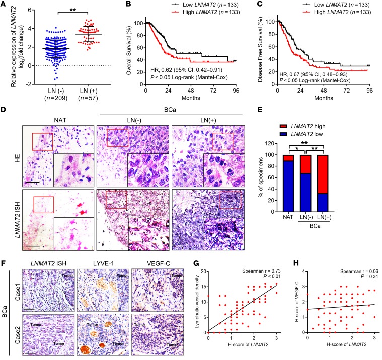
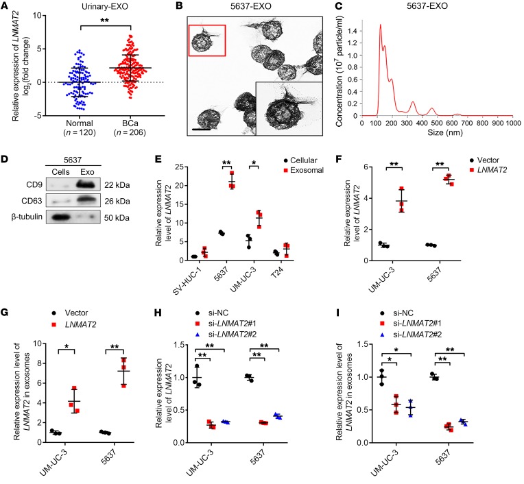
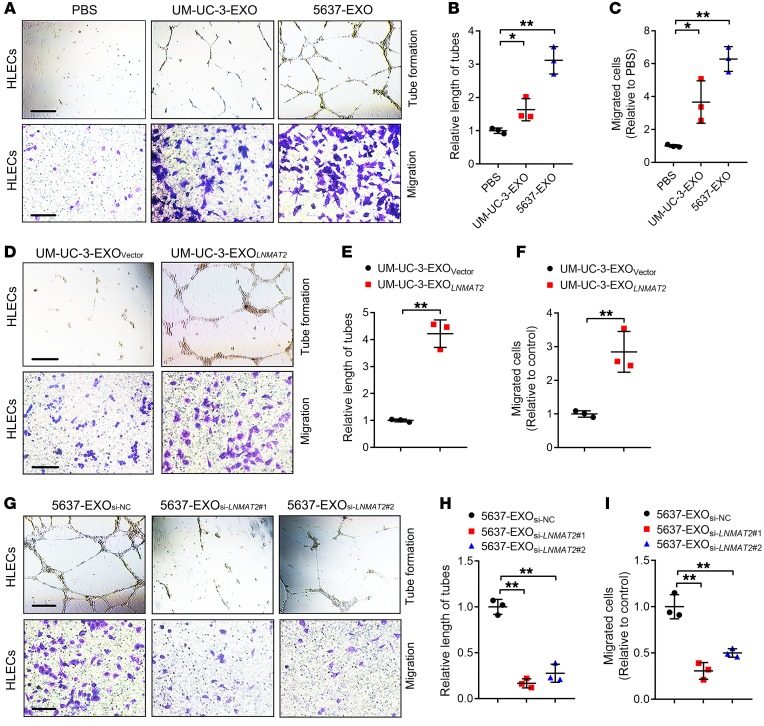
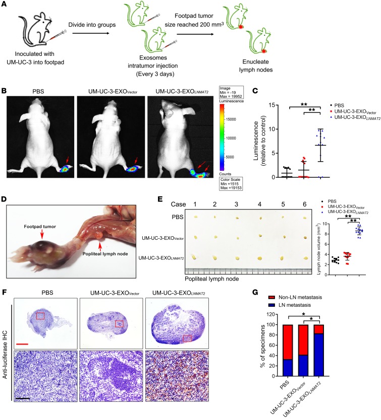
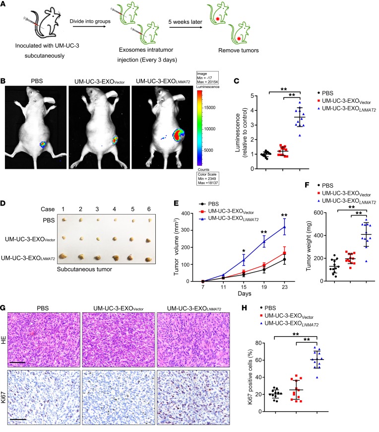
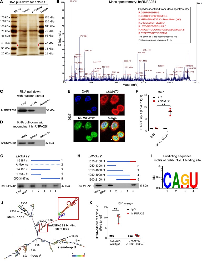
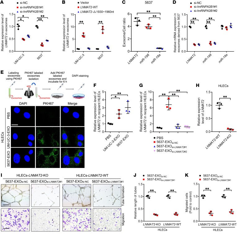
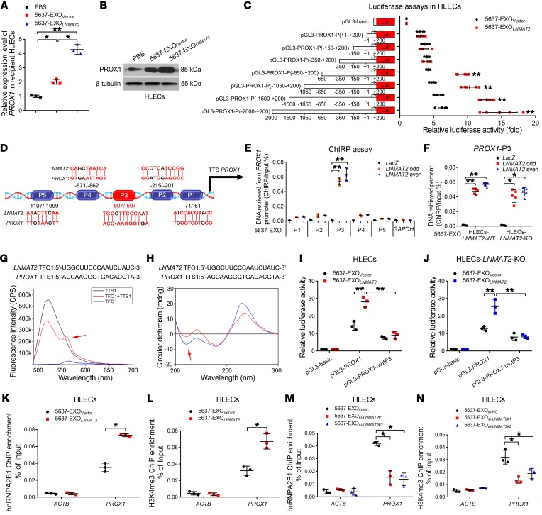
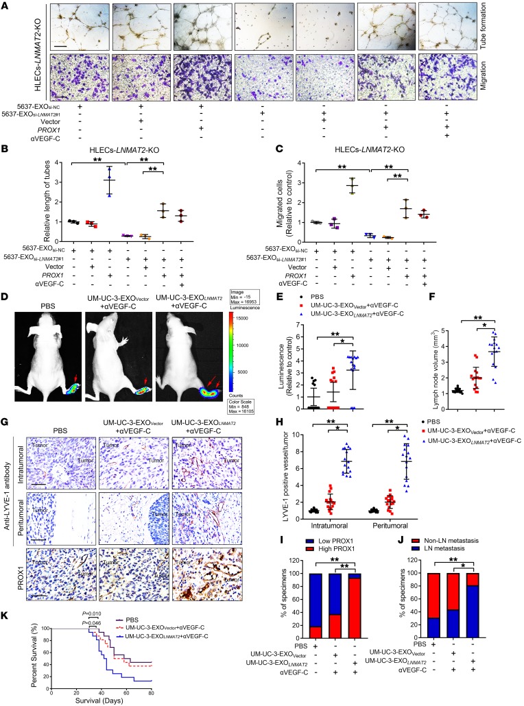
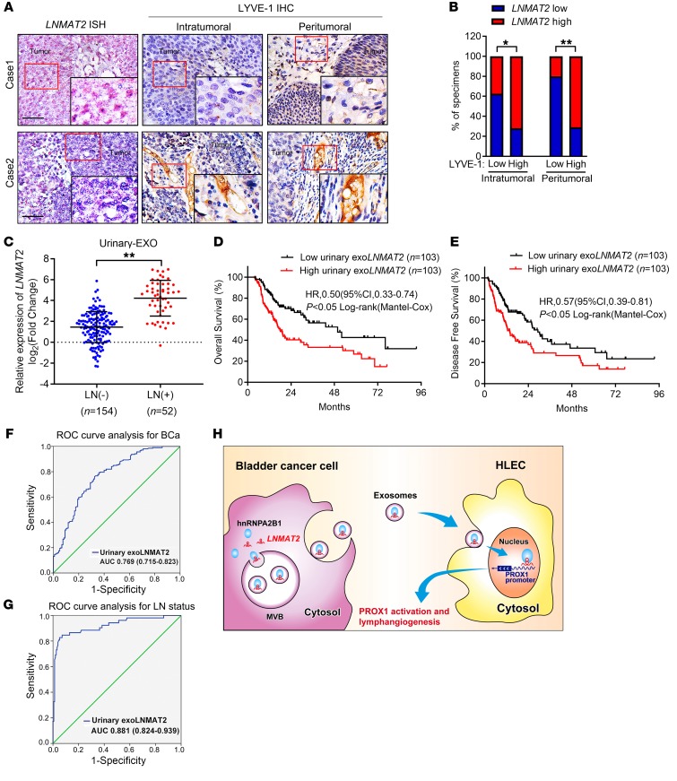
Patients with bladder cancer (BCa) with clinical lymph node (LN) metastasis have an extremely poor prognosis. VEGF-C has been demonstrated to play vital roles in LN metastasis in BCa. However, approximately 20% of BCa with LN metastasis exhibits low VEGF-C expression, suggesting a VEGF-C-independent mechanism for LN metastasis of BCa. Herein, we demonstrate that BCa cell-secreted exosome-mediated lymphangiogenesis promoted LN metastasis in BCa in a VEGF-C-independent manner. We identified an exosomal long noncoding RNA (lncRNA), termed lymph node metastasis-associated transcript 2 (LNMAT2), that stimulated human lymphatic endothelial cell (HLEC) tube formation and migration in vitro and enhanced tumor lymphangiogenesis and LN metastasis in vivo. Mechanistically, LNMAT2 was loaded to BCa cell-secreted exosomes by directly interacting with heterogeneous nuclear ribonucleoprotein A2B1 (hnRNPA2B1). Subsequently, exosomal LNMAT2 was internalized by HLECs and epigenetically upregulated prospero homeobox 1 (PROX1) expression by recruitment of hnRNPA2B1 and increasing the H3K4 trimethylation level in the PROX1 promoter, ultimately resulting in lymphangiogenesis and lymphatic metastasis. Therefore, our findings highlight a VEGF-C-independent mechanism of exosomal lncRNA-mediated LN metastasis and identify LNMAT2 as a therapeutic target for LN metastasis in BCa.

Keywords: Lymph; Noncoding RNAs; Oncology; Urology.


Conflict of interest statement

Conflict of interest: The authors have declared that no conflict of interest exists.

Figures

Figure 1
Figure 1. LNMAT2 overexpression is associated with BCa lymphatic metastasis.
(A) qRT-PCR analysis of LNMAT2 expression in a cohort of 266 BCa patients according to LN status. Groups were compared using the nonparametric Mann-Whitney U test. GAPDH was used as an internal control. The OS (B) and DFS (C) of patients with BCa with lower vs. higher LNMAT2 expression were estimated using Kaplan-Meier curves. The median expression was used as the cut-off value. Representative ISH images (D) and percentages (E) of LNMAT2 expression (blue) in paraffin-embedded NAT and BCa tissue with or without LN metastasis (n = 266). Scale bars: 50 μm. Statistical significance was assessed by χ2 test. Representative images (F) and correlation analysis (G and H) of ISH and IHC staining showing positive correlation between LNMAT2 expression and lymphatic vessel density indicated by anti–LYVE-1 staining, and that LNMAT2 expression was not correlated with VEGF-C levels in the BCa tissues (n = 266). Scale bars: 50 μm. *P < 0.05; **P < 0.01.
Figure 2
Figure 2. LNMAT2 is upregulated in BCa cell–secreted exosomes.
(A) qRT-PCR analysis of LNMAT2 expression in urinary-EXO from 206 patients with BCa and 120 healthy participants. GAPDH was used as an internal control. Groups were compared using the nonparametric Mann-Whitney U test. Purified 5637-EXO was identified by TEM (B) and NanoSight (C). Scale bars: 100 nm. (D) Western blot analysis of exosomal protein markers in 5637 cell lysates or 5637-EXO. (E) qRT-PCR analysis of LNMAT2 expression levels in bladder cell lines and in their corresponding exosomes. GAPDH was used as an internal control. Statistical significance was assessed using 1-way ANOVA followed by Dunnett’s tests. qRT-PCR analysis of LNMAT2 expression in LNMAT2-overexpressing control BCa cells (F) and their corresponding exosomes (G). GAPDH was used as an internal control. Statistical significance was assessed using 2-tailed Student’s t test followed by Dunnett’s tests for multiple comparisons. qRT-PCR detection of LNMAT2 expression in LNMAT2 knockdown control BCa cells (H) and their corresponding exosomes (I). GAPDH was used as an internal control. Statistical significance was assessed using 1-way ANOVA followed by Dunnett’s tests for multiple comparisons. Error bars represent the SD of 3 independent experiments. *P < 0.05; **P < 0.01.
Figure 3
Figure 3. Exosomal LNMAT2 promotes lymphangiogenesis in vitro.
Representative images (A) and quantification of tube formation (B) and Transwell migration (C) by HLECs treated with PBS, 5637-EXO, or UM-UC-3-EXO. Scale bars: 100 μm. Statistical significance was assessed using 1-way ANOVA followed by Dunnett’s tests. Representative images (D) and quantification of tube formation (E) and Transwell migration (F) by HLECs treated with UM-UC-3-EXOVector or UM-UC-3-EXOLNMAT2. Scale bars: 100 μm. Statistical significance was assessed using 2-tailed Student’s t test. Representative images (G) and quantification of tube formation (H) and Transwell migration (I) by HLECs treated with 5637-EXOsi-NC, 5637-EXOsi-LNMAT2#1, or 5637-EXOsi-LNMAT2#2. Scale bars: 100 μm. Statistical significance was assessed using 1-way ANOVA followed by Dunnett’s tests. Error bars represent the SD of 3 independent experiments. *P < 0.05; **P < 0.01.
Figure 4
Figure 4. Exosomal LNMAT2 promotes lymphatic metastasis in vivo.
(A) Schematic representation of establishment of the nude mouse model of popliteal LN metastasis. Representative bioluminescence images (B) and histogram analysis (C) of popliteal metastatic LN from nude mice treated with PBS, UM-UC-3-EXOVector, or UM-UC-3-EXOLNMAT2 after UM-UC-3 cells had been inoculated into the footpad (n = 12). Red arrow indicates footpad tumor and metastatic popliteal LN. Statistical significance was assessed using 1-way ANOVA followed by Dunnett’s tests. (D) Representative image of the popliteal LN metastasis model. (E) Representative images of enucleated popliteal LNs (left) and histogram analysis (right) of the LN volume of all groups (n = 12). Statistical significance was assessed using 1-way ANOVA followed by Dunnett’s tests. (F) Representative images of IHC staining with anti-luciferase antibody (n = 12). Scale bars: 500 μm (red) or 50 μm (black). (G) Percentages of LN status in all groups (n = 12). Statistical significance was assessed by χ2 test. Error bars represent the SD of 3 independent experiments. *P < 0.05; **P < 0.01.
Figure 5
Figure 5. Exosomal LNMAT2 promotes BCa tumorigenesis in vivo.
(A) Schematic representation of the establishment of the xenograft model. Representative bioluminescence images (B) and histogram analysis (C) of subcutaneous tumors from nude mice treated with PBS, UM-UC-3-EXOVector, or UM-UC-3-EXOLNMAT2 (n = 12). Statistical significance was assessed using 1-way ANOVA followed by Dunnett’s tests. (D) Representative images of gross appearance of subcutaneous tumors from nude mice treated with PBS, UM-UC-3-EXOVector, or UM-UC-3-EXOLNMAT2 (n = 12). Measured tumor volumes (E) and weights (F) (n = 12). Statistical significance was assessed using 1-way ANOVA followed by Dunnett’s tests. Representative images (G) and histogram analysis (H) of IHC staining for Ki67 expression (n = 12). Scale bars: 50 μm. Statistical significance was assessed using 1-way ANOVA followed by Dunnett’s tests. Error bars represent the SD of 3 independent experiments. *P < 0.05; **P < 0.01.
Figure 6
Figure 6. Direct interaction of LNMAT2 with hnRNPA2B1.
(A) RNA pull-down assay using LNMAT2 sense and antisense RNAs in 5637 cells, followed by silver staining. Red arrows indicate hnRNPA2B1. (B) MS identification of LNMAT2-binding proteins. RNA pull-down and Western blot with 5637 cell nuclear extract (C) or purified recombinant hnRNPA2B1 (D) confirmed that LNMAT2 was associated with hnRNPA2B1. (E) Fluorescence assessment of LNMAT2 and hnRNPA2B1 colocalization in 5637 cells. Scale bar: 5 μm. (F) RIP analysis using the anti-hnRNPA2B1 antibody revealing that LNMAT2 interacted with hnRNPA2B1 in 5637 cells. Negative control, IgG; nonspecific control, U1. Statistical significance was assessed using 2-tailed Student’s t test. (G and H) Serial deletions of LNMAT2 were used in RNA pull-down assays to identify regions required for LNMAT2 and hnRNPA2B1 interaction. (I) POSTAR2 prediction of sequence motifs of hnRNPA2B1 binding sites. (J) RNAalifold predicted that LNMAT2 would have 4 stable stem-loop structures. The inset (framed in red) indicates the hnRNPA2B1 binding stem-loop structures in LNMAT2. (K) RIP assays performed after site-directed mutagenesis of 1930–1960 nt of LNMAT2 in 5637 cells. Statistical significance was assessed using 2-tailed Student’s t test. Error bars represent the SD of 3 independent experiments. *P < 0.05; **P < 0.01.
Figure 7
Figure 7. LNMAT2 is packaged into exosomes in an hnRNPA2B1-dependent manner and transported to HLECs.
(A) qRT-PCR analysis of LNMAT2 expression in exosomes secreted by hnRNPA2B1 knockdown cells. Statistical significance was assessed using 1-way ANOVA followed by Dunnett’s tests. (B) qRT-PCR analysis of LNMAT2 expression in BCa cell–secreted exosomes. Statistical significance was assessed using 1-way ANOVA followed by Dunnett’s tests. (C) The exosome/cell ratio of RNAs in 5637 cells obtained by qRT-PCR. Statistical significance was assessed using 1-way ANOVA followed by Dunnett’s tests. (D) qRT-PCR analysis of RNA levels in exosomes secreted by hnRNPA2B1 knockdown 5637 cells. Statistical significance was assessed using 1-way ANOVA followed by Dunnett’s tests. (E) Schematic illustration of exosome internalization assays and representative images of HLEC fluorescence after incubation with PKH67-labeled (green) BCa cell exosomes. Scale bar: 5 μm. qRT-PCR analysis of LNMAT2 expression in HLECs treated with PBS, 5637-EXO, UM-UC-3-EXO (F) or 5637-EXOsi-NC, 5637-EXOsi-LNMAT2#1, or 5637-EXOsi-LNMAT2#2 (G). Statistical significance was assessed using 1-way ANOVA followed by Dunnett’s tests for multiple comparisons. (H) qRT-PCR confirming the LNMAT2 knockout. Statistical significance was assessed using 2-tailed Student’s t test. Representative images (I) and quantification of tube formation (J) and Transwell migration (K) by HLECs (LNMAT2-KO or LNMAT2-WT) after treating with 5637-EXOsi-NC or 5637-EXOsi-LNMAT2#1. Scale bars: 100 μm. Statistical significance was assessed using 2-tailed Student’s t test. GAPDH was used as an internal control for qRT-PCR analysis in AH. Error bars represent the SD of 3 independent experiments. *P < 0.05; **P < 0.01.
Figure 8
Figure 8. Exosomal LNMAT2 forms a DNA-RNA triplex with the PROX1 promoter.
qRT-PCR (A) and Western blot (B) of PROX1 expression in HLECs treated with PBS, 5637-EXOVector, or 5637-EXOLNMAT2. GAPDH was used as an internal control in qRT-PCR. Statistical significance was assessed using 1-way ANOVA followed by Dunnett’s tests. (C) Sequential deletions for evaluating the transcriptional activity of the PROX1 promoter in HLECs treated with 5637-EXOVector or 5637-EXOLNMAT2. Statistical significance was assessed using 1-way ANOVA followed by Dunnett’s tests. (D) Schematic presentation of the predicted LNMAT2 binding sites in the PROX1 promoter. (E) ChIRP of LNMAT2-associated chromatin in HLECs treated with 5637-EXO. Statistical significance was assessed using 2-tailed Student’s t test. (F) ChIRP of LNMAT2-associated chromatin in LNMAT2-WT or LNMAT2-KO HLECs treated with 5637-EXO. Statistical significance was assessed using 2-tailed Student’s t test. (G) FRET of a 1:5 mixture (red) of TFO (black) in LNMAT2 with TTS (blue) in the PROX1 promoter. (H) CD spectrum of a 1:1 mixture of TFO in LNMAT2 with TTS in the PROX1 promoter (red). The sum of the TFO and TTS is shown in blue. Evaluation of WT or LNMAT2 binding site mutated PROX1 promoter in LNMAT2-WT (I) or LNMAT2-KO (J) HLECs, respectively, treated with 5637-EXOVector or 5637-EXOLNMAT2. Statistical significance was assessed using 1-way ANOVA followed by Dunnett’s tests. ChIP-qPCR of hnRNPA2B1 occupancy (K and M) and H3K4me3 (L and N) status in the PROX1 promoter after HLEC incubation with indicated exosomes. Statistical significance was assessed using 2-tailed Student’s t test and 1-way ANOVA followed by Dunnett’s tests for multiple comparisons. Error bars represent the SD of 3 independent experiments. *P < 0.05; **P < 0.01.
Figure 9
Figure 9. PROX1 is required for exosomal LNMAT2-mediated lymphangiogenesis.
Representative images (A) and histogram analysis of tube formation (B) and Transwell migration (C) by LNMAT2-KO HLECs treated with 5637-EXOsi-NC or 5637-EXOsi-LNMAT2#1, transfected with vector or PROX1 plasmid, or in which VEGF-C was inhibited. Scale bars: 100 μm. Statistical significance was assessed using 1-way ANOVA followed by Dunnett’s tests. Representative bioluminescence images (D) and histogram analysis (E) of popliteal metastatic LN from nude mice treated with PBS, UM-UC-3-EXOVector, or UM-UC-3-EXOLNMAT2 with or without coinjection of VEGF-C neutralizing antibody after UM-UC-3 cells had been inoculated into the footpad (n = 16). Red arrow indicates footpad tumor and metastatic popliteal LN. Statistical significance was assessed using 1-way ANOVA followed by Dunnett’s tests. (F) Histogram analysis of the LN volume (n = 16). Statistical significance was assessed using 1-way ANOVA followed by Dunnett’s tests. Representative images (G) and histogram analysis of IHC staining evaluating lymphatic vessel density based on LYVE-1 (H) and PROX1 expression (I) in footpad tumors (n = 16). Scale bars: 50 μm. Statistical significance was assessed using 1-way ANOVA followed by Dunnett’s tests and the χ2 test. (J) The percentages of LN status in all groups (n = 16). Statistical significance was assessed by χ2 test. (K) Kaplan-Meier survival curve for control, UM-UC-3-EXOVector, or UM-UC-3-EXOLNMAT2 groups with or without inhibition of VEGF-C (n = 16). Error bars represent the SD of 3 independent experiments. *P < 0.05; **P < 0.01.
Figure 10
Figure 10. Exosomal LNMAT2 is associated with BCa lymphatic metastasis.
Representative images (A) and percentages (B) of BCa tissues (n = 206) with high and low LYVE-1 levels in the intratumoral and peritumoral lymphatic vessels in patients with different expression of LNMAT2. Scale bars: 50 μm. Statistical significance was assessed by χ2 test. (C) qRT-PCR analysis of LNMAT2 expression in a 206-patient cohort of urinary-EXO from BCa patients with or without LN metastasis. GAPDH was used as an internal control. Groups were compared using the nonparametric Mann-Whitney U test. Kaplan-Meier curves of OS (D) and DFS (E) of patients with BCa according to the relative urinary exosomal LNMAT2 expression. The median expression was used as the cut-off value (n = 206). ROC curve analyses for evaluating the diagnostic potential of urinary exosomal LNMAT2 for BCa (F) and LN metastasis (G). (H) Proposed model of BCa cell–secreted exosomal LNMAT2-mediated PROX1 activation in HLECs for promoting lymphangiogenesis and LN metastasis. *P < 0.05; **P < 0.01.

Similar articles

Cited by

References

    1. Bray F, Ferlay J, Soerjomataram I, Siegel RL, Torre LA, Jemal A. Global cancer statistics 2018: GLOBOCAN estimates of incidence and mortality worldwide for 36 cancers in 185 countries. CA Cancer J Clin. 2018;68(6):394–424. doi: 10.3322/caac.21492. - DOI - PubMed
    1. Hautmann RE, de Petriconi RC, Pfeiffer C, Volkmer BG. Radical cystectomy for urothelial carcinoma of the bladder without neoadjuvant or adjuvant therapy: long-term results in 1100 patients. Eur Urol. 2012;61(5):1039–1047. doi: 10.1016/j.eururo.2012.02.028. - DOI - PubMed
    1. He W, et al. Long noncoding RNA BLACAT2 promotes bladder cancer-associated lymphangiogenesis and lymphatic metastasis. J Clin Invest. 2018;128(2):861–875. doi: 10.1172/JCI96218. - DOI - PMC - PubMed
    1. Chen C, et al. LNMAT1 promotes lymphatic metastasis of bladder cancer via CCL2 dependent macrophage recruitment. Nat Commun. 2018;9(1):3826. doi: 10.1038/s41467-018-06152-x. - DOI - PMC - PubMed
    1. Zu X, Tang Z, Li Y, Gao N, Ding J, Qi L. Vascular endothelial growth factor-C expression in bladder transitional cell cancer and its relationship to lymph node metastasis. BJU Int. 2006;98(5):1090–1093. doi: 10.1111/j.1464-410X.2006.06446.x. - DOI - PubMed

Publication types

MeSH terms