. 2020 Feb 20;22(2):253-266.
doi: 10.1093/neuonc/noz189.

CCL5 of glioma-associated microglia/macrophages regulates glioma migration and invasion via calcium-dependent matrix metalloproteinase 2

Affiliations

CCL5 of glioma-associated microglia/macrophages regulates glioma migration and invasion via calcium-dependent matrix metalloproteinase 2

Caren Yu-Ju Wu et al. Neuro Oncol. .

Abstract

Background: Glioma-associated microglia/macrophages (GAMs) comprise macrophages of peripheral origin and brain-intrinsic microglia, which support tumor progression. Chemokine C-C ligand 5 (CCL5) is an inflammatory mediator produced by immune cells and is involved in tumor growth and migration in several cancers, including glioma. However, the mechanisms detailing how CCL5 facilitates glioma invasion remain largely unresolved.

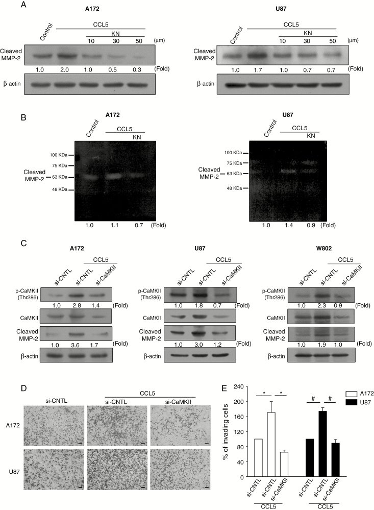
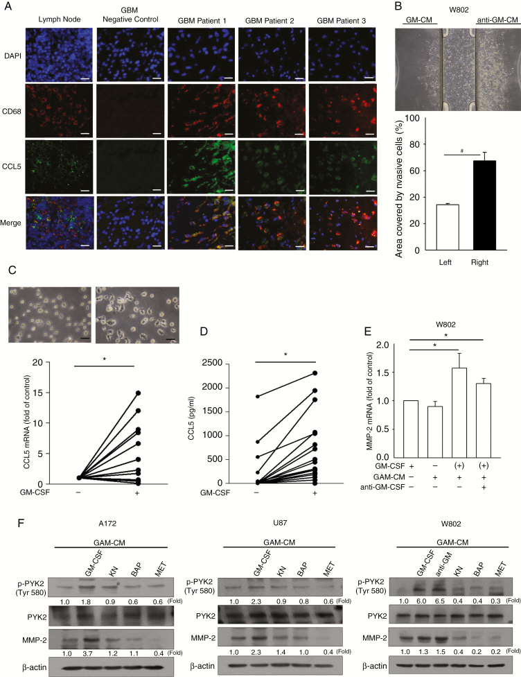
Methods: Glioma migration and invasion were determined by wound healing, transwell assay, and 3D µ-slide chemotaxis assay. The expression levels of CCL5, CD68, matrix metalloproteinase 2 (MMP2), phosphorylated Ca2+/calmodulin-dependent protein kinase II (p-CaMKII), p-Akt, and phosphorylated proline-rich tyrosine kinase 2 were determined by cytokine array, quantitative PCR, western blot, or immunohistochemistry. Zymography and intracellular calcium assays were used to analyze MMP2 activity and intracellular calcium levels, respectively.

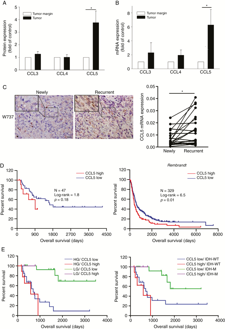
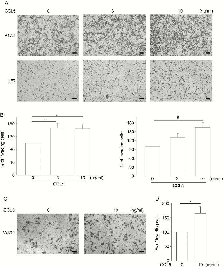
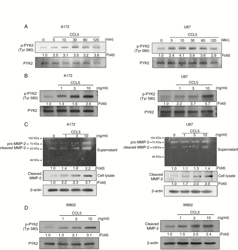
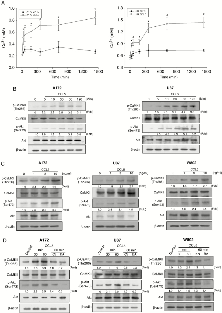
Results: CCL5 modulated the migratory and invasive activities of human glioma cells in association with MMP2 expression. In response to CCL5, glioma cells underwent a synchronized increase in intracellular calcium levels and p-CaMKII and p-Akt expression levels. CCL5-directed glioma invasion and increases in MMP2 were suppressed after inhibition of p-CaMKII. Glioma cells tended to migrate toward GAM-conditioned media activated by granulocyte-macrophage colony-stimulating factor (GM-CSF) in which CCL5 was abundant. This homing effect was associated with MMP2 upregulation, and could be ameliorated either by controlling intracellular and extracellular calcium levels or by CCL5 antagonism. Clinical results also revealed the associations between CCL5 and GAM activation.

Conclusion: Our results suggest that modulation of glioma CaMKII may restrict the effect of CCL5 on glioma invasion and could be a potential therapeutic target for alleviating glioma growth.

Keywords: CCL5; MMP2; glioma; invasion; microglia.


Figures

Fig. 1.
Fig. 1.
Survival analysis results for CCL5 in newly diagnosed glioma. (A) CCL5 expression quantified from 4 newly diagnosed GBM tissues. (B) The CCL5 mRNA expression of newly diagnosed gliomas (n = 15) relative to the tumor margin was determined. (C) IHC staining of CCL5 (brown) in paired newly diagnosed and recurrent GBM tumor sections. CCL5 mRNA identified in paired newly diagnosed (n = 20) and recurrent GBM (n = 20) tumors. (D) Kaplan–Meier survival plots for glioma patients with a low CCL5 level (low = 12, high = 35) showed a longer OS. Using the REMBRANDT database, Kaplan–Meier survival plots confirmed a survival advantage in the patients with a low CCL5 level (low = 83, high = 246). (E) Glioma patients (n = 47) were stratified based on CCL5, grading, and IDH status. LG: low-grade; HG: high-grade; IDH-M: mutant IDH. Bar: 100 µm. All data are presented as means ± SE. *P < 0.05, #P < 0.01.
Fig. 2.
Fig. 2.
CCL5 promotes heterogeneous glioma invasive activity. (A, C) The effect of CCL5 on glioma invasion was analyzed. The numbers of invading cells were increased in CCL5-treated A172, U87, and W802 cells after 24 h. (B, D) The number of invading cells are presented as percent of invading cells. Bar: 100 µm. All data are presented as means ± SE. *P < 0.05, #P < 0.01.
Fig. 3.
Fig. 3.
CCL5 induces PYK2 phosphorylation and MMP2 activation in glioma cells. (A) PYK2 phosphorylation in A172 and U87 cells was induced by CCL5 (10 ng/mL) in a time-dependent manner. (B) CCL5 (0–10 ng/mL) induced a concentration-dependent increase in PYK2 phosphorylation in A172 and U87 cells. (C) MMP2 protease activity and protein expression were induced by 24 h of CCL5 treatment (10 ng/mL) in both A172 and U87 cells. (D) CCL5 (0–10 ng/mL) induced an increase in PYK2 phosphorylation and cleaved MMP2 expression in W802 cells. Blots are quantified as fold of control.
Fig. 4.
Fig. 4.
CCL5 induces a calcium-dependent signaling pathway in a concentration- and time-dependent manner in glioma cells. (A) CCL5 (10 ng/mL) induced an increase in intracellular calcium from 60 to 1440 min in A172 and U87 cells. (B) CCL5 (10 ng/mL) induced CaMKII and Akt phosphorylation over the indicated time periods. (C) CaMKII and Akt phosphorylation was increased in a concentration-dependent manner by CCL5 (0–10 ng/mL). (D) CaMKII and Akt phosphorylation was determined after cells pretreated with KN (20 µM) or BAP (20 µM) followed by CCL5 (10 ng/mL) treatment. The data are presented as means ± SE. *P < 0.05, #P < 0.01 compared with control. Blots are quantified as fold of control.
Fig. 5.
Fig. 5.
Inhibition of CaMKII phosphorylation downregulates CCL5-induced MMP2 expression. (A, B) Cells pretreated with KN (10–50 µM) followed by CCL5 treatment showed inhibition of cleaved MMP2 expression. (C) Glioma cells transfected with CaMKII siRNA followed by CCL5 (10 ng/mL) treatment showed inhibited CaMKII phosphorylation and cleaved MMP2 protein expression. (D) Invasive activity was inhibited in the glioma cells with si-CaMKII followed by CCL5 (10 ng/mL) treatment. (E) The invading cells in (D) were quantified and presented as percent of invading cells. Bar: 100 µm. The data are presented as means ± SE. *P < 0.05, #P < 0.01. Blots are quantified as fold of control.
Fig. 6.
Fig. 6.
The increased MMP2 expression by GM-CM is suppressed by inhibition of calcium-related signaling pathways. (A) IHC staining of CD68 (red), CCL5 (green), and DAPI (blue) in newly diagnosed GBM tumor sections (n = 3). (B) W802 cells invaded toward the reservoir containing anti–GM-CM. Percentages of areas covered by invading W802 cells are shown. (C) Representative images of GAMs stimulated with or without GM-CSF, which showed that the CCL5 mRNA level was increased in GM-CSF–stimulated GAMs (n = 21). (D) The amounts of CCL5 in CM and GM-CM were Figure 6 Continued.determined (n = 21). (E) MMP2 mRNA expression in glioma cells was determined after 24 h of treatment with CM or GM-CM. (F) Glioma cells pretreated with KN, BAP, or MET prior to GM-CM or CM treatment showed downregulation of PYK2 phosphorylation and cleaved MMP2 protein expression. “(+)” represents CM collected from GM-CSF–activated GAMs. Bar: 100 µm. All data are presented as means ± SE. *P < 0.05, #P < 0.01. Blots are quantified as fold of control.

References

    1. Hsieh WT, Yeh WL, Cheng RY, et al. . Exogenous endothelin-1 induces cell migration and matrix metalloproteinase expression in U251 human glioblastoma multiforme. J Neurooncol. 2014;118(2):257–269. - PubMed
    1. Chen J, Li Y, Yu TS, et al. . A restricted cell population propagates glioblastoma growth after chemotherapy. Nature. 2012;488(7412):522–526. - PMC - PubMed
    1. Osswald M, Jung E, Sahm F, et al. . Brain tumour cells interconnect to a functional and resistant network. Nature. 2015;528(7580):93–98. - PubMed
    1. Galvao RP, Kasina A, McNeill RS, et al. . Transformation of quiescent adult oligodendrocyte precursor cells into malignant glioma through a multistep reactivation process. Proc Natl Acad Sci U S A. 2014;111(40):E4214–E4223. - PMC - PubMed
    1. Hambardzumyan D, Gutmann DH, Kettenmann H. The role of microglia and macrophages in glioma maintenance and progression. Nat Neurosci. 2016;19(1):20–27. - PMC - PubMed

Publication types

MeSH terms