

Near infrared light examination as part of the management of sporadic pancreatic head insulinoma: Case report
- PMID: 31593916
- PMCID: PMC6796722
- DOI: 10.1016/j.ijscr.2019.09.024
Near infrared light examination as part of the management of sporadic pancreatic head insulinoma: Case report
Abstract
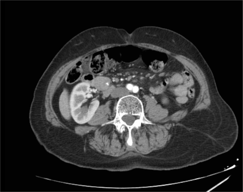
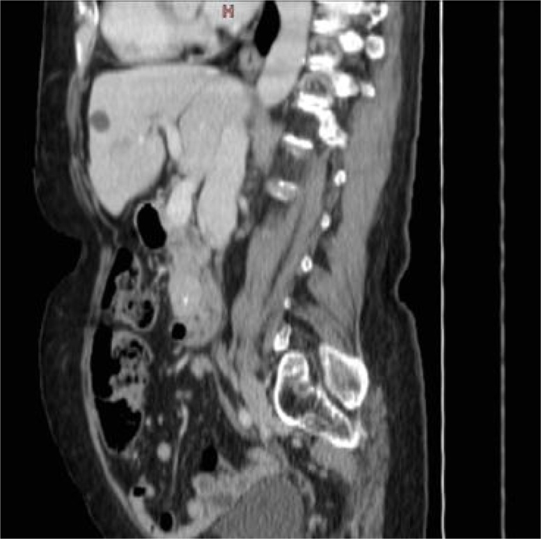
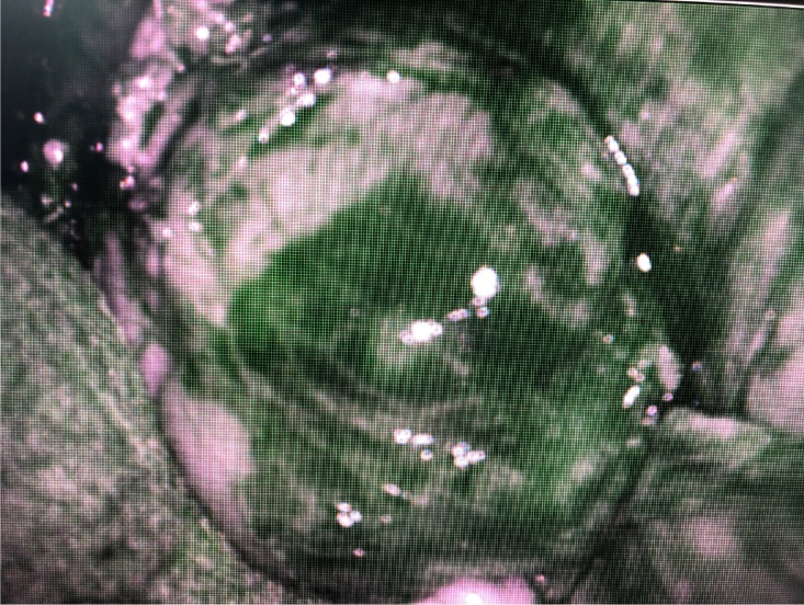
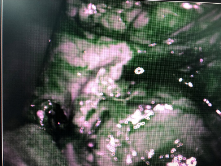
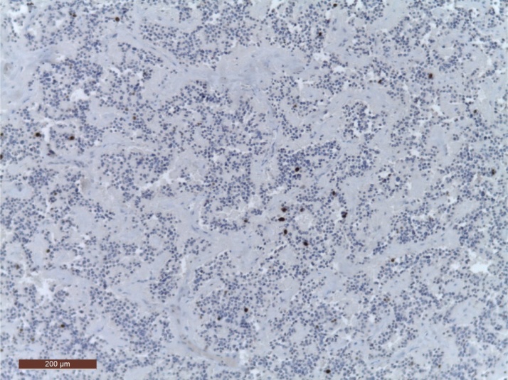
Introduction: We report the case of a 77-year-old female patient with the diagnosis of pancreatic head insulinoma, in whom we used near infrared light (NIR) to detect synchronous pancreatic tumors and potential secondary lymph node or liver involvement. The patient presented with hypoglycemia manifesting by lipothymia. With the diagnosis of secretory neuroendocrine tumor (insulinoma) of the pancreatic head, cephalic pancreatoduodenectomy with the preservation of the pylorus was performed after NIR visualization of the pancreatic tumor mass. At 6, 12, 18 months postoperatively, the patient no longer had hypoglycemia and her general state was good.
Conclusion: NIR with indocyanine green (ICG) evidences pancreatic neuroendocrine tumors, as well as possible synchronous tumors and secondary lymph node or liver involvement.
Keywords: Case report; Indocyanine green; Insulinoma; Near infrared light; Pancreatic neuroendocrine tumors.
Copyright © 2019 The Authors. Published by Elsevier Ltd.. All rights reserved.
Conflict of interest statement
None of the authors have any conflict of interest.
Figures
References
-
- Deguelte S., Mestier L., Hentic O., Cros J., Lebtahi R., Hammel P., Kianmanesh R. Preoperativ imaging and pathologic classifiaction for pancreatic neuroendocrine tumors. J. Visc. Surg. 2018;155:117–125. - PubMed
-
- Agha R.A., Borrelli M.R., Farwana R., Koshy K., Fowler A., Orgill D.P., For the SCARE Group The SCARE 2018 statement: updating consensus Surgical CAse REport (SCARE) guidelines. Int. J. Surg. 2018;60:132–136. - PubMed
-
- Shirata C., Kawaguchi Y., Kobayashi K. Usefulness of indocyanine green-fluorescence imaging for real-time visualization of pancreas neuroendocrine tumor and cystic neoplasm. J. Surg. Oncol. 2018;118:1012–1020. - PubMed
-
- TNCD: The Version Finale 2016. http://snfge.org/sites/default/files/SNFGE/TNCD/tncdtnefinale10-3-16.pdf.
LinkOut - more resources
Full Text Sources
Miscellaneous
