

Application of optical coherence tomography in clinical diagnosis
- PMID: 31594279
- PMCID: PMC7029333
- DOI: 10.3233/XST-190559
Application of optical coherence tomography in clinical diagnosis
Abstract
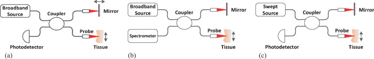
Background: Optical coherence tomography (OCT) is a non-invasive diagnosing tool used in clinics. Due to its high resolution (<10um), it is appropriate for the early detection of tiny infections. It has been widely used in diagnosis and treatment of diseases, evaluation of therapeutic efficacy, and monitoring of various physiological and pathological processes.
Objective: To systemically review literature to summarize the clinic application of OCT in recent years.
Methods: For clinic applications that OCT has been applied, we selected studies that describe the most relevant works. The discussion included: 1) which tissue could be used in the OCT detection, 2) which character of different tissue could be used as diagnosing criteria, 3) which diseases and pathological process have been diagnosed or monitored using OCT imaging, and 4) the recent development of clinic OCT diagnosing.
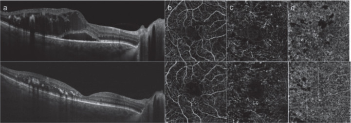
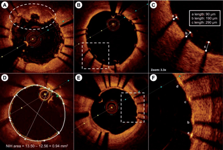
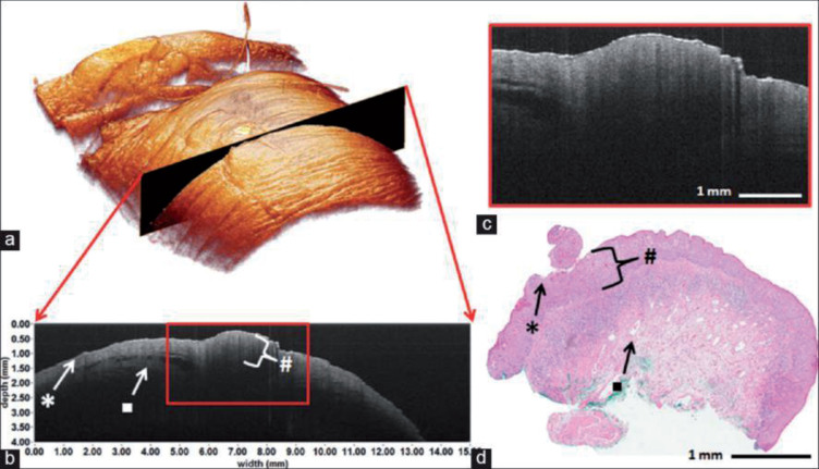
Results: The literature showed that the OCT had been listed as a routine test choice for ophthalmic diseases, while the first commercial product for cardiovascular OCT detection had gotten clearance. Meanwhile, as the development of commercial benchtop OCT equipment and tiny fiber probe, the commercial application of OCT in dermatology, dentistry, gastroenterology and urology also had great potential in the near future.
Conclusions: The analysis and discussions showed that OCT, as an optical diagnosing method, has been used successfully in many clinical fields, and has the potential to be a standard inspection method in several clinic fields, such as dermatology, dentistry and cardiovascular.
Keywords: Optical coherence tomography (OCT); clinical application of OCT.
Figures
References
-
- Pan Y., Birngruber R., Rosperich J., et al., Low-coherence optical tomography in turbid tissue: Theoretical analysis, Applied Optics 34 (1995), 6564. - PubMed
-
- Leitgeb R., Wojtkowski M., Kowalczyk A., et al., Spectral measurement of absorption by spectroscopic frequency-domain optical coherence tomography, Optics Letters 25 (2000), 820–822. - PubMed
Publication types
MeSH terms
LinkOut - more resources
Full Text Sources
