. 2019 Oct 8;10(5):e02169-19.
doi: 10.1128/mBio.02169-19.

A Genome-Wide Knockout Screen in Human Macrophages Identified Host Factors Modulating Salmonella Infection

Affiliations

A Genome-Wide Knockout Screen in Human Macrophages Identified Host Factors Modulating Salmonella Infection

Amy T Y Yeung et al. mBio. .

Abstract

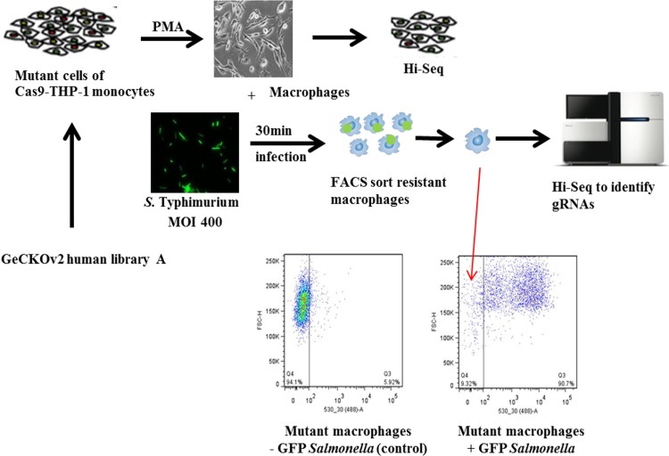
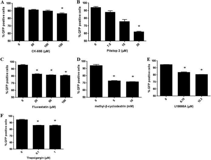
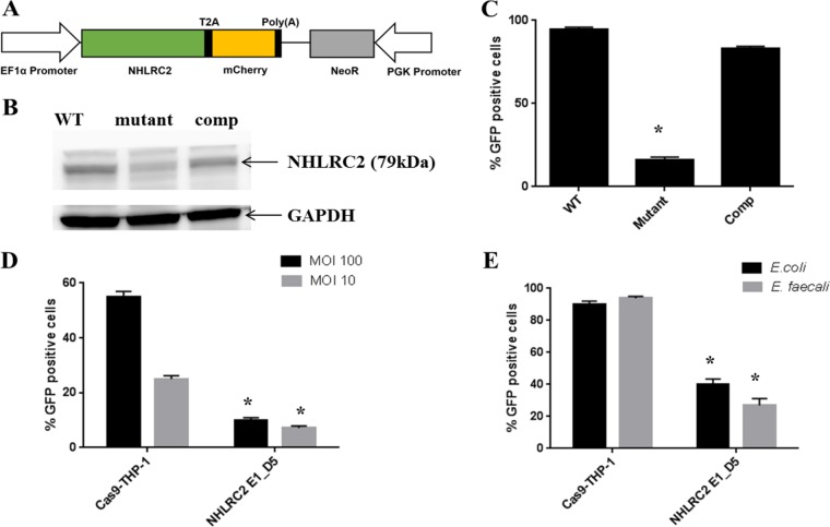
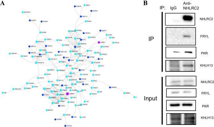
A genome-scale CRISPR knockout library screen of THP-1 human macrophages was performed to identify loss-of-function mutations conferring resistance to Salmonella uptake. The screen identified 183 candidate genes, from which 14 representative genes involved in actin dynamics (ACTR3, ARPC4, CAPZB, TOR3A, CYFIP2, CTTN, and NHLRC2), glycosaminoglycan metabolism (B3GNT1), receptor signaling (PDGFB and CD27), lipid raft formation (CLTCL1), calcium transport (ATP2A2 and ITPR3), and cholesterol metabolism (HMGCR) were analyzed further. For some of these pathways, known chemical inhibitors could replicate the Salmonella resistance phenotype, indicating their potential as targets for host-directed therapy. The screen indicated a role for the relatively uncharacterized gene NHLRC2 in both Salmonella invasion and macrophage differentiation. Upon differentiation, NHLRC2 mutant macrophages were hyperinflammatory and did not exhibit characteristics typical of macrophages, including atypical morphology and inability to interact and phagocytose bacteria/particles. Immunoprecipitation confirmed an interaction of NHLRC2 with FRYL, EIF2AK2, and KLHL13.IMPORTANCESalmonella exploits macrophages to gain access to the lymphatic system and bloodstream to lead to local and potentially systemic infections. With an increasing number of antibiotic-resistant isolates identified in humans, Salmonella infections have become major threats to public health. Therefore, there is an urgent need to identify alternative approaches to anti-infective therapy, including host-directed therapies. In this study, we used a simple genome-wide screen to identify 183 candidate host factors in macrophages that can confer resistance to Salmonella infection. These factors may be potential therapeutic targets against Salmonella infections.

Keywords: CRISPR; Salmonella; bacteria; genome-wide screen; macrophages.


Figures

FIG 1
FIG 1
Schematic of a CRISPR/Cas9 screen setup to identify host factors involved in early Salmonella-macrophage interaction. Cas9-THP-1 monocytes were transduced with GeCKO human pooled gRNA library A at an MOI of 0.3 and at 100× coverage. The mutant library cells were selected in puromycin for 2 weeks. After drug selection, genomic DNA was extracted from 1 × 107 mutant cells. Genomic DNA was used as the template for PCR and barcoding for Hi-Seq to evaluate sgRNA coverage in the THP-1-GeCKO library. The remaining mutant cells were differentiated into macrophages using PMA, and genomic DNAs were extracted from a subset of the mutant macrophages. S. Typhimurium expressing constitutive GFP was used to infect the mutant library macrophages for 30 min. The infected cells were washed and sorted on a flow cytometer for GFP-negative macrophages. Genomic DNA was extracted from the resultant sorted cells and used as the template for PCR and barcoding for Hi-Seq to identify enriched gRNAs. Three biological replicates were performed.
FIG 2
FIG 2
Interaction network of Salmonella-resistant mutants and relative uptake of Salmonella for mutant versus WT THP-1 macrophages. (A) One hundred eighty-three candidate genes with Salmonella-resistant phenotypes (nodes highlighted in blue, with NHLRC2 highlighted in magenta) were submitted to NetworkAnalyst to identify known first-order protein-protein interactors (gray nodes) using the IMEx Interactome database. The observed protein-protein interaction network indicates functional interactions between the individual proteins that when deleted demonstrate Salmonella resistance. (B) Selected homozygous, compound heterozygous, or heterozygous clonal mutants, generated from the same gRNA, that displayed the strongest Salmonella-resistant phenotypes are shown. The mutant and WT Cas9-THP-1 macrophages were infected with S. Typhimurium constitutively expressing GFP for 30 min at an MOI of 400. Infected macrophages were washed, and GFP was measured using a flow cytometer. Results are averages from at least 3 independent experiments. For the labeling of each mutant cell line, the name of the gene is followed by the gRNA used (Table S3A) and the position of the clone picked.
FIG 3
FIG 3
Effect of inhibitors on S. Typhimurium infection of THP-1 macrophages. THP-1 macrophages were infected with S. Typhimurium constitutively expressing GFP at an MOI of 400 in the presence or absence of the indicated inhibitor for 30 min. Postinfection, the cells were washed, and GFP was measured using a flow cytometer. Results are the average of 3 independent experiments ± standard deviation (SD). * indicates statistically significant difference (P < 0.05) between untreated and treated as determined using Student's t test.
FIG 4
FIG 4
NHLRC2 complementation vector and S. Typhimurium infection of WT and NHLRC2 mutant THP-1 macrophages. (A) Schematic view of NHLRC2 complementation vector comprised of EF1α promoter, human NHLRC2 open reading frame (ORF) clone, T2A-mCherry, neomycin-resistant gene, and a PGK promoter. (B) WT, NHLRC2_E1_D5 mutant, and NHLRC2 complemented mutant samples were probed with anti-NHLRC2 primary antibody (HPA038493) and horseradish peroxidase (HRP)-conjugated anti-rabbit IgG secondary antibody. The resultant bands were visualized on an ImageQuant Las 4000. Anti-GAPDH antibody was used as a loading control. (C and D) WT, NHLRC2_E1_D5 mutant, and NHLRC2 complemented macrophages were infected with S. Typhimurium constitutively expressing GFP for 30 min at MOI of (C) 400 and (D) 100 or 10. (E) WT and NHLRC2_E1_D5 mutant macrophages were infected with carboxyfluorescein succinimidyl ester (CFSE)-stained E. coli or E. faecalis for 30 min at an MOI of 400. Uninfected macrophages were used as a control. Postinfection, the cells were washed, and GFP was measured using a flow cytometer. The results shown are the average of 3 independent experiments ± SD. * indicates a statistically significant difference (P < 0.05) between the WT and NHLRC2 mutant as determined using Student's t test.
FIG 5
FIG 5
Characterization of WT and NHLRC2 mutant macrophages. (A) Cell size comparison for WT and NHLRC2 mutant macrophages. Phase-contrast microscopy at a resolution of ×20 magnification was used to image WT and NHLRC2_E1_D5 macrophages. Results are the average of 50 cells ± SD. * indicates statistically significant difference (P < 0.05) between the WT and each clonal mutant as determined using Student's t test. (B and C) Representative SEM images of (B) naive and (C) S. Typhimurium-infected WT, NHLRC2 mutant, and NHLRC2 complemented (comp) macrophages.
FIG 6
FIG 6
Evaluation of cell surface markers and inflammatory cytokines produced by WT and NHLRC2 mutant macrophages. (A) Expression of selected surface markers on WT and NHLRC2 mutant macrophages was determined using a flow cytometer. Results shown are the mean percentages of fold changes from 3 independent experiments ± SD. (B) Expression of TNF-α, IL-6, and IL-1β in supernatants of WT and NHLRC2 mutant macrophages stimulated with LPS, flagellin, or live S. Typhimurium cells was determined using a flow cytometer. The results shown are the averages from 3 independent experiments ± SD.
FIG 7
FIG 7
Protein-protein interaction networks and co-IP of NHLRC2 and its interactors. (A) NetworkAnalyst was utilized to construct a zero-order (direct interactors only) protein-protein interaction (based on the InnateDB database) network from 82 of the 191 proteins (turquoise nodes) identified to interact with NHLRC2 and 30 of the 183 host factors (dark blue) identified by CRISPR/Cas9 to influence Salmonella infection of macrophages. There were 3 overlapping proteins common to both data sets (NHLRC2, PSMA3, and LAMP1 [magenta nodes]); NHLRC2 does not appear but interacts with all turquoise nodes shown. (B) A co-IP experiment was performed to confirm protein interactors of NHLRC2 identified from the IP-MS experiment. IgG antibody was used as a control.

Similar articles

Cited by

References

    1. Eng S-K, Pusparajah P, Ab Mutalib N-S, Ser H-L, Chan K-G, Lee L-H. 2015. Salmonella: a review on pathogenesis, epidemiology and antibiotic resistance. Front Life Sci 8:284–293. doi:10.1080/21553769.2015.1051243. - DOI
    1. Crump JA, Sjölund-Karlsson M, Gordon MA, Parry CM. 2015. Epidemiology, clinical presentation, laboratory diagnosis, antimicrobial resistance, and antimicrobial management of invasive Salmonella infections. Clin Microbiol Rev 28:901–937. doi:10.1128/CMR.00002-15. - DOI - PMC - PubMed
    1. Karkey A, Thwaites GE, Baker S. 2018. The evolution of antimicrobial resistance in Salmonella Typhi. Curr Opin Gastroenterol 34:25–30. doi:10.1097/MOG.0000000000000406. - DOI - PubMed
    1. Watson KG, Holden DW. 2010. Dynamics of growth and dissemination of Salmonella in vivo. Cell Microbiol 12:1389–1397. doi:10.1111/j.1462-5822.2010.01511.x. - DOI - PubMed
    1. Ruby T, McLaughlin L, Gopinath S, Monack D. 2012. Salmonella’s long-term relationship with its host. FEMS Microbiol Rev 36:600–615. doi:10.1111/j.1574-6976.2012.00332.x. - DOI - PubMed

Publication types

Substances