. 2020 Jan;30(1):5-20.
doi: 10.1038/s41422-019-0236-6. Epub 2019 Oct 8.

UFMylation of RPL26 links translocation-associated quality control to endoplasmic reticulum protein homeostasis

Affiliations

UFMylation of RPL26 links translocation-associated quality control to endoplasmic reticulum protein homeostasis

Lihui Wang et al. Cell Res. 2020 Jan.

Abstract

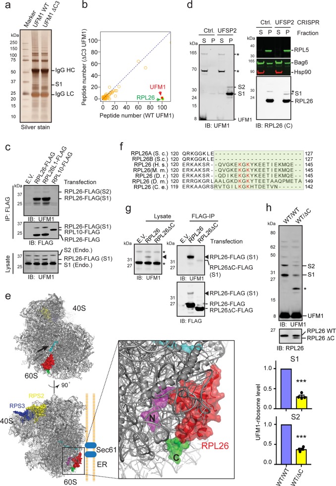
Protein biogenesis at the endoplasmic reticulum (ER) in eukaryotic cells is monitored by a protein quality control system named ER-associated protein degradation (ERAD). While there has been substantial progress in understanding how ERAD eliminates defective polypeptides generated from erroneous folding, how cells remove nascent chains stalled in the translocon during co-translational protein insertion into the ER is unclear. Here we show that ribosome stalling during protein translocation induces the attachment of UFM1, a ubiquitin-like modifier, to two conserved lysine residues near the COOH-terminus of the 60S ribosomal subunit RPL26 (uL24) at the ER. Strikingly, RPL26 UFMylation enables the degradation of stalled nascent chains, but unlike ERAD or previously established cytosolic ribosome-associated quality control (RQC), which uses proteasome to degrade their client proteins, ribosome UFMylation promotes the targeting of a translocation-arrested ER protein to lysosomes for degradation. RPL26 UFMylation is upregulated during erythroid differentiation to cope with increased secretory flow, and compromising UFMylation impairs protein secretion, and ultimately hemoglobin production. We propose that in metazoan, co-translational protein translocation into the ER is safeguarded by a UFMylation-dependent protein quality control mechanism, which when impaired causes anemia in mice and abnormal neuronal development in humans.


Conflict of interest statement

The authors declare no competing interests.

Figures

Fig. 1
Fig. 1
RPL26 is UFMylated at two conserved lysine residues in its carboxyl tail. a Proteins purified following the scheme in Supplementary information, Fig. S2a were analyzed by SDS-PAGE and silver staining. HC heavy chain, LC light chain. b A scatter plot shows the peptide count for proteins identified by mass spectrometry from gel slices at the S1 position in a. c UFSP2 knockout HEK293T cells transfected with the indicated plasmids were lysed. Lysates were subjected to immunoprecipitation (IP) by FLAG antibodies under denaturing conditions. Precipitated proteins and a fraction of the lysates were analyzed by immunoblotting (IB). E.V. empty vector. Endo. endogenous S1, S2. d Immunoblotting of ribosome pellet (P) and ribosome-free supernatant (S) fractions (see “MATERIALS AND METHODS” section) from the indicated cells. RPL26(C), antibodies recognizing C-terminus of RPL26, which might have lower affinity to modified RPL26 than the unmodified species, causing an underestimate of S1 and S2. Asterisks, non-specific bands. e Structural analysis of the UFMylation site on ribosome. The model is based on PDB 5LKS and 5AJ0. Red, RPL26; arrow, peptide exiting tunnel. The COOH- (contains Lys132 and Lys134) and NH2-terminus of RPL26 are labeled in green and magenta, respectively. The small ribosomal subunits that undergo ubiquitination in cytosolic RQC are labeled in blue and yellow. f Sequence alignment of RPL26 COOH-tails. S.c. Saccharomyces cerevisiae, H.s. Homo sapiens, M.m. Mus musculus, D.r. Danio Rerio, D.m. Drosophila melanogaster, C.e. Caenorhabditis elegans. g RPL26-FLAG and RPL26∆C-FLAG immunoprecipitated (IP) from transfected UFSP2 knockout HEK293T cells by FLAG antibodies under denaturing conditions were analyzed by immunoblotting together with a fraction of cell lysates. The arrowheads indicate UFMylated RPL26-FLAG (S1). Asterisks, endogenous S1 and S2; E.V. empty vector. h Whole cell extracts from WT or RPL26∆C heterozygous (WT/∆C) K562 cells were analyzed by immunoblotting (asterisk, a protein that becomes UFMylated when endogenous target sites are missing). The graphs show quantification of S1 and S2 band intensity (error bars, SEM, n= 4, ***P< 0.001, by paired two-tailed Student's t test)
Fig. 2
Fig. 2
Ribosome stalling during translation elongation induces RPL26 UFMylation. a Immunoblotting analysis of whole cell extracts from HEK293T cells treated with anisomycin (ANS) (200 nM) for the indicated time periods. b Ribosomes from control or ANS-treated HEK293T cells were subjected to fractionation by continuous sucrose gradient. The collected fractions were analyzed by immunoblotting. The graphs show ribosome profiles revealed by absorbance 254 nm after ultracentrifugation. Asterisk, non-specific band. c Lysates from HEK293T cells treated for 1 h with ANS (200 nM), harringtonine (HTN) (5 µM), or Puromycin (Puro.) (10 µg/mL) either individually or in combinations were analyzed by immunoblotting as in a. Asterisk, non-specific band. d Quantification of experiments in c (error bars, SEM, n= 4, ns not significant, *P< 0.05; **P< 0.01 by two-tailed Student's t test). Fold change (FC) is normalized by S1 level in control cells. e Immunoblotting analysis of whole cell extracts from HEK293T cells treated with the indicated compounds for 3 h. Tg. Thapsigargin, Rapa. Rapamycin, Baf Bafilomycin A1, BFA Brefeldin A, LPS Lipopolysaccharides. f The graph shows the quantification of S1 level in e and in CHX-treated cells in Supplementary information, Fig. S5b. error bars, SEM, n is indicated by the dots, *P< 0.05, ***P< 0.001 by two-tailed Student's t test
Fig. 3
Fig. 3
Translation stalling during co-translational translocation at the ER is a specific trigger of RPL26 UFMylation. a A model of regulated ribosome UFMylation. b A schematic diagram showing the constructs used in cg. SS signal sequence, CHO glycosylation site, X stalling sequence. c Radiolabeling analysis of nascent polypeptides in cells transfected with the indicated constructs. Asterisk, full-length (FL) proteins, AP arrested product. Sample in lane 5 was also treated with Endo H (lane 7) or untreated (lane 6). d Nascent AP_ER is protected by membranes. ER_K20-transfected cells were radiolabeled, permeabilized, and fractionated into a cytosol and membrane fraction (lanes 1 and 2). Where indicated (lanes 5–8), the membrane fraction was treated with proteinase K (PK). Samples were subjected to immunoprecipitation with GFP antibodies to purify ER_K20. A fraction of the precipitated samples were Endo H-treated prior to SDS-PAGE (lanes 4, 6, and 8). e Ribosome purification by sucrose cushion shows that newly synthesized AP_ER is mostly co-sedimented with ribosomes (Ribo). Cells transfected with the indicated constructs were radiolabeled and lysed. The lysates were subjected to centrifugation through a sucrose cushion to isolate ribosome (Ribo) and ribosome-free fractions followed by immunoprecipitation with FLAG antibodies. f RNase treatment revealed a fraction of tRNA-linked AP_ER that is associated with ribosomes. Cells transfected with ER_K20 were radiolabeled and lysed. The resulting lysates were fractionated and processed as in e except that a fraction of ribosomes were treated with RNase before immunoprecipitation (lane 4). g ER_K20 overexpression induces ribosome UFMylation. Cells transfected as indicated were subjected to immunoblotting analysis. The graph shows the quantification of the experiments (error bars, sem, n= 3; *P< 0.05 and **P< 0.01 by paired, two-tailed Student's t test). FC fold change
Fig. 4
Fig. 4
RPL26 UFMylation promotes the turnover of a translation-arrested ER substrate in a lysosome-dependent manner. a Pulse-chase analysis of cells transfected with ER_K20 showed that RPL26 C-tail is required for efficient degradation of AP_ER, but not AP_Cyto. b Quantification of the experiments in a. c, d UFM1 is required for degradation of AP_ER. c As in a, except that control or UFM1 CRISPR knockout (KO) cells transfected with ER_K20 were used. d Quantification of the experiments in c. e The degradation of AP_ER and AP_Cyto was analyzed by pulse-chase using ER_K20-transfected cells treated with DMSO (control), CQ (Chloroquine, 100 µM), or MG132 (20 µM). f, g Quantification of the experiments in e. Error bars represent SEM, n= 3; *P< 0.05; **P< 0.01; ***P< 0.001 by two-tailed Student's t test in (b, d, f, g). h As in e, except that cells treated with DMSO and Brefeldin A (BFA, 10 µg/mL) were analyzed (lanes 2–9) and that a sample from radiolabeled cells stably expressing ER_K20 (C20) was analyzed in lane 1. i Quantification of the experiments in h. Error bars represent data from two independent experiments
Fig. 5
Fig. 5
RPL26 UFMylation promotes the targeting of translation-arrested ER substrate to lysosomes. a FACS analysis of GFP fluorescence in C20 cells that had been transfected with control (Ctrl.), UFM1, or UBA5 siRNA. A fraction of the cells were analyzed by immunoblotting to verify knockdown efficiency (right panels). b C20 cells treated with the indicated drugs were analyzed by FACS. c Confocal fluorescence microscopy analyses of ER_K20 localization in drug-treated C20 cells (panels 1–4) or Bafilomycin A1 (Baf. A1)-treated UFM1 null cells stably expressing ER_K20 (panel 5). Arrowheads in panel 1 indicate vesicle-localization of ER_K20. Arrows in panel 2 indicate ER_K20-bearing vesicles clustered in peri-nuclear regions. The graph in panel 6 shows the percentages of lysosome-localized cells in collected images (each contains 8–20 cells) from three independent experiments. d Co-localization of ER_K20 in C20 cells with the lysosome marker LysoTracker (LysoT, top panels) or mCherry-LAMP1 (bottom panels). The arrow indicates ER_K20 co-localized with LAMP1 in a peri-nuclear region. The insets show enlarged view of the indicated area. scale bars, 5 µm
Fig. 6
Fig. 6
UFMylation promotes ER protein biogenesis in erythroid differentiation models. a Ribosome UFMylation is induced in hemin-treated K562 cells. b RNA-seq analysis of ribosome-associated mRNAs in hemin-treated vs. untreated K562 cells. Yellow circles indicate genes enriched by >threefold after hemin treatment. c Gene ontology analysis of the yellow-labeled transcripts in b shows an enrichment of membrane/glycoproteins. d UFM1 KO K562 cells are defective in protein secretion during differentiation. Control or UFM1 KO K562 cells were treated with hemin (50 µM) for 24 h or left untreated. Cells were incubated in fresh medium. Medium harvested at the indicated time points (top panels) or cell lysates prepared at the last time point (lower panels) were analyzed by immunoblotting (IB). The graph shows the quantification of Clusterin secretion. A.U. arbitrary units. Error bars, SEM, n= 3, *P< 0.05, **P< 0.01, ***P< 0.001 by two-tailed Student's t test. e RPL26 C-tail is required for hemoglobin α production in differentiating K562 cells. Immunoblotting analysis of control (WT/WT) or K562 cells bearing one allele of RPL26∆C (WT/∆C) that were both treated with hemin as indicated. The graph shows the quantification of hemoglobin α (Hb α) levels (error bars, SEM, n= 4, **P< 0.01; ***P< 0.001 by two-tailed Student's t test). A.U. arbitrary units. f A model of ER-associated quality control of translocation-stalled proteins. Ribosome stalling during protein translocation induces RPL26 UFMylation, which facilitates the trafficking of stalled nascent chains to lysosomes for degradation

Comment in

References

    1. Christianson JC, Ye Y. Cleaning up in the endoplasmic reticulum: ubiquitin in charge. Nat. Struct. Mol. Biol. 2014;21:325–335. - PMC - PubMed
    1. Ruggiano A, Foresti O, Carvalho P. Quality control: ER-associated degradation: protein quality control and beyond. J. Cell Biol. 2014;204:869–879. - PMC - PubMed
    1. Needham PG, Guerriero CJ, Brodsky JL. Chaperoning endoplasmic reticulum-associated degradation (ERAD) and protein conformational diseases. Cold Spring Harb. Perspect. Biol. 2019;11:a033928. - PMC - PubMed
    1. Klauer AA, van Hoof A. Degradation of mRNAs that lack a stop codon: a decade of nonstop progress. Wiley Interdiscip. Rev. RNA. 2012;3:649–660. - PMC - PubMed
    1. Brandman O, Hegde RS. Ribosome-associated protein quality control. Nat. Struct. Mol. Biol. 2016;23:7–15. - PMC - PubMed

Publication types