Review
. 2020 Feb;57(2):910-925.
doi: 10.1007/s12035-019-01775-7. Epub 2019 Oct 8.

A Perspective: Active Role of Lipids in Neurotransmitter Dynamics

Affiliations
Review

A Perspective: Active Role of Lipids in Neurotransmitter Dynamics

Pekka A Postila et al. Mol Neurobiol. 2020 Feb.

Abstract

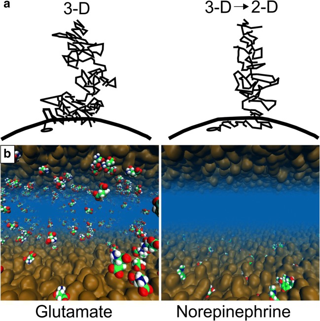
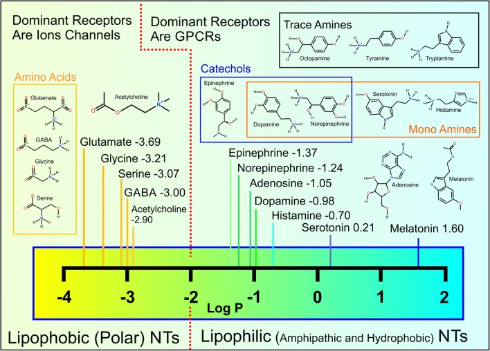
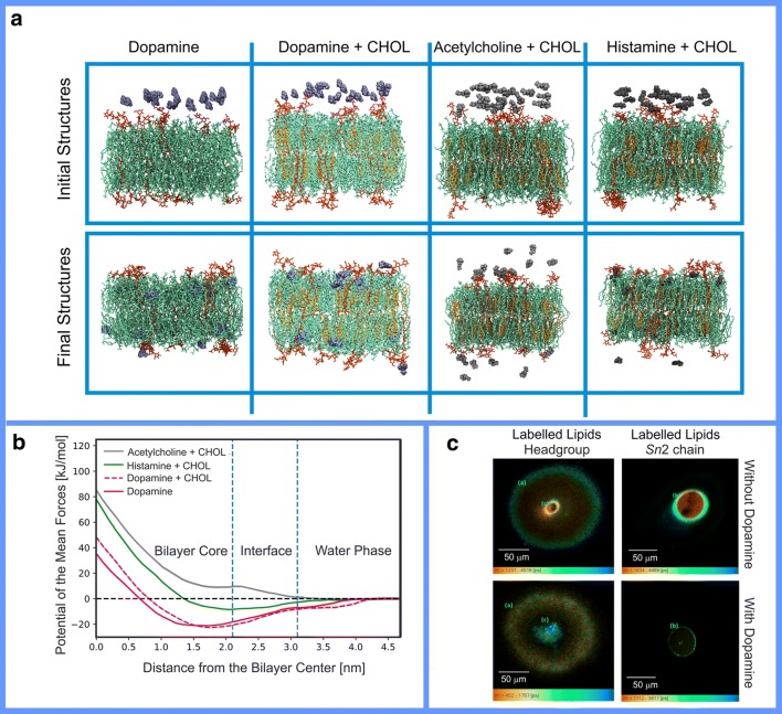
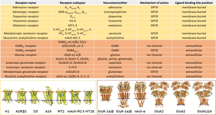
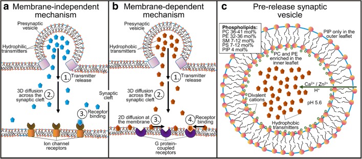
Synaptic neurotransmission is generally considered as a function of membrane-embedded receptors and ion channels in response to the neurotransmitter (NT) release and binding. This perspective aims to widen the protein-centric view by including another vital component-the synaptic membrane-in the discussion. A vast set of atomistic molecular dynamics simulations and biophysical experiments indicate that NTs are divided into membrane-binding and membrane-nonbinding categories. The binary choice takes place at the water-membrane interface and follows closely the positioning of the receptors' binding sites in relation to the membrane. Accordingly, when a lipophilic NT is on route to a membrane-buried binding site, it adheres on the membrane and, then, travels along its plane towards the receptor. In contrast, lipophobic NTs, which are destined to bind into receptors with extracellular binding sites, prefer the water phase. This membrane-based sorting splits the neurotransmission into membrane-independent and membrane-dependent mechanisms and should make the NT binding into the receptors more efficient than random diffusion would allow. The potential implications and notable exceptions to the mechanisms are discussed here. Importantly, maintaining specific membrane lipid compositions (MLCs) at the synapses, especially regarding anionic lipids, affect the level of NT-membrane association. These effects provide a plausible link between the MLC imbalances and neurological diseases such as depression or Parkinson's disease. Moreover, the membrane plays a vital role in other phases of the NT life cycle, including storage and release from the synaptic vesicles, transport from the synaptic cleft, as well as their synthesis and degradation.

Keywords: Membrane lipid composition (MLC); Membrane-based sorting; Molecular dynamics (MD); Neurotransmitter; Synaptic neurotransmission; Synaptic receptor.


Conflict of interest statement

The authors declare that they have no conflict of interest.

Figures

Fig. 1
Fig. 1
The effect of the membrane on small molecule diffusion and receptor entry. a The ligands can either diffuse in 3D towards their membrane-bound receptors (black dot; left) or the 3D diffusion can transform into 2D diffusion along the membrane plane (curved line) prior to the receptor binding (right). Reproduced with the permission from ref. [40]. Copyright 2009 Elsevier. b The membrane (brown opaque surface) adherence (lipophilicity) or repulsion (lipophobicity) of neurotransmitters (CPK models) is demonstrated for the norepinephrine (right) and glutamate (left), respectively, using atomistic molecular dynamics simulations. Reproduced with modifications from ref. [42]. Copyright 2016 Postila et al. (https://creativecommons.org/licenses/by/4.0/legalcode)
Fig. 2
Fig. 2
Chemical structures and log P values of non-peptidic neurotransmitters. The given log P values are experimental except for acetylcholine (https://pubchem.ncbi.nlm.nih.gov/). The log P values suggest that the neurotransmitters (NTs) belong to either lipophobic (− 3.69 to − 2.90) or lipophilic (− 1.37 to 1.60) categories. In reality, most of the NTs are amphipathic molecules with both hydrophilic (“water-loving”) or hydrophobic (“water-hating”) groups whose combined effect determines how likely they are to remain in the water phase or to aggregate on the membrane, respectively.
Fig. 3
Fig. 3
a Snapshots of the initial (0 ns) and final configurations (500 ns) obtained in molecular dynamics (MD) simulations of lipid bilayers (stick models) composed of POPC (1-palmitoyl-2-oleoyl-sn-glycero-3-phosphocholin; green), CHOL (cholesterol; orange), ganglioside GM1 (monosialotetrahexosylganglioside; red), in the presence of dopamine, acetylcholine, and histamine (blue or gray CPK models). Water is omitted for clarity. Reproduced with the permission from ref. [57]. Copyright 2018 Elsevier. b Free energy profiles of the neurotransmitters translocating thorough the lipid bilayer indicate that dopamine preferred a location at the core of the bilayer below the lipid headgroup region. The center of mass of the bilayer is at 0 nm. Vertical blue dashed lines show approximate regions of the bilayer hydrocarbon core, the membrane-water interface, and water. Reproduced with the permission from ref. [57]. Copyright 2018 Elsevier. c Fluorescence lifetime imaging microscopy images of giant lipid vesicles containing nitrobenzoxadiazole (NBD)-labeled lipids at the headgroup or last carbon of the Sn2 chain in the absence and presence of dopamine. The images show dopamine aggregation at the membrane interior. Reproduced with the permission from ref. [64]. Copyright 2017 the American Chemical Association
Fig. 4
Fig. 4
The positions of ligand-binding sites of conventional synaptic receptors in the postsynaptic membrane. The approximate positions of the ligand-binding sites of G protein-coupled receptors (GPCRs) (yellow) and ion channel-forming receptors (orange) are circled green in the protein 3D structures (cartoon models). The inner and outer bilayer leaflets are shown with blue and red lines, respectively. Reproduced with modification from ref. [42]. Copyright 2016 Postila et al. (https://creativecommons.org/licenses/by/4.0/legalcode)
Fig. 5
Fig. 5
Synaptic neurotransmission models. Left panel—membrane-dependent model: (1) release of lipophilic neurotransmitters (NTs), (2) diffusion across the synaptic cleft, (3) binding onto the postsynaptic membrane surface and 2D diffusion on the membrane plane, and, finally, binding into the receptors. Middle panel—membrane-independent model: (1) release of lipophobic NTs, (2) diffusion across the synaptic cleft, and binding into the receptors. Right panel—the presynaptic vesicle with its known lipid composition [77, 78]. Reproduced with the permission from ref. [58]. Copyright 2017 American Chemical Society
Fig. 6
Fig. 6
Chemical structures of the most common lipids. PC, phosphatidylcholine; PE, phosphatidylethanolamine; SPH, sphingomyelin; CHOL, cholesterol; LPC, lysoPC; DAG, diacylglycerol; PS, phosphatidylserine; GM1, monosialotetrahexosylganglioside; PG, phosphatidylglycerol; PIP2, phosphatidylinositol bisphosphate; PIP3, phosphatidylinositol triphosphate; PA, phosphatidic acid
Fig. 7
Fig. 7
The pivotal role of the membrane in membrane-bound catechol-o-methyl transferase catalysis and selective inhibition. a Steps of catalytic mechanism of the membrane-bound catechol-o-methyl transferase (MB-COMT): (A1) the catalytic domain interacts weakly with membrane in the apo form; (A2) the cofactor S-adenosyl-l-methionine (ADOMET) binds to the catalytic site of MB-COMT; (A3) MB-COMT in complex with ADOMET opens the catalytic site towards the membrane, which, in turn, allows the protein to bind to the membrane surface; (A4), finally, the MB-COMT binds an Mg2+ ion that is already present at the membrane surface. b The behavior of MB-COMT selective vs. non-selective inhibitors in the membrane: selective inhibitors orient catechol group towards the water phase and, in contrast, non-selective inhibitors could be oriented less optimally in relation to the MB-COMT catalytic site. c The estimations of interactions of the ADOMET and catalytic domain of COMT in complex and separately with lipids indicate that the catalytic domain is preferably membrane-oriented: (C1) the free energy changes when protein is pulled away from the lipid bilayer; (C2) quartz crystal microbalance (QCM) frequency changes during interaction with the lipid bilayer; (C3) dissociation constant (inverse of affinity) from lipid bilayer (vesicle) determined by isothermal calorimetry. Reproduced with permission from ref. [146]. Copyright 2018 the Royal Society of Chemistry

References

    1. Zucker RS, Kullmann DM, Kaeser PS (2014) Release of Neurotransmitters. In: Byrne JH, Heidelberger R, Waxham MN (eds) From Molecules to Networks. Elsevier Inc., pp 443–488
    1. Deutch AY, Roth RH (2014) Pharmacology and Biochemistry of Synaptic Transmission Classical Transmitters. In: Byrne JH, Heidelberger R, Waxham MN (eds) From Molecules to Networks. Elsevier Inc., pp 207–237
    1. Chua JJE, Kindler S, Boyken J, Jahn R. The architecture of an excitatory synapse. J Cell Sci. 2010;123:819–823. doi: 10.1242/jcs.052696. - DOI - PubMed
    1. Dieterich DC, Kreutz MR. Proteomics of the synapse – a quantitative approach to neuronal plasticity. Mol Cell Proteomics. 2016;15:368–381. doi: 10.1074/mcp.R115.051482. - DOI - PMC - PubMed
    1. Takahashi T. Strength and precision of neurotransmission at mammalian presynaptic terminals. Proc Jpn Acad Ser B Phys Biol Sci. 2015;91:305–320. doi: 10.2183/pjab.91.305. - DOI - PMC - PubMed