Review
. 2020 Mar;225(6):2283-2300.
doi: 10.1111/nph.16240. Epub 2019 Nov 2.

Phytochrome evolution in 3D: deletion, duplication, and diversification

Affiliations
Review

Phytochrome evolution in 3D: deletion, duplication, and diversification

Nathan C Rockwell et al. New Phytol. 2020 Mar.

Abstract

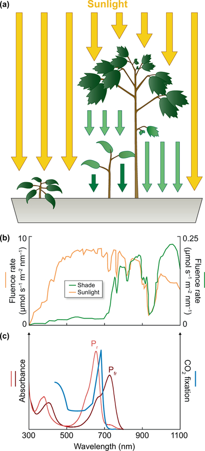
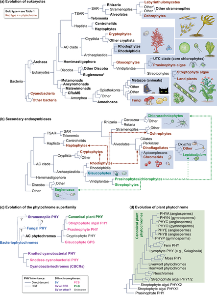
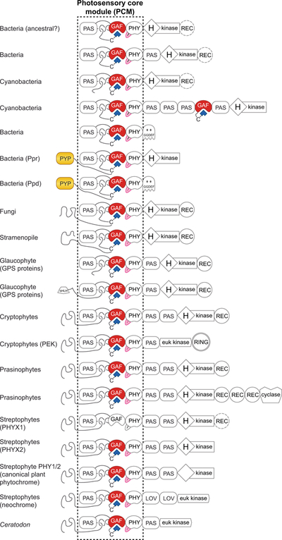
Canonical plant phytochromes are master regulators of photomorphogenesis and the shade avoidance response. They are also part of a widespread superfamily of photoreceptors with diverse spectral and biochemical properties. Plant phytochromes belong to a clade including other phytochromes from glaucophyte, prasinophyte, and streptophyte algae (all members of the Archaeplastida) and those from cryptophyte algae. This is consistent with recent analyses supporting the existence of an AC (Archaeplastida + Cryptista) clade. AC phytochromes have been proposed to arise from ancestral cyanobacterial genes via endosymbiotic gene transfer (EGT), but most recent studies instead support multiple horizontal gene transfer (HGT) events to generate extant eukaryotic phytochromes. In principle, this scenario would be compared to the emerging understanding of early events in eukaryotic evolution to generate a coherent picture. Unfortunately, there is currently a major discrepancy between the evolution of phytochromes and the evolution of eukaryotes; phytochrome evolution is thus not a solved problem. We therefore examine phytochrome evolution in a broader context. Within this context, we can identify three important themes in phytochrome evolution: deletion, duplication, and diversification. These themes drive phytochrome evolution as organisms evolve in response to environmental challenges.

Keywords: endosymbiosis; evolution; light harvesting; photosynthesis; shade avoidance.


Figures

Figure 1.
Figure 1.. Phytochrome and shade sensing.
(a) Plants growing in direct sunlight or under shade from competitors are exposed to different light environments, leading to distinct morphology and pigment development. The plant on the left is in direct sunlight, but the lower plant on the right is shaded by a competing plant of a different species. Scattering is omitted for clarity. (b) A competing canopy depletes light for shaded plants. In the case of tropical rain forests, attenuation is approximately 40-fold for the near-infrared and greater for photosynthetically useful red light. Units are μmol s−1 m−2 nm−1 for both axes. The red:far-red ratio (R:FR) is 1.25 in direct sunlight and 0.31 in shade (Lee & Graham, 1986). (c) The action spectrum for carbon fixation in the green alga Hydrodictyon is compared to absorption spectra for the red-absorbing Pr and far-red-absorbing Pfr photostates of wheat phytochrome C (Raven, 1969; Chen et al., 2014).
Figure 2.
Figure 2.. Structure of the knotted phytochrome PCM.
(a) Cyanobacterial phytochrome Cph1 from Synechocystis sp. PCC 6803 (Essen et al., 2008) is shown color-coded by sequence similarity, calculated using a published sequence alignment (Rockwell et al., 2006) and the BLOSUM62 matrix as implemented in homolmapper (Rockwell & Lagarias, 2007). (b) Topology cartoon for the knotted PAS-GAF region of the PCM with the knot in orange, the PAS domain in green, and the GAF domain in blue. Cys residues used for chromophore ligation are indicated in purple. Secondary structure elements are numbered from the N-terminus of each domain for the PAS and GAF domains, with the domain indicated by P and G, respectively. 1-turn 310 helices and short β-strands in the knot are indicated but not numbered. Domain names: GAF, cGMP phosphodiesterase/Adenylate cyclase/FhlA; PAS, Per/ARNT/Sim; PHY, phytochrome-specific. BV, biliverdin; N-term, N-terminus; tongue, conserved insertion loop specific to the PHY domain.
Figure 3.
Figure 3.. Phytochrome and the evolution of eukaryotes.
(a) A simplified working view of the tree of life is shown, drawing on recent studies (Duanmu et al., 2014; Wickett et al., 2014; Burki et al., 2016; Derelle et al., 2016; Brown et al., 2018; Lax et al., 2018; Gawryluk et al., 2019; Strassert et al., 2019). Bold type indicates taxa assessed for phytochrome and bilin biosynthesis in Table 1. Lineages containing at least some organisms in which phytochromes are present are indicated in red. Representative organisms are shown for selected lineages (not to scale; enclosed in dotted boxes). Branch lengths are arbitrary. Asterisk, see legend to Table 1 for possible phytochromes in Euglenozoa. CRuMS, supergroup containing collodictyonids, rigifilids, and Mantamonas; AC clade, Archaeplastida + Cryptista; UTC clade, Ulvophyceae, Trebouxiophyceae and Chlorophyceae; TSAR, Telonemia + SAR; SAR, supergroup containing Stramenopiles, Alveolates, and Rhizaria. (b) Secondary endosymbioses in eukaryotes. Acquisition of photosynthesis in cryptophytes, haptophytes, ochrophytes, and photosynthetic alveolates (chromerids and dinoflagellates) occurred via secondary endosymbiosis of a rhodophyte (dashed red lines). Acquisition of photosynthesis in euglenozoa and chlorarachniophytes occurred via secondary endosymbiosis of prasinophyte or chlorophyte algae (green dashed lines). “Green dinoflagellates” of the genus Lepidodinium have replaced the ancestral rhodophyte-derived plastid via secondary endosymbiosis of a prasinophyte or chlorophyte alga (green dotted lines). Representative organisms are shown for selected lineages as in (a). Colors indicate plastid lineage; nonphotosynthetic plastids are not colored. (c) A schematic view of phytochrome evolution is shown. Colors indicate chromophore precursors incorporated by phytochromes from different taxa: BV only, blue; BV or PCB, purple; BV or unknown bilin, deep purple (Rockwell et al., 2014); PCB, pink; PΦB, green; unknown, grey. Dashed lines indicate inferred HGT events. PHY, phytochrome; BV, biliverdin IXα; PCB, phycocyanobilin; PΦB, phytochromobilin. (d) A schematic view of plant phytochrome evolution is shown, demonstrating that canonical plant phytochromes are part of a larger clade of streptophyte phytochromes using the nomenclature of (Li et al., 2015).
Figure 4.
Figure 4.. Biosynthesis of bilin chromophores.
The pathway from heme to biliverdin IXα (BV) and reduced phycobilins (PCB and PΦB) is shown. Free bilins are then assembled with apophytochrome to yield covalent adducts, examples of which are shown on the right in the C5–Z,syn, C10–Z,syn, C15–Z,anti configuration (Wagner et al., 2005; Essen et al., 2008; Burgie et al., 2014a). The numbering system is indicated for BV. For simplicity, a single structure is shown; however, other BV adducts have been observed (Salewski et al., 2013). For BV the photoactive 15,16–double bond is highlighted in red. For PCB and PΦB, sites of reduction relative to BV are highlighted in red. The degree of reduction relative to BV is indicated (e, electron).
Figure 5.
Figure 5.. Diverse domain architectures in knotted phytochromes.
Jellybean domain diagrams are shown for diverse prokaryotic and eukaryotic phytochromes that use the canonical knotted PCM (Duanmu et al., 2014; Li et al., 2015). Bilin-binding GAF domains are red, the tongue region of the PHY domain is pink, bilin chromophores are shown as blue polygons, and chromophore-binding PYP domains are yellow. REC domains that are present in only some cases are dashed, as in PHYX1. Domain names: GAF, cGMP phosphodiesterase/Adenylate cyclase/FhlA; PAS, Per/ARNT/Sim; PHY, phytochrome-specific; (H)kinase, histidine kinase bidomain, with the presence of the His indicated by H; REC, response regulator receiver; RING, really interesting new gene; euk kinase, eukaryotic protein kinase; cyclase, eukaryotic adenylate/guanylate cyclase; GGDEF, diguanylate cyclase; PYP, photoactive yellow protein; bHLH, basic helix-loop-helix; LOV, PAS domain belonging to the light/oxygen/voltage lineage. Two-Cys phytochromes from cyanobacteria and glaucophytes are not indicated.
Figure 6.
Figure 6.. Structures of knotless phytochromes and CBCRs.
(a) The GAF-PHY PCM of Cph2 from Synechocystis sp. PCC 6803 is shown (Anders et al., 2013), color-coded by sequence similarity using a published sequence alignment (Gan et al., 2014) as in Fig. 2(a). (b) Detailed view of the PHY domain of Cph1, colored as in (a); the tongue region and the long helices connecting to adjacent domains are omitted for clarity. (c) Detailed view of the PHY domain of Cph2, colored as in (a) and presented as in (b). (d) The bilin-binding GAF domain of AnPixJg2 (red) from Nostoc sp. PCC 7120 (sometimes designated Anabaena sp. PCC 7120) is compared to that of Cph2 (gold) from (a) and that of Cph1 (blue) from Fig. 2 (Anders et al., 2013; Narikawa et al., 2013), with PCB coordinates used for superposition. (e) The PCB chromophores and ligated Cys residues are shown superposed for Cph1 (red), Cph2 (blue), and AnPixJg2 (grey). All three cases correspond to dark-adapted, red-absorbing photostates in the C5–Z,syn C10–Z,syn C15–Z,anti configuration. Domain names: GAF, cGMP phosphodiesterase/Adenylate cyclase/FhlA; PHY, phytochrome-specific. PCB, phycocyanobilin; tongue, conserved insertion loop specific to the PHY domain.
Figure 7.
Figure 7.. Diversification of the phytochrome superfamily in cyanobacteria.
Jellybean domain architectures and photocycles are shown for selected photoreceptors (Wu & Lagarias, 2000; Rockwell et al., 2011; Rockwell et al., 2012a; Rockwell et al., 2012b; Savakis et al., 2012; Hirose et al., 2013; Gan et al., 2014; Rockwell et al., 2016). Photocycles are shown for heterologously expressed GAF-only constructs that are highlighted with blue shading in the domain architectures. In each case, the 15Z configuration is colored blue and the 15E configuration is colored orange; peak wavelengths and domain names are indicated. In the domain architectures, bilin-binding GAF domains are colored by photocycle, with the dark-adapted state on the left and the photoproduct on the right. Bilin chromophores are shown as blue polygons; domains that do not photoconvert (Rockwell et al., 2012b) are indicated with a white “photoproduct” state. The bilin-binding GAF lineage defining the phytochrome superfamily is only a subset of all GAF domains, so non-photosensory GAF domains are indicated in white. Domain names: GAF, cGMP phosphodiesterase/Adenylate cyclase/FhlA; PAS, Per/ARNT/Sim; PHY, phytochrome-specific; (H)kinase, histidine kinase bidomain, with the presence of the His indicated by H; GGDEF, diguanylate cyclase; HAMP, histidine kinase/adenylate cyclase/methyl-accepting chemotaxis protein/phosphatase; CBS, cystathionine β-synthase domain; EAL, diguanylate phosphodiesterase; MA-MCP, methyl-accepting domain of methylated chemotaxis proteins. Vertical rectangles on TePixJ are predicted transmembrane helices. DXCF, ins-C are different second Cys residues.

References

    1. Anders K, Daminelli-Widany G, Mroginski MA, von Stetten D, Essen LO. 2013. Structure of the cyanobacterial phytochrome 2 photosensor implies a tryptophan switch for phytochrome signaling. Journal of Biological Chemistry 288(50): 35714–35725. - PMC - PubMed
    1. Armbrust EV, Berges JA, Bowler C, Green BR, Martinez D, Putnam NH, Zhou S, Allen AE, Apt KE, Bechner M, et al. 2004. The genome of the diatom Thalassiosira pseudonana: ecology, evolution, and metabolism. Science 306(5693): 79–86. - PubMed
    1. Auldridge ME, Forest KT. 2011. Bacterial phytochromes: more than meets the light. Critical Reviews in Biochemistry and Molecular Biology. 46(1): 67–88. - PubMed
    1. Ballaré CL, Pierik R. 2017. The shade-avoidance syndrome: multiple signals and ecological consequences. Plant Cell and Environment 40(11): 2530–2543. - PubMed
    1. Beisser D, Graupner N, Bock C, Wodniok S, Grossmann L, Vos M, Sures B, Rahmann S, Boenigk J. 2017. Comprehensive transcriptome analysis provides new insights into nutritional strategies and phylogenetic relationships of chrysophytes. PeerJ 5: e2832. - PMC - PubMed

Publication types