. 2019 Oct 9:25:7581-7590.
doi: 10.12659/MSM.916522.

miR-149-5p Inhibits Vascular Smooth Muscle Cells Proliferation, Invasion, and Migration by Targeting Histone Deacetylase 4 (HDAC4)

Affiliations

miR-149-5p Inhibits Vascular Smooth Muscle Cells Proliferation, Invasion, and Migration by Targeting Histone Deacetylase 4 (HDAC4)

Boya Zhang et al. Med Sci Monit. .

Abstract

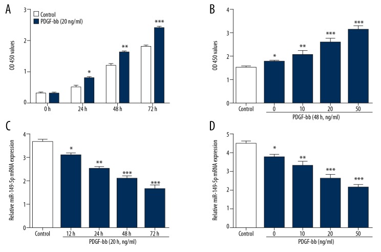
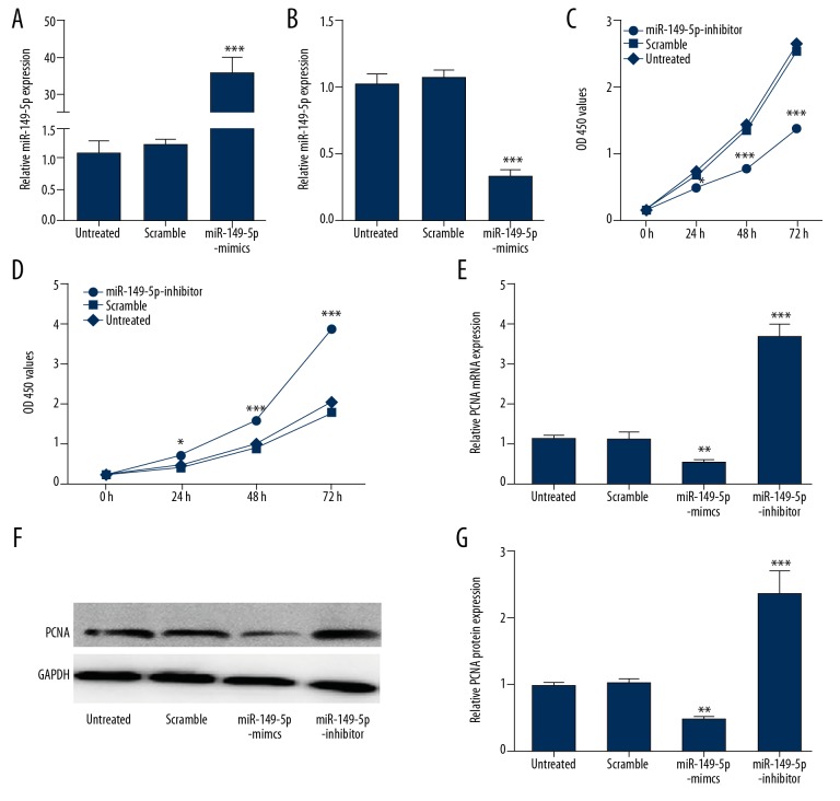
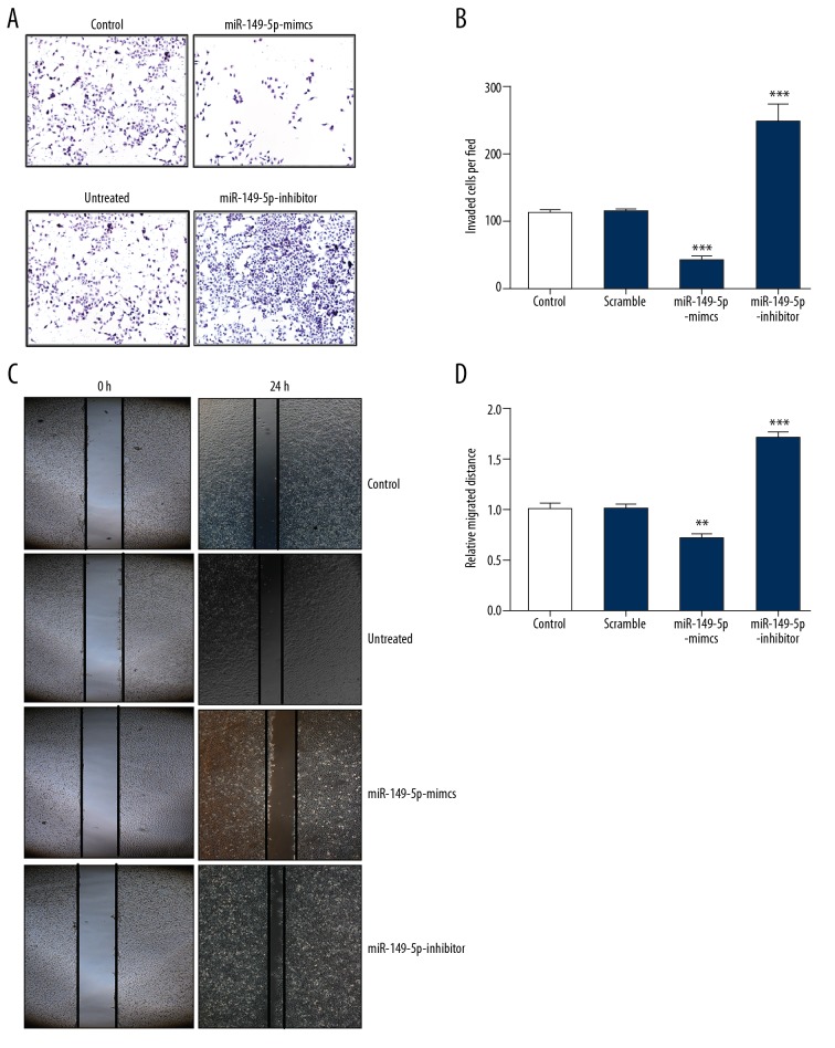
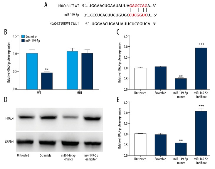
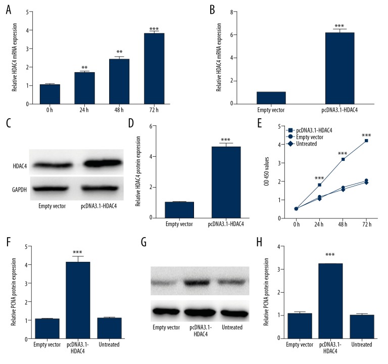
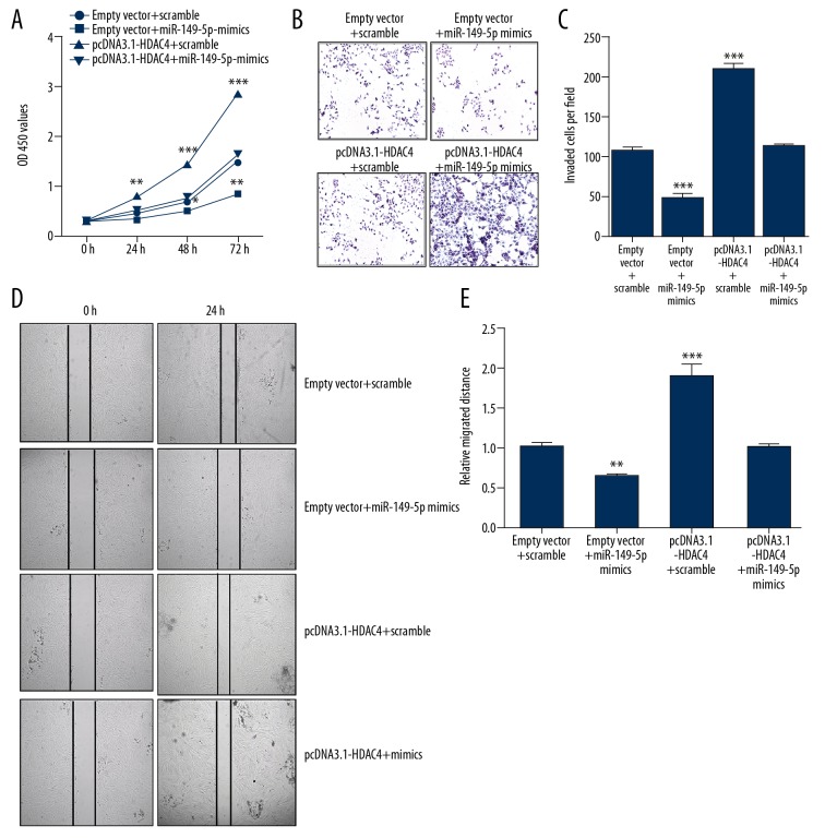
BACKGROUND Studies have demonstrated that microRNAs (miRNAs) have essential roles in biological functions of vascular smooth muscle cells (VSMCs). However, the function and related molecular mechanism of miR-149-5p in VSMCs remains unclear. MATERIAL AND METHODS We used MTT assay, Transwell assay, and wound-healing assay to measure the proliferation, invasion, and migration of VSMCs transfected with miR-149-5p mimics or inhibitors, respectively. Bioinformatics tools and luciferase assay were used to validate the relationship between miR-149-5p and histone deacetylase 4 (HDAC4). Rescue experiments were used to confirm the interaction of miR-149-5p and HDAC4 in regulating biological functions in VSMCs. RESULTS miR-149-5p was downregulated in PDGF-bb-induced VSMCs. It was also found that miR-149-5p overexpression suppressed proliferation, invasion, and migration of VSMCs, while miR-149-5p knockdown showed the opposite effects. Furthermore, HDAC4 was found to be a potential target of miR-149-5p, which rescued miR-149-5p-mediated proliferation, invasion, and migration in VSMCs. CONCLUSIONS We demonstrated that miR-149-5p can suppress biological functions of VSMCs by regulating HDAC4, which might provide a potent therapeutic target for VSMC growth-related diseases.


Conflict of interest statement

Conflicts of interest

None.

Figures

Figure 1
Figure 1
miR-149-5p is downregulated in PDGF-bb-induced VSMCs. (A, B) The growth ability of VSMCs simulated by different doses of PDGF-bb at various time-points. (C, D) The expression of miR-149-5p in VSMCs simulated by different doses and at time-points of PDGF-bb. * p<0.05 and ** p<0.01 vs. control.
Figure 2
Figure 2
Proliferation of VSMCs is inhibited by miR-149-5p. (A, B) The overexpression or down-expression of miR-149-5p in VSMCs transfected with miR-149-5p mimics or inhibitor verified by qRT-PCR (C, D). The MTT assay was performed to detect proliferation of VSMCs transfected with miR-149-5p mimics, inhibitor, or control. (E–G) The mRNA and protein level of PCNA in VSMCs transfected with miR-149-5p mimics, inhibitor, or control. * p<0.05 and ** p<0.01 vs. control.
Figure 3
Figure 3
Migration and invasion of VSMCs are suppressed by miR-149-5p. (A, B) Transwell assay was used to evaluate invasion ability of VSMCs transfected with miR-149-5p mimics, inhibitor, or control. (C, D). The cell migration of VSMCs transfected with miR-149-5p mimics, inhibitor, or control was measured by wound-healing assay. * p<0.05 and ** p<0.01 vs. control.
Figure 4
Figure 4
miR-149-5p targets HDAC4 in VSMCs. (A) Bioinformatics analysis predicted putative miR-149-5p target sites of HDAC4 3′UTR. (B) Luciferase activity of luciferase reporter plasmid containing wild-type or mutant HDAC4 3′UTR co-transfected with miR-149-5p. (C–E) RT-PCR analysis and Western blot were conducted to assess HDAC4 expression in VSMCs transfected with miR-149-5p mimics, inhibitor, or control. * p<0.05 and ** p<0.01 vs. control.
Figure 5
Figure 5
HDAC4 takes part in regulating proliferation of VSMC. (A) The expression of HDAC4 in PDGF-bb-stimulated VSMCs was measured by qRT-PCR. (B–D). qRT-PCR and Western blot analysis were were conducted to detect the overexpression efficiency of pcDNA3.1-HDAC4. (E) The proliferation of VSMCs transfected with pcDNA3.1-HDAC4, empty vector, or control were detected by MTT assay. (F–H) The mRNA and protein level of PCNA in VSMCs transfected with miR-149-5p mimics, inhibitor, or control. * p<0.05 and ** p<0.01 vs. control.
Figure 6
Figure 6
HDAC4 restoration rescued miR-149-5p-mediated growth, invasion, and migration. (A) The proliferation of VSMCs co-transfected with miR-149-5p mimics, pcDNA3.1-HDAC4, or corresponding controls. (B, C) Transwell assay was performed to detect cell invasion in VSMCs co-transfected with miR-149-5p mimics, pcDNA3.1-HDAC4, or corresponding controls. (D, E) Cell migration in VSMCs co-transfected with miR-149-5p mimics, pcDNA3.1-HDAC4, or corresponding controls. * p<0.05 and ** p<0.01 vs. control.

References

    1. Wang G, Jacquet L, Karamariti E, Xu Q. Origin and differentiation of vascular smooth muscle cells. J Physiol. 2015;593(14):3013–30. - PMC - PubMed
    1. Montezano AC, Lopes RA, Neves KB, et al. Isolation and culture of vascular smooth muscle cells from small and large vessels. Methods Mol Biol. 2017;1527:349–54. - PubMed
    1. Schlosser A, Pilecki B, Hemstra LE, et al. MFAP4 promotes vascular smooth muscle migration, proliferation and accelerates neointima formation. Arterioscler Thromb Vasc Biol. 2016;36(1):122–33. - PubMed
    1. Fabian MR, Sonenberg N, Filipowicz W. Regulation of mRNA translation and stability by microRNAs. Ann Rev Biochem. 2010;79(1):351–79. - PubMed
    1. Ha M, Kim VN. Regulation of microRNA biogenesis. Nat Rev Mol Cell Biol. 2014;15(8):509–24. - PubMed

MeSH terms