

doi: 10.1039/c9cc06831g.
A NIR-emitting cyanine with large Stokes shifts for live cell imaging: large impact of the phenol group on emission
Affiliations
- PMID: 31595909
- PMCID: PMC6918678
- DOI: 10.1039/c9cc06831g
Item in Clipboard
A NIR-emitting cyanine with large Stokes shifts for live cell imaging: large impact of the phenol group on emission
Chem Commun (Camb).
.
Abstract
There are a limited number of near-infrared (NIR) emitting (λem = 700-900 nm) molecular probes for imaging applications. A NIR-emitting probe that exhibits emission at ∼800 nm with a large Stokes shift was synthesized and found to exhibit excellent selectivity towards mitochondria for live-cell imaging. The photophysical properties were attributed to an excited "cyanine structure" via intramolecular charge transfer (ICT) involving a phenol group.
Conflict of interest statement
Conflicts of interest
“There are no conflicts to declare”.
Figures
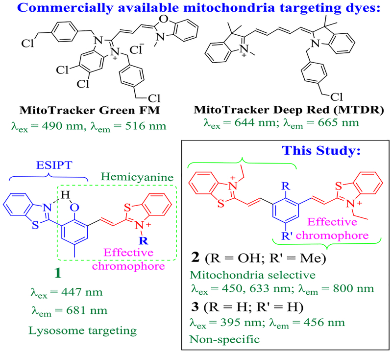
Chemical structures of probes 1-3 and some commercial mitochondria staining dyes.
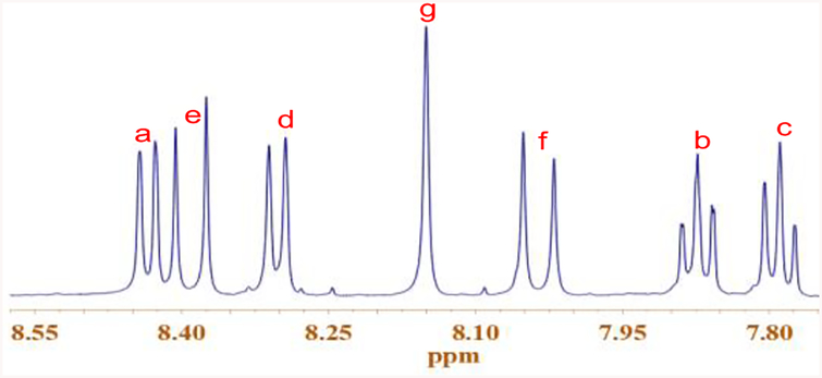
1H NMR of 2 in DMSO-d6, showing the resonance signals of aromatic protons (the alkyl region is omitted for clarity).
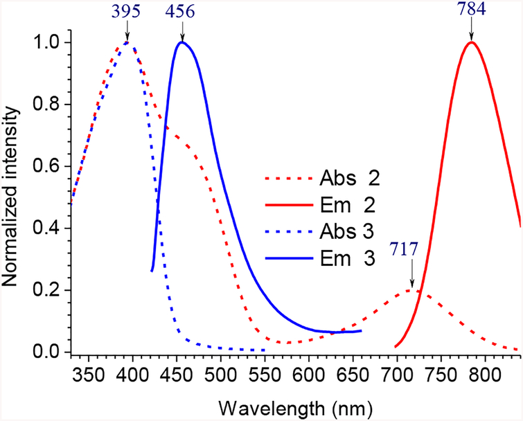
UV-vis absorption (broken line) and emission spectra (solid line) of 2 and 3 in CH2Cl2. The excitation wavelengths were 687 nm for 2 and 395 nm for 3.
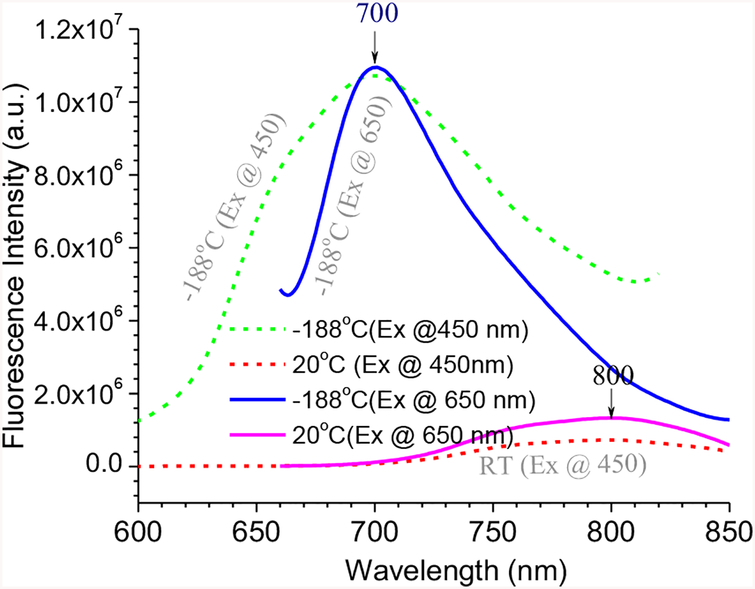
Fluorescence spectra of 2 at room temperature and low temperature, with λex at 450 nm (dotted lines) and 650 nm (solid lines).
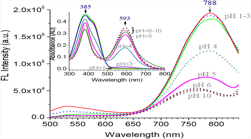
Fluorescence spectra of 2 (10 μM) in different pH buffer in water (excitation at 448 nm). Inset is the UV-absorption of the corresponding solution.
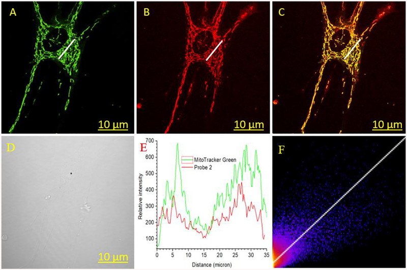
Normal human lungs fibroblast (NHLF) cells co-stained with 2 (1 μM) and MitoTracker green (200 nM), viewed in green (A) and NIR channel (B). (E) The plot of relative intensity vs. distance for probe 2 and MitoTracker Green for the region in white line in A and B, and (F) correlation plot between MitoTracker Green and 2. Excitation/emission λex/λem=488/525 nm for MitoTracker Green, and 640/(680 −735) nm for 2.
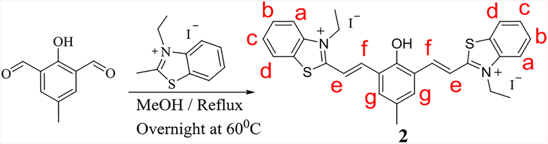
Synthesis of compound 2.
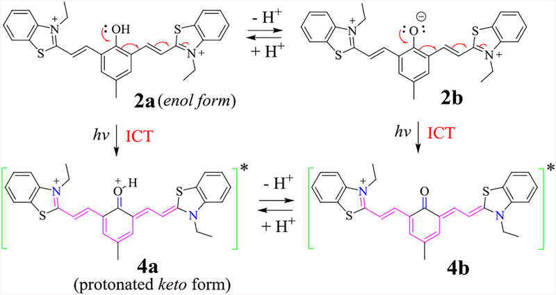
The proposed transformation of excited 2 to its keto form 4, and their equilibrium via proton dissociation.
References
MeSH terms
Substances
Grants and funding
LinkOut - more resources
Full Text Sources
Miscellaneous