

Myocardial Perfusion by Coronary Computed Tomography in the Evaluation of Myocardial Ischemia: Simultaneous Stress Protocol with SPECT
- PMID: 31596324
- PMCID: PMC7021272
- DOI: 10.5935/abc.20190201
Myocardial Perfusion by Coronary Computed Tomography in the Evaluation of Myocardial Ischemia: Simultaneous Stress Protocol with SPECT
Abstract
Background: Functional assessment to rule out myocardial ischemia using coronary computed tomography angiography (CCTA) is extremely important and data on the Brazilian population are still limited.
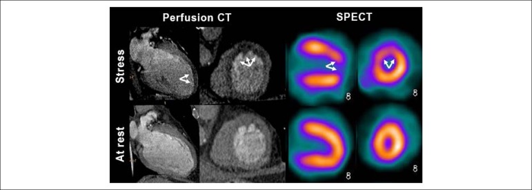
Objective: To assess the diagnostic performance of myocardial perfusion by CCTA in the detection of severe obstructive coronary artery disease (CAD) compared with single-photon emission computerized tomography (SPECT). To analyze the importance of anatomical knowledge to understand the presence of myocardial perfusion defects on SPECT imaging that is not identified on computed tomography (CT) scan.
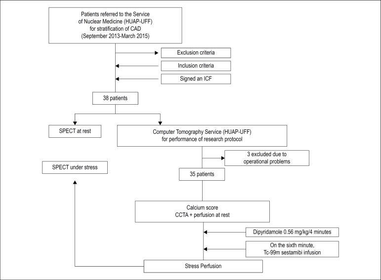
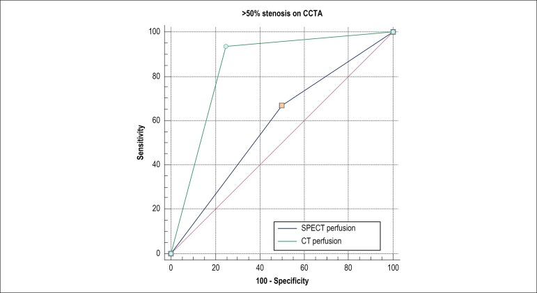
Method: A total of 35 patients were evaluated by a simultaneous pharmacologic stress protocol. Fisher's exact test was used to compare proportions. The patients were grouped according to the presence or absence of significant CAD. The area under the ROC curve was used to identify the diagnostic performance of CCTA and SPECT in perfusion assessment. P < 0.05 values were considered statistically significant.
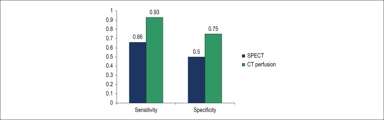
Results: For detection of obstructive CAD, CT myocardial perfusion analysis yielded an area under the ROC curve of 0.84 [a 95% confidence interval (CI95%): 0.67-0.94, p < 0.001]. SPECT myocardial perfusion imaging, on the other hand, showed an AUC of 0.58 (95% CI 0.40 - 0.74, p < 0.001). In this study, false-positive results with SPECT are described.
Conclusion: Myocardial perfusion analysis by CTA displays satisfactory results compared to SPECT in the detection of obstructive CAD. CCTA can rule out false-positive results of SPECT.
Conflict of interest statement
No potential conflict of interest relevant to this article was reported.
Figures
Comment in
-
Myocardial Computed Tomography Perfusion: One More Piece on The Board.Arq Bras Cardiol. 2019 Dec;113(6):1102-1103. doi: 10.36660/abc.20190671. Arq Bras Cardiol. 2019. PMID: 31800685 Free PMC article. No abstract available.
Similar articles
-
The diagnostic performance of SPECT-MPI to predict functional significant coronary artery disease by fractional flow reserve derived from CCTA (FFRCT): sub-analysis from ACCURACY and VCT001 studies.Int J Cardiovasc Imaging. 2017 Dec;33(12):2067-2072. doi: 10.1007/s10554-017-1207-y. Epub 2017 Jul 11. Int J Cardiovasc Imaging. 2017. PMID: 28699019
-
Early resting myocardial computed tomography perfusion for the detection of acute coronary syndrome in patients with coronary artery disease.Circ Cardiovasc Imaging. 2015 Mar;8(3):e002404. doi: 10.1161/CIRCIMAGING.114.002404. Circ Cardiovasc Imaging. 2015. PMID: 25752898 Free PMC article.
-
Diagnostic Performance of Coronary CT Angiography and Myocardial Perfusion Imaging in Kidney Transplantation Candidates.JACC Cardiovasc Imaging. 2015 May;8(5):553-562. doi: 10.1016/j.jcmg.2014.12.028. Epub 2015 Apr 10. JACC Cardiovasc Imaging. 2015. PMID: 25869350
-
Diagnostic Performance of Hybrid Cardiac Imaging Methods for Assessment of Obstructive Coronary Artery Disease Compared With Stand-Alone Coronary Computed Tomography Angiography: A Meta-Analysis.JACC Cardiovasc Imaging. 2018 Apr;11(4):589-599. doi: 10.1016/j.jcmg.2017.05.020. Epub 2017 Aug 16. JACC Cardiovasc Imaging. 2018. PMID: 28823745 Free PMC article.
-
Diagnostic accuracy of stress myocardial perfusion imaging compared to invasive coronary angiography with fractional flow reserve meta-analysis.Circ Cardiovasc Imaging. 2015 Jan;8(1):e002666. doi: 10.1161/CIRCIMAGING.114.002666. Circ Cardiovasc Imaging. 2015. PMID: 25596143 Review.
Cited by
-
Myocardial Computed Tomography Perfusion: One More Piece on The Board.Arq Bras Cardiol. 2019 Dec;113(6):1102-1103. doi: 10.36660/abc.20190671. Arq Bras Cardiol. 2019. PMID: 31800685 Free PMC article. No abstract available.
References
-
- Moran AE, Roth GA, Narula J, Mensah GA. 1990-2010 global cardiovascular disease atlas. Glob Heart. 2014;9(1):3–16. - PubMed
-
- Miller JM, Rochitte CE, Dewey M, Arbab-Zadeh A, Niinuma H, Gottlieb I, et al. Diagnostic performance of coronary angiography by 64-row CT. N Engl J Med. 2008;359(22):2324–2336. - PubMed
-
- Pugliese F, Mollet NR, Runza G, van Mieghem C, Meijboom WB, Malagutti P, et al. Diagnostic accuracy of non-invasive 64-slice CT coronary angiography in patients with stable angina pectoris. Eur Radiol. 2006;16(3):575–582. - PubMed
-
- Mollet NR, Cademartiri F, van Mieghem CA, Runza G, McFadden EP, Baks T, et al. High-resolution spiral computed tomography coronary angiography in patients referred for diagnostic conventional coronary angiography. Circulation. 2005;112(15):2318–2323. - PubMed
Publication types
MeSH terms
LinkOut - more resources
Full Text Sources
Medical
Miscellaneous
