. 2020 Apr;75(4):921-932.
doi: 10.1111/all.14081. Epub 2019 Oct 31.

ILC3 deficiency and generalized ILC abnormalities in DOCK8-deficient patients

Affiliations

ILC3 deficiency and generalized ILC abnormalities in DOCK8-deficient patients

Ahmet Eken et al. Allergy. 2020 Apr.

Abstract

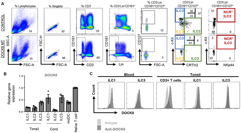
Background: Dedicator of cytokinesis 8 (DOCK8) deficiency is the main cause of the autosomal recessive hyper-IgE syndrome (HIES). We previously reported the selective loss of group 3 innate lymphoid cell (ILC) number and function in a Dock8-deficient mouse model. In this study, we sought to test whether DOCK8 is required for the function and maintenance of ILC subsets in humans.

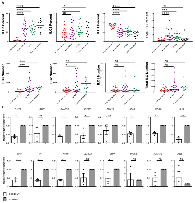
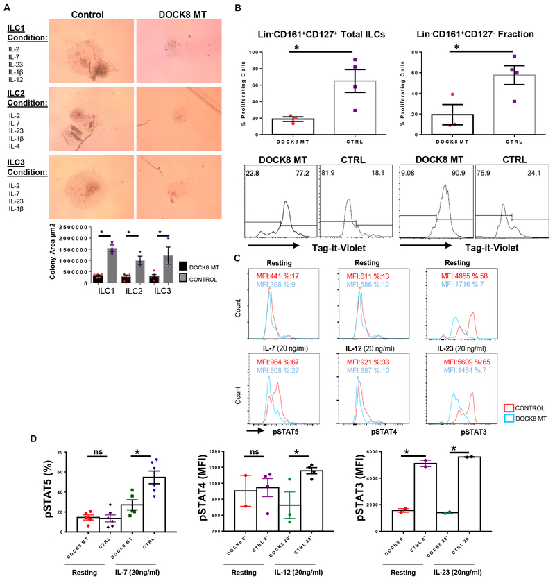
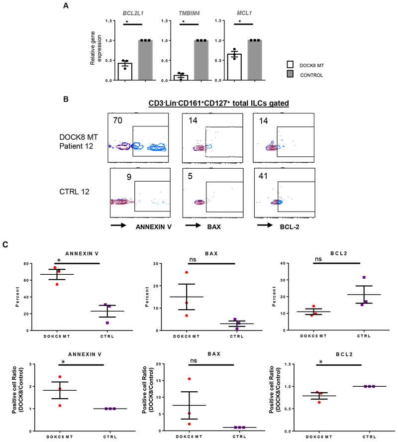
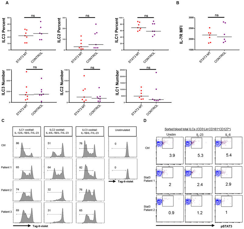
Methods: Peripheral blood ILC1-3 subsets of 16 DOCK8-deficient patients recruited at the pretransplant stage, and seven patients with autosomal dominant (AD) HIES due to STAT3 mutations, were compared with those of healthy controls or post-transplant DOCK8-deficient patients (n = 12) by flow cytometry and real-time qPCR. Sorted total ILCs from DOCK8- or STAT3-mutant patients and healthy controls were assayed for survival, apoptosis, proliferation, and activation by IL-7, IL-23, and IL-12 by cell culture, flow cytometry, and phospho-flow assays.

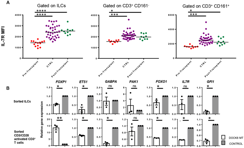
Results: DOCK8-deficient but not STAT3-mutant patients exhibited a profound depletion of ILC3s, and to a lesser extent ILC2s, in their peripheral blood. DOCK8-deficient ILC1-3 subsets had defective proliferation, expressed lower levels of IL-7R, responded less to IL-7, IL-12, or IL-23 cytokines, and were more prone to apoptosis compared with those of healthy controls.

Conclusion: DOCK8 regulates human ILC3 expansion and survival, and more globally ILC cytokine signaling and proliferation. DOCK8 deficiency leads to loss of ILC3 from peripheral blood. ILC3 deficiency may contribute to the susceptibility of DOCK8-deficient patients to infections.

Keywords: DOCK8; Hyper-IgE syndrome (HIES); ILC; ILC3; STAT3.


Conflict of interest statement

Disclosure of potential conflict of interest: The authors declare no conflict of interest.

Figures

Figure 1
Figure 1
Human ILCs express DOCK8. A) Gating strategy for human ILCs, representative plots for control and patient blood Blue, green, orange and red gates indicate ILC3, ILC2, ILC1 and natural cytotoxicity receptor (NCR)+ ILC3s, respectively. B) DOCK8 gene expression by sorted human ILC subsets or monocyte derived dendritic cells (moDC) or naïve T cells. DOCK8 expression was quantified as fold expression over that of T cells. C) DOCK8 intracellular staining in ILC1 and ILC3 or CD3+ T cells sorted from human peripheral blood and tonsils.
Figure 2
Figure 2
ILC3s are reduced in DOCK8-deficient HIES patients. A) PBMCs of DOCK8-deficient patients (pre and post-transplant), mothers and fathers of patients, and control subjects were stained and gated as shown in Figure 1. CD3LinCD161+CD127+ cells were gated as total ILCs and analyzed by c-kit and CRTH2 expression. Percent of ILC subsets among total ILCs, and total ILCs among CD3LinCD161+ cells were shown in the top panel. Absolute number of ILC subsets or total ILCs per ml blood were shown in the bottom panel. Individual patient plots were shown in Figure S1. DOCK8-deficient patients (n=16), Healthy controls (n=14-17), Parents (n=22), post-transplant DOCK8 patients (n=10). In absolute number graphs, CTRL includes healthy controls and healthy parents. B) Reduced human blood ILC3-associated gene expression levels in DOCK8-deficient patients. CD3 LinCD161+CD127+ cells were sorted from peripheral blood of control and DOCK8-deficient patients (n=3-4 per group). Expression of indicated genes were assessed via real time qPCR. Results are expressed as fold change over the average of related mRNA levels in controls. Sorted and lysed cells for each group were pooled, five technical replicates run for each group. * p<0.05, ** p<0.01, *** p<0.001, ns: not significant.
Figure 3
Figure 3
Impaired proliferation and cytokine signaling of DOCK8-deficient human ILCs. A) Equal number of sorted total ILCs (CD3LinCD161+CD127+) from DOCK8-deficient patients and healthy controls were cultured for 21 days with indicated cytokine ILC subset polarizing cocktails, proliferation of ILCs were quantified by area of growth. (n= 3-5 per group). B) Sorted total ILCs (CD3LinCD161+CD127+) or CD3LinCD161+CD127 cells were labeled with Tag-it-violet and cultured with same ILC3 subset polarizing cytokine cocktail for 5 days. * indicates p-value <0.05. DOCK8-deficient patients (n=3), healthy controls (n=4). C) IL-7R, IL-12R and IL-23R signaling in DOCK8-deficient human ILCs are impaired. Sorted ILCs (CD3LinCD161+CD127+) from control and DOCK8 patients were cultured overnight and stimulated with indicated cytokines for 20 minutes. Respective STAT phosphorylation was examined via phospho-flow. Percent or MFI indicates mean fluorescence intensity. Representative histograms belong to one patient. D) Percentages of pSTAT5 in sorted total ILCs of five (5) DOCK8 MT patients and six (6) controls upon IL-7 stimulation for 20 min (left); STAT4 phosphorylation mean fluorescent intensity (MFI) upon IL-12 stimulation for 20 min (middle), total ILCs from 2 different patients used (2 technical replicates for one patient); MFI of STAT3 phosphorylation upon IL-23 stimulation for 20 min (right), (technical replicates of one patient, the other patient’s histogram was presented in Figure S3 in the Online Repository). * indicates p-value <0.05, ns: not significant.
Figure 4
Figure 4
DOCK8-deficient human ILCs are more prone to apoptosis. A) Sorted total ILCs (CD3LinCD161+CD127+) from DOCK8-deficient and sufficient donors were used to assess the expression of anti-apoptotic genes. Results are expressed as fold change over the average of related mRNA levels in controls. Sorted total ILCs from three patients or controls were pooled. B) Sorted ILCs (CD3LinCD161+CD127+) from control and DOCK8 MT HIES patients were stained for ANNEXIN V, BAX or BCL2, a representative plot per patient is shown. C) Quantification of ANNEXIN V, BAX or BCL2 staining in patients and controls (three-patients per group). * p<0.05 ns: not significant.
Figure 5
Figure 5
Reduced IL-7Rα expression on ILC surface in DOCK8 mutant patients. A) Blood samples from DOCK8-deficient patients, controls and patients after transplantation were stained and gated as shown in Figure 1. CD3LinCD161+CD127+ total ILCs or CD3+ T cells with or without CD161 expression were gated and mean fluorescent intensity (MFI) of IL-7Rα was analyzed. Individual plots were shown in Figure S4. DOCK8-deficient patients (n=16); CTRL includes both healthy controls (n=12) and healthy parents (n=22); post-transplant DOCK8 patients (n=10). B) Sorted total ILCs (CD3LinCD161+CD127+) and CD3+ T cells from DOCK8-deficient patients and control donors were used to assess the expression of various transcription factors that regulate IL-7Rα expression. Results are expressed as fold change over the average of related mRNA levels in controls. * p<0.05, ** p<0.01, *** p<0.001, ns: not significant.
Figure 6
Figure 6
Blood ILC3 numbers and IL-7Rα levels are not altered in STAT3 MT HIES patients. A) PBMCs from STAT3 mutant patients and controls were stained and gated as shown in Supplemental Figure 1. The percentage (upper panel) and absolute number (lower panel) of ILC subsets among CD3LinCD161+CD127+ cells. B) Mean fluorescent intensity of IL-7Rα protein expression by ILCs obtained from controls or STAT3 MT HIES patient blood. C) Sorted total ILCs (CD3LinCD161+CD127+) obtained from controls or STAT3 MT HIES patient blood were cultured in ILC1, ILC2 and ILC3 conditions after labeling with tag-it-violet. At day 7 they were examined by flow cytometry. D) Sorted total ILCs from controls or two STAT3 MT HIES patients were stimulated 20 min with media, IL-23 or IL-6 and pSTAT3 levels were measured by phospho-flow.

References

    1. Biggs CM, Keles S, Chatila TA. DOCK8 deficiency: Insights into pathophysiology, clinical features and management. Clin Immunol 2017;181:75–82. - PMC - PubMed
    1. Zhang Q, Davis JC, Lamborn IT, Freeman AF, Jing H, Favreau AJ et al. Combined Immunodeficiency Associated with DOCK8 Mutations. N Engl J Med 2009;361:2046–2055. - PMC - PubMed
    1. Engelhardt KR, McGhee S, Winkler S, Sassi A, Woellner C, Lopez-Herrera G et al. Large deletions and point mutations involving the dedicator of cytokinesis 8 (DOCK8) in the autosomal-recessive form of hyper-IgE syndrome. J Allergy Clin Immunol 2009;124:1289–1302.e4. - PMC - PubMed
    1. Aydin SE, Kilic SS, Aytekin C, Kumar A, Porras O, Kainulainen L et al. DOCK8 Deficiency: Clinical and Immunological Phenotype and Treatment Options - a Review of 136 Patients. J Clin Immunol 2015;35:189–198. - PubMed
    1. Harada Y, Tanaka Y, Terasawa M, Pieczyk M, Habiro K, Katakai T et al. DOCK8 is a Cdc42 activator critical for interstitial dendritic cell migration during immune responses. Blood 2012;119:4451–4461. - PMC - PubMed

Publication types