. 2019 Oct 9;14(10):e0215789.
doi: 10.1371/journal.pone.0215789. eCollection 2019.

Neuronal network remodeling and Wnt pathway dysregulation in the intra-hippocampal kainate mouse model of temporal lobe epilepsy

Affiliations

Neuronal network remodeling and Wnt pathway dysregulation in the intra-hippocampal kainate mouse model of temporal lobe epilepsy

Kunal Gupta et al. PLoS One. .

Abstract

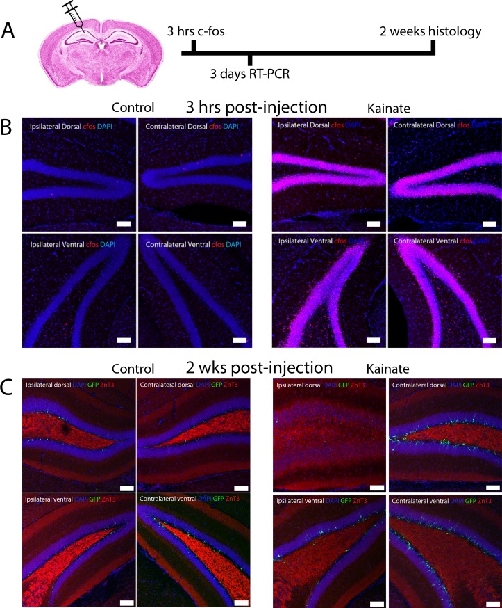
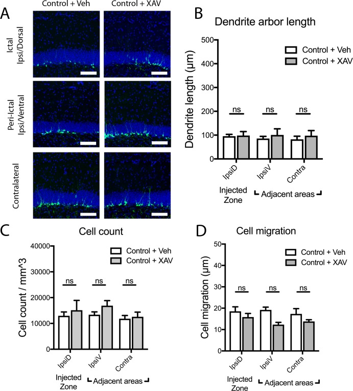
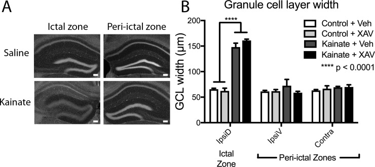
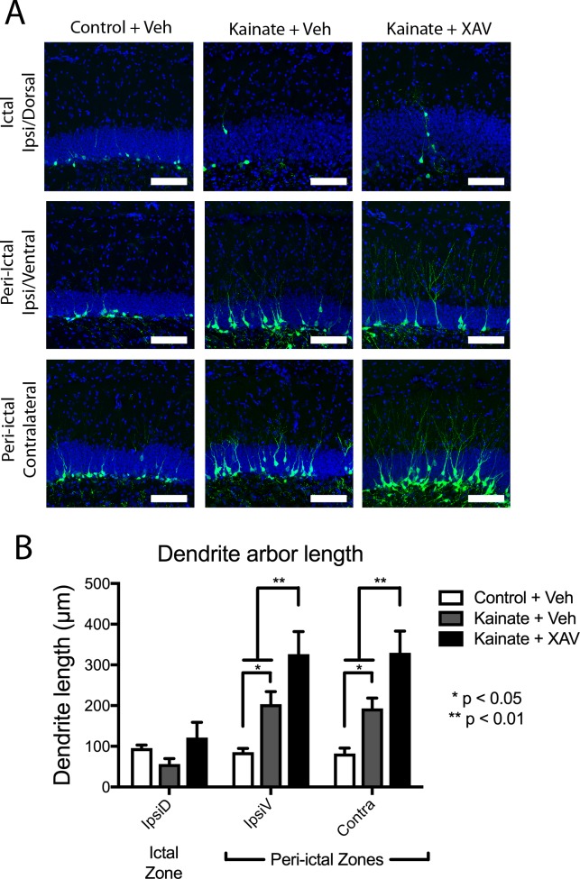
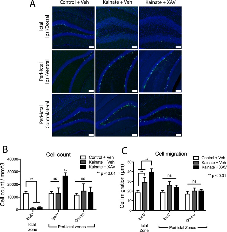
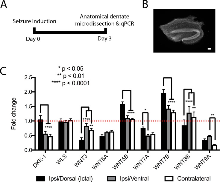
Mouse models of mesial temporal lobe epilepsy recapitulate aspects of human epilepsy, which is characterized by neuronal network remodeling in the hippocampal dentate gyrus. Observational studies suggest that this remodeling is associated with altered Wnt pathway signaling, although this has not been experimentally examined. We used the well-characterized mouse intrahippocampal kainate model of temporal lobe epilepsy to examine associations between hippocampal neurogenesis and altered Wnt signaling after seizure induction. Tissue was analyzed using immunohistochemistry and confocal microscopy, and gene expression analysis was performed by RT-qPCR on RNA extracted from anatomically micro-dissected dentate gyri. Seizures increased neurogenesis and dendritic arborization of newborn hippocampal dentate granule cells in peri-ictal regions, and decreased neurogenesis in the ictal zone, 2-weeks after kainate injection. Interestingly, administration of the novel canonical Wnt pathway inhibitor XAV939 daily for 2-weeks after kainate injection further increased dendritic arborization in peri-ictal regions after seizure, without an effect on baseline neurogenesis in control animals. Transcriptome analysis of dentate gyri demonstrated significant canonical Wnt gene dysregulation in kainate-injected mice across all regions for Wnt3, 5a and 9a. Intriguingly, certain Wnt genes demonstrated differential patterns of dysregulation between the ictal and peri-ictal zones, most notably Wnt5B, 7B and DKK-1. Together, these results demonstrate regional variation in Wnt pathway dysregulation early after seizure induction, and surprisingly, suggest that some Wnt-mediated effects might actually temper aberrant neurogenesis after seizures. The Wnt pathway may therefore provide suitable targets for novel therapies that prevent network remodeling and the development of epileptic foci in high-risk patients.


Conflict of interest statement

The authors have declared that no competing interests exist.

Figures

Fig 1
Fig 1. Intrahippocampal kainate injection causes widespread bilateral dentate granule cell activation, followed by delayed focal mossy fiber sprouting.
(A) Experimental timeline after intrahippocampal injection. (B) c-Fos expression (red) in the dentate gyri from hippocampal quadrants 3hrs after saline (control; left) or kainate (seizure; right) injection into the ipsilateral/dorsal region CA1. Images show extensive bilateral c-Fos expression 3hrs after kainate in the dentate gyrus. (C) The mossy fiber terminal marker ZnT3 is localized to the denate hilus as expected 2wks after saline injection (control; left), and localizes to the granule cell and molecular layers adjacent to the site of kainate injection in post-kainate POMC-EGFP animals. Scale bar 100μm.
Fig 2
Fig 2. Treatment with novel Wnt antagonist XAV939 does not alter newborn dentate granule cell maturation in control animals.
(A) Images demonstrate eGFP+ newborn dentate granule cells two weeks after intrahippocampal saline injection (control) in the ipsilateral dorsal, ipsilateral ventral, and contralateral dentate gyrus, of mice treated with vehicle vs. XAV939 daily for two weeks after injection. Scale bars 100μm. (B-D) In saline-injected control animals, continuous Wnt inhibition by XAV939 treatment does not alter newborn dentate granule cell arbor length (B), cell density (C), or cell migration (D), when compared with vehicle-treated control mice (ns = not significant).
Fig 3
Fig 3. Granule cell dispersion in the ictal zone is not affected by XAV939 treatment.
(A). DAPI-stained images of coronal whole dentate gyrus after saline (control) and kainate injection in the ictal (injected) and peri-ictal (non-injected) regions of the dentate gyrus. Scale bar 100μm. (B). Dentate granule cell layer dispersion is only seen in the ictal zone after seizure induction by kainate, and this was not affected by daily administration of Wnt antagonist XAV939 for two weeks after kainate.
Fig 4
Fig 4. Post-kainate increase in newborn granule cell dendrite length is augmented by Wnt inhibition.
(A) Images of POMC-EGFP+ adult-born dentate granule cell neurons from indicated hippocampal regions two weeks after intrahippocampal kainate or saline injection, followed by daily systemic treatment with XAV939 vs. vehicle control. Scale bars 100μm. (B) In peri-ictal regions, mean dendritic arbor length per eGFP+ cell increased significantly after kainate-induced seizure in peri-ictal zones, and was further increased by XAV939 treatment.
Fig 5
Fig 5. Modulation of neurogenesis and cell migration by the Wnt antagonism.
(A) POMC-EGFP+ newborn dentate granule cells 2wks after kainate-induced seizure, followed by vehicle or XAV939 treatment, in ictal and peri-ictal regions. Scale bars 100μm. (B) Newly born (POMC-EGFP+) granule cell density decreased in the ictal zone, and was not rescued by XAV939 treatment. In the ipsilateral peri-ictal region, cell density increased after XAV939 treatment only in kainate-treated mice. No XAV939-induced change in cell density was observed in the contralateral dentate. (C) Newborn dentate granule cell migration increased in the ictal zone 2wks after seizure induction, which was further increased by XAV939 treatment. Cell migration was unchanged after kainate in the peri-ictal zones.
Fig 6
Fig 6. Transcriptional profiling of the whole dentate gyrus 3 days after seizure induction.
(A) Timeline of transcriptional analysis. (B) Image of a representative DAPI-labeled cross-section of micro-dissected dentate gyrus used for transcriptional analysis. Scale bar 100μm. (C) Relative Wnt pathway gene transcription in the ictal and peri-ictal regions 3d after seizure induction. Various patterns are observed, whereby certain genes are selectively upregulated in the ictal zone (Wnt5b and Wnt7b), selectively downregulated in ictal (Wnt3) or peri-ictal regions (Dkk-1), or relatively unchanged from baseline (WLS, Wnt8b).

Similar articles

Cited by

References

    1. Frey LC. Epidemiology of posttraumatic epilepsy: a critical review. Epilepsia. 2003;44(s10):11–7. 10.1046/j.1528-1157.44.s10.4.x - DOI - PubMed
    1. Ramantani G, Holthausen H. Epilepsy after cerebral infection: review of the literature and the potential for surgery. Epileptic disorders: international epilepsy journal with videotape. 2017;19(2):117–36. 10.1684/epd.2017.0916 - DOI - PubMed
    1. Myint PK, Staufenberg EF, Sabanathan K. Post-stroke seizure and post-stroke epilepsy. Postgraduate medical journal. 2006;82(971):568–72. 10.1136/pgmj.2005.041426 - DOI - PMC - PubMed
    1. French JA. Febrile seizures: possible outcomes. Neurology. 2012;79(9):e80–2. Epub 2012/08/29. 10.1212/WNL.0b013e31826aa902 . - DOI - PubMed
    1. French JA. Refractory epilepsy: clinical overview. Epilepsia. 2007;48 Suppl 1:3–7. 10.1111/j.1528-1167.2007.00992.x - DOI - PubMed

Publication types

MeSH terms