. 2019 Oct 9;14(10):e0223725.
doi: 10.1371/journal.pone.0223725. eCollection 2019.

Defective base excision repair in the response to DNA damaging agents in triple negative breast cancer

Affiliations

Defective base excision repair in the response to DNA damaging agents in triple negative breast cancer

Kevin J Lee et al. PLoS One. .

Abstract

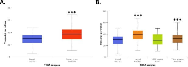
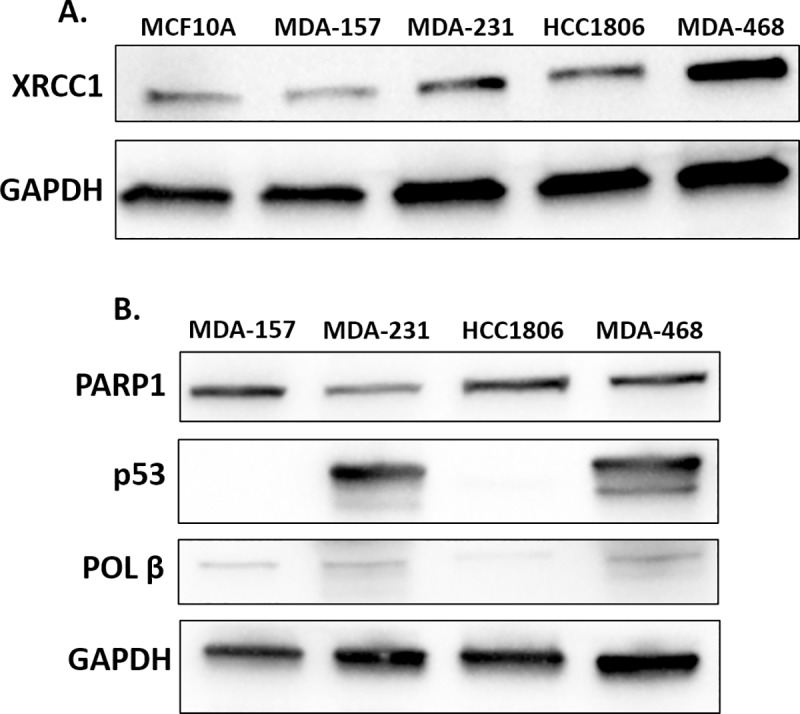
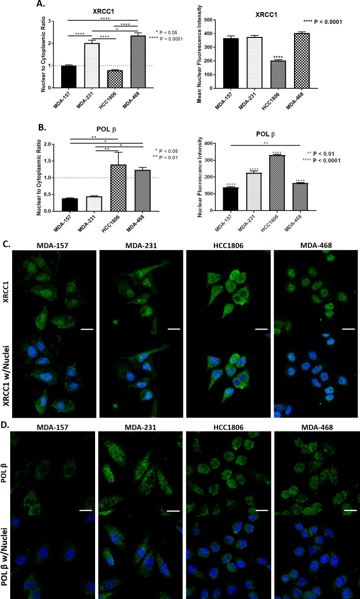
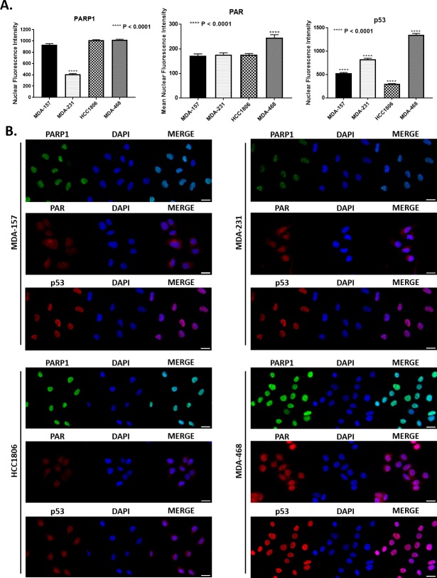
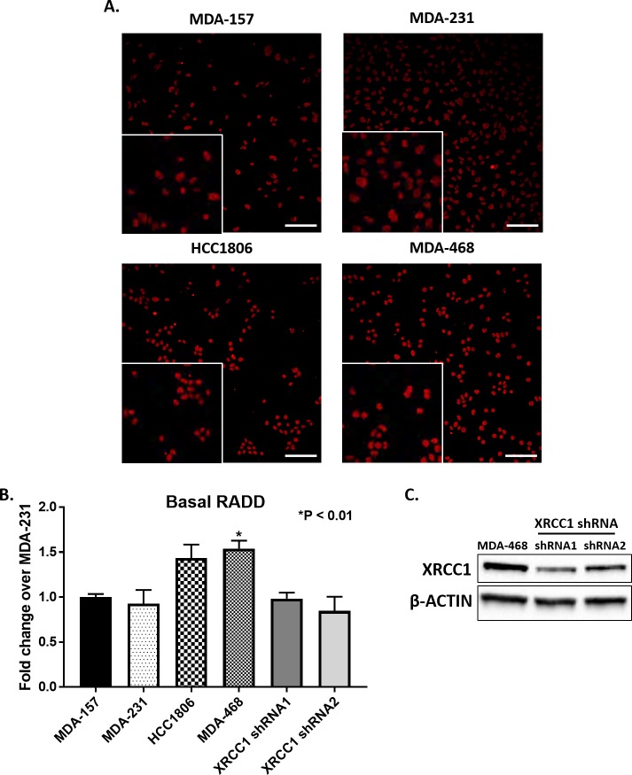
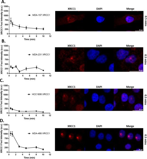
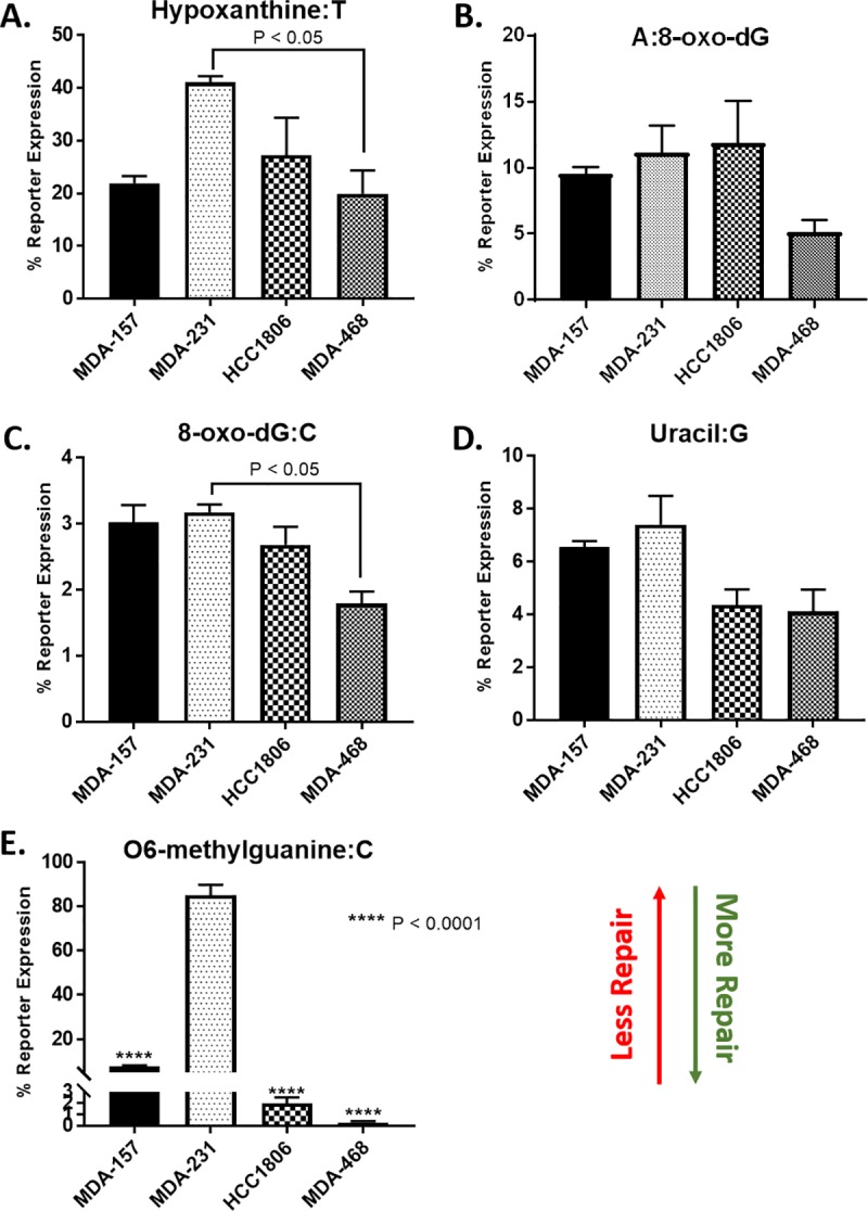
DNA repair defects have been increasingly focused on as therapeutic targets. In hormone-positive breast cancer, XRCC1-deficient tumors have been identified and proposed as targets for combination therapies that damage DNA and inhibit DNA repair pathways. XRCC1 is a scaffold protein that functions in base excision repair (BER) by mediating essential interactions between DNA glycosylases, AP endonuclease, poly(ADP-ribose) polymerase 1, DNA polymerase β (POL β), and DNA ligases. Loss of XRCC1 confers BER defects and hypersensitivity to DNA damaging agents. BER defects have not been evaluated in triple negative breast cancers (TNBC), for which new therapeutic targets and therapies are needed. To evaluate the potential of XRCC1 as an indicator of BER defects in TNBC, we examined XRCC1 expression in the TCGA database and its expression and localization in TNBC cell lines. The TCGA database revealed high XRCC1 expression in TNBC tumors and TNBC cell lines show variable, but mostly high expression of XRCC1. XRCC1 localized outside of the nucleus in some TNBC cell lines, altering their ability to repair base lesions and single-strand breaks. Subcellular localization of POL β also varied and did not correlate with XRCC1 localization. Basal levels of DNA damage correlated with observed changes in XRCC1 expression, localization, and measure repair capacity. The results confirmed that XRCC1 expression changes indicate DNA repair capacity changes but emphasize that basal DNA damage levels along with protein localization are better indicators of DNA repair defects. Given the observed over-expression of XRCC1 in TNBC preclinical models and tumors, XRCC1 expression levels should be assessed when evaluating treatment responses of TNBC preclinical model cells.


Conflict of interest statement

The authors have declared that no competing interests exist.

Figures

Fig 1
Fig 1. TCGA analysis of XRCC1.
A) TCGA analysis using the UALCAN web interface [23] revealed XRCC1 transcript expression to be significantly higher in breast tumor tissue over normal tissue (p = 1.6 x 10⁻¹², Primary tumor to Normal). B) XRCC1 was increased in Luminal (p < 1 x 10⁻¹², Luminal to Normal) and TNBC (p < 0.001, TNBC to Normal) tumor types compared to normal tissue using the same analysis interface. *** P < 0.001.
Fig 2
Fig 2. Base excision repair proteins in TNBC cell lines.
A) Lysates of MCF10A, MDA-157, MDA-231, HCC1806, and MDA-468 were probed by immunoblot for the expression of XRCC1 with GAPDH serving as loading control. B) Lysates of MDA-MB-157 (MDA-157), MDA-MB-231 (MDA-231), HCC1806, and MDA-MB-468 (MDA-468) were probed by immunoblot for the expression of PARP1, p53, POL β, and GAPDH as a loading control.
Fig 3
Fig 3. Subcellular localization and quantification of BER proteins.
A) Left, XRCC1 nuclear to cytoplasmic ratio (N/C) is reported as 1 representing equal distribution (MDA-157), > 1 representing nuclear localization (MDA-231 and MDA-468), and < 1 representing nuclear exclusion (HCC1806). (**** P < 0.0001, MDA-157 to MDA-231, MDA-157 to MDA-468, MDA-231 to HCC1806, HCC1806 to MDA-468; * P < 0.05, MDA-231 to MDA-468). Right, mean fluorescence intensity of XRCC1. (**** P < 0.0001, HCC1806 to MDA-157, MDA-231, and MDA-468) B) Left, N/C ratio of POL β. (** P < 0.01, MDA-157 to HCC1806, MDA-231 to HCC1806; * P < 0.05, MDA-157 to MDA-468, MDA-231 to MDA-468) Right, mean fluorescence intensity of POL β. (**** P < 0.0001, MDA-157 to MDA-231, MDA-157 to HCC1806, MDA-231 to HCC1806, MDA-231 to MDA-468, HCC1806 to MDA-468; ** P < 0.01, MDA-157 to MDA-468) C) Representative images of localization of XRCC1 in TNBC cell lines. D) Representative images of localization of POL β in TNBC cell lines. Scale bar = 20 μm.
Fig 4
Fig 4. Nuclear intensity of key BER proteins.
A) Quantification of mean nuclear fluorescence intensity is shown for PARP1 (**** P < 0.0001, MDA-231 to MDA-157, HCC1806, and MDA-468), PAR (**** P < 0.0001, MDA-468 to MDA-157, MDA-231, and HCC1806), and p53 (**** P < 0.0001, MDA-157 to MDA-231, MDA-157 to MDA-468, MDA-157 to HCC1806, MDA-231 to MDA-468, MDA-231 to HCC1806, and MDA-468 to HCC1806). B) Representative fluorescent images used for quantification in A for PARP1, PAR, and p53 for all cell lines tested. Scale bar = 20 μm.
Fig 5
Fig 5. Basal DNA damage in TNBC cell lines.
A) Representative images of basal levels of DNA damage as measured by RADD in MDA-157, MDA-231, HCC1806, MDA-468 with insets showing blown up cropped images. Scale bar = 100 μm. B) Quantification of images in A normalized to MDA-231 show an increase in DNA damage in HCC1806, and a significantly higher amount (P < 0.01, MDA-468 to MDA-231) of DNA damage for MDA-468. MDA-468 cells with XRCC1 knockdown show decreased levels of basal DNA damage. C) Expression of XRCC1 in MDA-468 and MDA-468 XRCC1 knockdown cells.
Fig 6
Fig 6. XRCC1 recruitment following 355 nm laser microirradiation in TNBC cell lines.
Microirradiation with a 355 nm laser occurred and cells were fixed at the indicated time points and processed for immunofluorescence of XRCC1. Quantification over time is shown on the left with a representative image from 0.5 min shown on the right for A) MDA-157, B) MDA-231, C) HCC1806, and D) MDA-468. A minimum of 18 cells were irradiated over three separate experiments and quantified as described in the materials and methods section and reported as XRCC1 Foci Intensity in arbitrary units (a.u.). Scale bar = 20 μm.
Fig 7
Fig 7. FM-HCR analysis of DNA repair capacity in TNBC cell lines.
Cells were transfected with fluorescent reporter plasmids containing the indicated DNA lesions, A) Hypoxanthine:T (P < 0.05, MDA-231 to MDA-468), B) A:8-oxo-dG, C) 8-oxo-dG:C (P < 0.05, MDA-231 to MDA-468), D) Uracil:G, E) O6-methylguanine:C (**** P < 0.0001, MDA-157 to MDA-231, HCC1806 to MDA-231, MDA-468 to HCC1806), as well as an undamaged plasmid to normalize for transfection efficiency. DNA repair capacity is inversely proportional to % reporter expression.

Similar articles

Cited by

References

    1. Li SX, Sjolund A, Harris L, Sweasy JB. DNA repair and personalized breast cancer therapy. Environ Mol Mutagen. 2010;51(8–9):897–908. 10.1002/em.20606 - DOI - PMC - PubMed
    1. Wolf DM, Yau C, Sanil A, Glas A, Petricoin E, Wulfkuhle J, et al. DNA repair deficiency biomarkers and the 70-gene ultra-high risk signature as predictors of veliparib/carboplatin response in the I-SPY 2 breast cancer trial. NPJ Breast Cancer. 2017;3:31 10.1038/s41523-017-0025-7 - DOI - PMC - PubMed
    1. Carter SL, Eklund AC, Kohane IS, Harris LN, Szallasi Z. A signature of chromosomal instability inferred from gene expression profiles predicts clinical outcome in multiple human cancers. Nat Genet. 2006;38(9):1043–8. 10.1038/ng1861 . - DOI - PubMed
    1. Karahalil B, Bohr VA, Wilson DM, 3rd. Impact of DNA polymorphisms in key DNA base excision repair proteins on cancer risk. Hum Exp Toxicol. 2012;31(10):981–1005. 10.1177/0960327112444476 - DOI - PMC - PubMed
    1. Abdel-Fatah TM, Russell R, Agarwal D, Moseley P, Abayomi MA, Perry C, et al. DNA polymerase beta deficiency is linked to aggressive breast cancer: a comprehensive analysis of gene copy number, mRNA and protein expression in multiple cohorts. Mol Oncol. 2014;8(3):520–32. 10.1016/j.molonc.2014.01.001 - DOI - PMC - PubMed

Publication types

MeSH terms

Substances